Journal of Rural Medicine
Original article
Characteristics of patients with a diagnosis of sarcoidosis:
a comparison of the 2006 and 2015 versions of diagnostic
criteria for sarcoidosis in Japan
Noritaka Sakamoto1, Michiru Sawahata1, Yoshitaka Yamanouchi1, Satoshi Konno2, Noriharu Shijubo3, Tetsuo Yamaguchi4, Yosikazu Nakamura5, Takuji Suzuki1, Koichi Hagiwara1, and Masashi Bando1
1Division of Pulmonary Medicine, Department of Medicine, Jichi Medical University, Japan
2Department of Respiratory Medicine, Faculty of Medicine and Graduate School of Medicine, Hokkaido University, Japan 3Department of Respiratory Medicine, JR Sapporo Hospital, Japan
4Shinjuku Tsurukame Clinic, Japan
5Department of Public Health, Jichi Medical University, Japan
Abstract
Objective: Histological verification of epithelioid cell granuloma is important in diagnosing sarcoidosis; tissue sampling is a
worldwide requirement. In 2006, to reduce medical expenses and avoid invasive procedures, diagnostic criteria without histological verification were permitted by the Japanese government. In 2015, new diagnostic criteria, allowed clinical diagnoses based on only respiratory, ocular, and cardiac systems with at least a two-system involvement, increasing the need to sample tissue from clinically unevaluable organs in suspected sarcoidosis. This study aimed to compare the characteristics of patients who were diagnosed with sarcoidosis according to the 2006 and 2015 criteria.
Materials and Methods: Using the 2015 version, we re-evaluated the characteristics of 264 patients with diagnosed or suspected
sarcoidosis according to the 2006 criteria, at Jichi Medical University Hospital between 2004 and 2012 (clinical diagnosis, 84; histological diagnosis, 117; suspected sarcoidosis 63).
Results: Thirty-nine patients were diagnosed with suspected sarcoidosis due to the absence of at least a two-system involvement;
two patients had insufficient laboratory data suggestive of sarcoidosis. Six patients moved from suspected sarcoidosis to a histologi-cal diagnosis because of a greater leniency in the criteria for supportive findings. The 2015 diagnostic criteria excluded patients with organ involvement without a requirement for systemic steroids from the clinical diagnosis group. A case of schwannoma, er-roneously placed in the clinical diagnosis group by the 2006 criteria, was reclassified according to the 2015 criteria.
Conclusion: The 2015 version is preferable for clinically diagnosing sarcoidosis, even without histological specimens, and provides
guidance for indications for systemic treatment.
Key words: sarcoidosis, diagnosis, treatment, epidemiology
(J Rural Med 2021; 16(2): 77–82)
Introduction
Sarcoidosis is a granulomatous disease that causes vary-ing types of lesions in different organs throughout the body, particularly in the respiratory organs. Some patients under-go spontaneous remission, but the prolongation and exac-erbation of symptoms are common concerns. Poor progno-sis is also associated with the progression of latent lesions, pulmonary fibrogenesis, and the involvement of the cardiac and central nervous system. Because of the wide range of organ involvement, severity of the disease, and its clinical course, it is difficult to define organ involvement by uniform approaches, and thus the establishment of reliable
diag-Received: May 8, 2020 Accepted: November 16, 2020
Correspondence: Michiru Sawahata, Division of Pulmonary Med-icine, Jichi Medical University, 3311-1 Yakushiji, Shimotsuke, Tochigi 329-0498, Japan
E-mail: sawahata@jichi.ac.jp
This is an open-access article distributed under the terms of the Creative Commons Attribution Non-Commercial No Derivatives (by-nc-nd) License <http://creativecommons.org/ licenses/by-nc-nd/4.0/>.
Journal of Rural Medicine
nostic criteria have been an important task in the clinical management of sarcoidosis. Histological verification of the epithelioid cell granuloma is important in the diagnosis of sarcoidosis, and tissue sampling is a diagnostic requirement worldwide. To assist with medical expenses and to avoid in-vasive experimental procedures, the Japanese government previously allowed the development of clinical diagnostic criteria without using any histological verification [Japan Society of Sarcoidosis and Other Granulomatous Disorders (JSSOG) 2006 Diagnostic Criteria and Guideline for
Sar-coidosis] (JSSOG 2006 criteria)1).
In 2015, a new version of diagnostic criteria was pro-posed by the Japanese Ministry of Health, Labour and Welfare [JSSOG 2015 Diagnostic Criteria and Guidelines
for Sarcoidosis] (JSSOG 2015 criteria)2). The new criteria,
allowed the clinical evaluation of only respiratory, ocular, and cardiac organs, and required the involvement of at least two of these for clinical diagnosis. The involvement of these three organs, from which it is difficult to collect tissue speci-mens, are relatively commonly seen in Japan. In a previ-ous epidemiologic study, we showed that in addition to lung involvement, which was present in almost all the patients, involvement of the eyes, extrathoracic lymph nodes, skin, and heart was observed in 68.0%, 19.6%, 8.8%, and 2.4% of
patients, respectively3). This change in the diagnostic
crite-ria of sarcoidosis increased the need to sample tissue from potentially involved organs that cannot be evaluated clini-cally in patients with suspected sarcoidosis.
Against this background, we aimed in the present study to compare the characteristics of patients who were diag-nosed with sarcoidosis according to both the JSSOG 2006 and JSSOG 2015 criteria.
Materials and Methods
Study population
We retrospectively reviewed the medical records of 264 inpatients and/or outpatients who were newly diagnosed or suspected to have sarcoidosis at Jichi Medical University Hospital between February 2004 and August 2012 (84 with clinical diagnosis, 117 with histological diagnosis, and 63 with suspected sarcoidosis) based on the JSSOG 2006
cri-teria1). Some of the patients were the same as those included
in our previous study3, 4). We re-evaluated these patients
ac-cording to the JSSOG 2015 criteria, and compared the dif-ferences in their characteristics between the two criteria. This was a retrospective study, and it was difficult to ob-tain consent from patients and family members. Therefore, this study was reviewed and approved by the Jichi Medical University Ethics Committee in the form of “Substituting an opt-out consent form (posted on the website)” (No. Rin-A18-190, February 13, 2019).
JSSOG 2006 Diagnostic criteria and guidelines for sarcoidosis
These criteria focus on definitive diagnoses based on
biopsies and clinical findings1). The criteria are consistent
with the concepts of the American Thoracic Society/Euro-pean Respiratory Society/World Association of Sarcoidosis and Other Granulomatous Disorders statement on
sarcoid-osis adopted in 19995), which describe the clinical features
of sarcoidosis, including radiological, laboratory, and path-ological findings (noncaseous epithelioid cell granuloma), and differential diagnoses. Without histological evidence, a clinically proven diagnosis was made based on: (1) clini-cal features suggesting sarcoidosis-like lesions in at least two organs; and (2) at least two clinical features suggest-ing a systemic reaction, such as bilateral hilar-mediastinal lymphadenopathy (BHL), elevated serum angiotensin-con-verting enzyme (ACE), hypercalcemia, negative tuberculin test, abnormal uptake on gallium-67 citrate scintigraphy, elevated lymphocyte count, and elevated CD4/CD8 ratio in bronchoalveolar lavage fluid.
JSSOG 2015 Diagnostic criteria and guidelines for sarcoidosis
The followings2) are the key changes made to the 2006
criteria:
(1) A clinically proven diagnosis with no histological evi-dence can be made based on the clinical evaluation of only respiratory, ocular, and cardiac organs, and it re-quires the involvement of at least two of these organs for a clinical diagnosis.
(2) There is a greater leniency for supportive findings (clini-cal and laboratory findings strongly suggestive of organ involvement), when a biopsy-proven diagnosis is ob-tained; and an increase in the need to sample tissue from the potentially involved organs other than the ocular and cardiac organs that are clinically unevaluable in patients with suspected sarcoidosis.
(3) The characteristic test results, in addition to BHL, are: elevated serum ACE activity, elevated serum lysozyme level, elevated serum soluble interleukin 2 receptor level, marked uptake on gallium-67 citrate scintigraphy or fluorine-18 fluorodeoxyglucose PET, and an elevated lymphocyte rate or elevated CD4/CD8 ratio ≥ 3.5. The requirement for a negative tuberculin test and hypercal-cemia was removed.
Organ involvement assessment
Respiratory, ocular, and cardiac involvement deter-mined at the time of clinical diagnosis by respiratory physi-cians, ophthalmologists, and cardiologists, respectively, was retrospectively evaluated in each patient, according to the
JSSOG 2015 criteria2) based on case history, physical
deter-Journal of Rural Medicine
mined from laboratory findings that were closest to the time of diagnosis during admission.
For the clinical evaluation of organ involvement other than respiratory, ocular, and cardiac involvement, the World Association of Sarcoidosis and Other Granulomatous
Dis-orders (WASOG) sarcoidosis organ assessment instrument6)
was used: organ involvement was confirmed when the cri-teria for “highly probable” or “at least probable” were met.
Results
Changes in the characteristics of subjects diagnosed with sarcoidosis according both the JSSOG 2006 and JSSOG 2015 criteria
When the 2015 criteria were applied, 43 patients had a clinical diagnosis, 123 with a histological diagnosis, and 98 with suspected sarcoidosis (Figure 1). Forty-one patients (39 patients with the absence of at least two involved organs, and
two patients without sufficient laboratory data suggestive of sarcoidosis), were moved from the clinical diagnosis group to the suspected sarcoidosis group; resulting in a decrease in the clinical diagnosis group from 84 cases to 43 cases. Six patients moved from the suspected sarcoidosis group to the histological diagnosis group, resulting in an increase in the histological diagnosis group from 117 cases to 123 cases.
Of the 123 patients in the histological diagnosis group based on the 2015 criteria, 49 (39.8%) had organ involve-ment other than respiratory organs.
Cases moved from the clinical diagnosis group to the suspected sarcoidosis group due to absence of at least two-system involvement
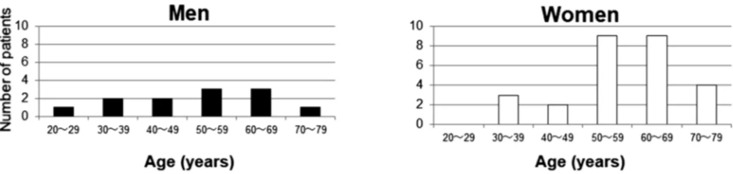
Figure 2 shows the age and sex distribution of the 39 cases that moved from the clinical diagnosis group to the suspected sarcoidosis group due to the absence of at least two-system involvement. They consisted of 12 men and
Figure 1 Changes in the characteristics of subjects diagnosed as having sarcoidosis according to the two versions of
diagnostic criteria in Japan, proposed in 2006 and 2015.
Re-evaluation revealed diagnoses consistent with clinical diagnosis in 43 patients, histological diagnosis in 123 patients, and suspected sarcoidosis in 98 patients. Thirty-nine patients moved from the clinical diagno-sis group to the suspected sarcoidodiagno-sis group due to the absence of at least two-system involvement; two pa-tients had insufficient laboratory data suggestive of sarcoidosis. Six papa-tients moved from the suspected sar-coidosis group to the histological diagnosis group due to greater leniency of criteria for supportive findings.
Figure 2 Distribution of 39 cases moved from the clinical diagnosis group to the suspected sarcoidosis group due to absence of at least
two-system involvement, by age group at diagnosis.
This figure shows the age and sex distribution of 39 cases that moved from the clinical diagnosis group to the suspected sarcoidosis group due to the absence of at least two-system involvement. They consisted of 12 men and 27 women.
Journal of Rural Medicine
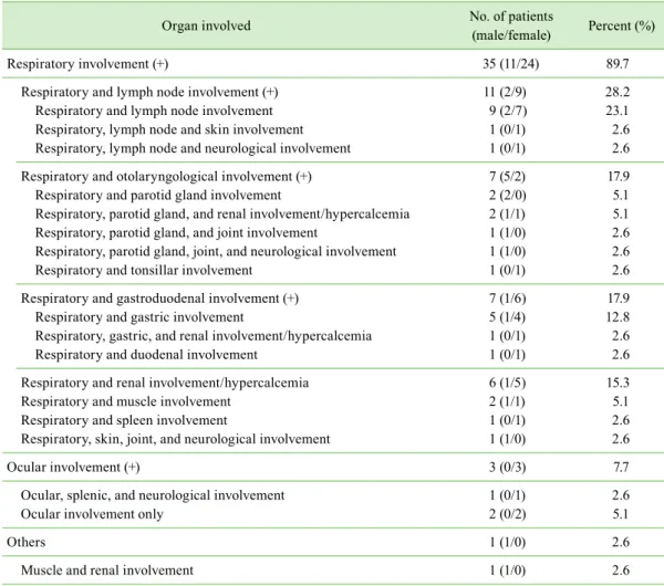
27 women. Of the 39 patients, 35 (89.7%) had respiratory involvement but no ocular or cardiac involvement, three (7.7%) had no respiratory involvement but had ocular or car-diac involvement, and one (2.6%) had no respiratory, ocular, or cardiac involvement (Table 1).
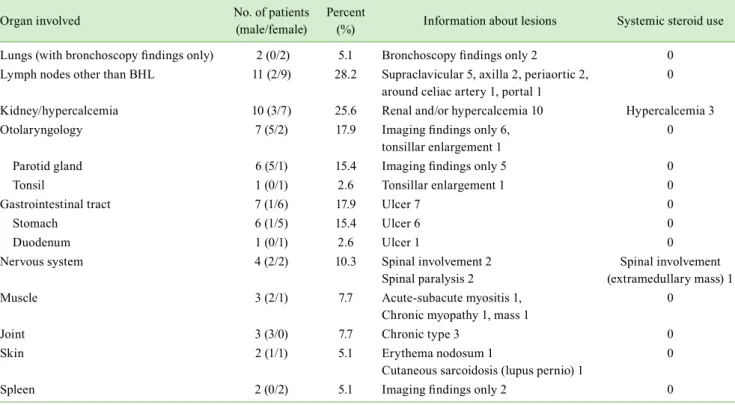
The frequency of the organs involved, which was not considered in the diagnosis of sarcoidosis in 39 patients, was as follows (Table 2): the lungs (no findings other than bronchoscopy findings strongly suggestive of sarcoidosis) in two cases (5.1%), lymph nodes other than BHL in 11 cases (28.2%), renal involvement/hypercalcemia in 10 cas-es (25.6%), otolaryngology organs in seven cascas-es (17.9%), stomach or duodenum in seven cases (17.9%), nervous system organ in four cases (10.3%), muscle in three cases (7.7%), joints in three cases (7.7%), skin in two cases (5.1%), and spleen in two cases (5.1%).
Most of the involved lesions ignored during clini-cal evaluation under the 2015 criteria did not require any treatment, whereas three patients with renal involvement/ hypercalcemia and one patient with neurological involve-ment were treated with systemic steroids. The latter was an
intradural extramedullary tumor, which was revealed to be a schwannoma after the biopsy following unresponsive ste-roid treatment.
Cases moved from the suspected sarcoidosis group to the histological diagnosis group
Six patients moved from the suspected sarcoidosis group to the histological diagnosis group owing to a greater leni-ency in the criteria for supportive findings (clinical and lab-oratory findings strongly suggestive of organ involvement).
Discussion
To the best of our knowledge, this is the first epidemio-logical study that compared the characteristics of subjects diagnosed with sarcoidosis according to the two diagnostic criteria in Japan, proposed in 2006 and 2015. There are three important observations from this study. First, use of the 2015 criteria instead of the 2006 criteria resulted in an increase in the number of patients in the histological diagnosis group with fewer patients in the clinical diagnosis group.
Thirty-Table 1 Thirty-nine cases moved from the clinical diagnosis group to the suspected sarcoidosis group due to
ab-sence of at least two-system involvement
Organ involved No. of patients (male/female) Percent (%)
Respiratory involvement (+) 35 (11/24) 89.7
Respiratory and lymph node involvement (+) 11 (2/9) 28.2
Respiratory and lymph node involvement 9 (2/7) 23.1
Respiratory, lymph node and skin involvement 1 (0/1) 2.6
Respiratory, lymph node and neurological involvement 1 (0/1) 2.6 Respiratory and otolaryngological involvement (+) 7 (5/2) 17.9
Respiratory and parotid gland involvement 2 (2/0) 5.1
Respiratory, parotid gland, and renal involvement/hypercalcemia 2 (1/1) 5.1 Respiratory, parotid gland, and joint involvement 1 (1/0) 2.6 Respiratory, parotid gland, joint, and neurological involvement 1 (1/0) 2.6
Respiratory and tonsillar involvement 1 (0/1) 2.6
Respiratory and gastroduodenal involvement (+) 7 (1/6) 17.9
Respiratory and gastric involvement 5 (1/4) 12.8
Respiratory, gastric, and renal involvement/hypercalcemia 1 (0/1) 2.6
Respiratory and duodenal involvement 1 (0/1) 2.6
Respiratory and renal involvement/hypercalcemia 6 (1/5) 15.3
Respiratory and muscle involvement 2 (1/1) 5.1
Respiratory and spleen involvement 1 (0/1) 2.6
Respiratory, skin, joint, and neurological involvement 1 (1/0) 2.6
Ocular involvement (+) 3 (0/3) 7.7
Ocular, splenic, and neurological involvement 1 (0/1) 2.6
Ocular involvement only 2 (0/2) 5.1
Others 1 (1/0) 2.6
Journal of Rural Medicine
nine patients had a clinical diagnosis of suspected sarcoid-osis due to the absence of at least a two-system involvement; two patients had insufficient laboratory data suggestive of sarcoidosis. Six patients moved from suspected sarcoidosis to histological diagnosis owing to a greater leniency in the criteria for supportive findings. Second, the 2015 diagnostic criteria successfully excluded patients with organ involve-ment that did not require the use of systemic steroids, from the clinical diagnosis group. Third, a case of schwannoma, erroneously placed in the clinical diagnosis group by the 2006 criteria, was successfully reclassified according to the 2015 criteria.
Sarcoidosis is regarded as an amplified and persistent granulomatous reaction to inhaled antigens that develops when an individual with a genetic predisposition encoun-ters environmental change. Defining organ involvement by unique detection approaches has been difficult because of considerable variations in clinical course, organ involve-ment, and severity, and thus establishing reliable diagnostic criteria has been an important task in the clinical manage-ment of sarcoidosis. Histological evidence for epithelioid cell granulomas is important in the diagnosis of sarcoidosis, and the collection of specimens is recommended as stated by the American Thoracic Society/European Respiratory
Society/WASOG in 19995). In Japan, on the other hand, the
clinical diagnosis group was added to the histological
di-agnosis group because sarcoidosis is a designated disease approved for medical expense subsidy, and patients who re-quire this support need to be clearly defined. The diagnostic criteria established by the Ministry of Health, Labour and Welfare in 1976 have not been revised, while the society’s
diagnostic criteria were revised in 20061). In January 2015,
new diagnostic criteria were established by JSSOG together with the Diffuse Lung Disease Group from the Ministry of
Health, Labour and Welfare2).
When considering the first observation made in this study, forty-one patients moved from the clinical diagnosis group of criteria 2006 to the suspected sarcoidosis group of criteria 2015, resulting in a decrease in the clinical diagnosis group from 84 cases to 43 cases. Six patients moved from the suspected sarcoidosis group of criteria 2006 to the his-tological diagnosis group of criteria 2015, resulting in an in-crease in the histological diagnosis group from 117 cases to 123 cases. This means that the goal for establishing the 2015 criteria, which recommends collection of specimens for his-tological diagnosis when involvement of organs is highly likely, was met. It is noteworthy that, of the 123 patients in the histological diagnosis group by the 2015 diagnostic criteria, 49 (39.8%) had involvement of organs other than respiratory organs, suggesting that proactive cooperation with physicians in corresponding departments is necessary to judge the possibility of a histological diagnosis.
Table 2 Organ involvements ignored in clinical evaluation under JSSOG 2015 criteria: examples of 39 cases moved from the clinical diagnosis
group to the suspected sarcoidosis group due to absence of at least two-system involvement
Organ involved No. of patients (male/female) Percent (%) Information about lesions Systemic steroid use Lungs (with bronchoscopy findings only) 2 (0/2) 5.1 Bronchoscopy findings only 2 0
Lymph nodes other than BHL 11 (2/9) 28.2 Supraclavicular 5, axilla 2, periaortic 2,
around celiac artery 1, portal 1 0 Kidney/hypercalcemia 10 (3/7) 25.6 Renal and/or hypercalcemia 10 Hypercalcemia 3
Otolaryngology 7 (5/2) 17.9 Imaging findings only 6,
tonsillar enlargement 1
0
Parotid gland 6 (5/1) 15.4 Imaging findings only 5 0
Tonsil 1 (0/1) 2.6 Tonsillar enlargement 1 0
Gastrointestinal tract 7 (1/6) 17.9 Ulcer 7 0
Stomach 6 (1/5) 15.4 Ulcer 6 0
Duodenum 1 (0/1) 2.6 Ulcer 1 0
Nervous system 4 (2/2) 10.3 Spinal involvement 2
Spinal paralysis 2
Spinal involvement (extramedullary mass) 1
Muscle 3 (2/1) 7.7 Acute-subacute myositis 1,
Chronic myopathy 1, mass 1 0
Joint 3 (3/0) 7.7 Chronic type 3 0
Skin 2 (1/1) 5.1 Erythema nodosum 1
Cutaneous sarcoidosis (lupus pernio) 1
0
Spleen 2 (0/2) 5.1 Imaging findings only 2 0
Journal of Rural Medicine
Concerning the second and third observations, out of the 39 patients moved from the clinical diagnosis group to the suspected sarcoidosis group due to the absence of at least two-system involvement, 35 patients did not require sys-temic steroid treatment. Only three patients with renal in-volvement/hypercalcemia and one patient with neurological involvement were treated with systemic steroids. The latter was an intradural extramedullary tumor, erroneously in-cluded as neurological involvement of the clinical diagnosis group by the 2006 criteria, which was successfully reclas-sified by the 2015 criteria. The improvement in quality of life and avoidance of hazardous prognosis are key factors
to consider when determining the need for treatment5). In
Japan, patients with a pulmonary lesion across a wide area with subjective symptoms, ocular involvement resistant to topical treatment, and cardiac and neurological involvement
are candidates for systemic steroids7). Histological
diagno-ses of ocular and cardiac involvement is particularly diffi-cult, and it is plausible that clinical diagnoses can be made for these involvements. Given the requirement for systemic steroids and difficulties in histological diagnosis, neurologi-cal involvement may also be better diagnosed clinineurologi-cally; this should be investigated in the future.
This study has some limitations, including potential se-lection bias because of the limited number of patients, all of whom visited the respiratory center of a single university hospital in the Tochigi Prefecture. Therefore, the results of this study might not be generalizable to the entire Japanese population. The subjects of this study included all patients with suspected sarcoidosis admitted to the respiratory medi-cine department or who underwent bronchoscopy between
2004 and 2013 at our university hospital, but did not include outpatients who underwent close examination.
Conclusion
The results of our current study indicate that the 2015 criteria may be more suitable for identifying patients with sarcoidosis based on clinical diagnoses, even without histo-logical specimens. The new criteria provides a useful guide for patients’ follow-up strategies, and for evaluating the in-dications for systemic treatment, when pathological speci-mens cannot be obtained. The validity of the 2015 criteria should be evaluated, and the epidemiology of sarcoidosis in Japan should be clarified by future studies.
Author contributions: NS and MS designed the study and wrote the initial draft of the manuscript. NS, MS, YY, and YN contributed to the collection of data. MB, KH, SK, and TY assisted the preparation of the manuscript. All au-thors contributed to data analysis and interpretation and critically reviewed the manuscript. All authors have read and approved the final manuscript.
Financial support: This study was supported by a grant to the Diffuse Lung Diseases Research Group from the Ministry of Health, Labour and Welfare, Japan. It was also partly supported by a grant from the Academic Award from the JSSOG in 2018 (MS), and by Jichi Medical University Alumni “2019 Itokai Research Project Grant” in 2018 (MS).
Conflicts of interest: We report no conflicts of interest.
References
1. [The Japanese Society of Sarcoidosis and Other Granulomatous Disorders (JSSOG) Diagnostic criteria and guidelines for sarcoidosis-2006.] Jpn J Sarcoid-osis/Other Granulomatous Disorders. 2007; 22: 89–101.
2. [The Japanese Society of Sarcoidosis and Other Granulomatous Disorders (JSSOG) Diagnostic Standard and Guideline for Sarcoidosis-2015]: <http:// jssog.com/www/top/shindan/shindan2-1new.html>.
3. Sawahata M, Sugiyama Y, Nakamura Y, et al. Age-related and historical changes in the clinical characteristics of sarcoidosis in Japan. Respir Med 2015; 109: 272–278. [Medline] [CrossRef]
4. Yamanouchi Y, Sawahata M, Sakamoto N, et al. Characteristics of 68 patients with clinically proven sarcoidosis based on the Japan Society of Sarcoidosis and Other Granulomatous Disorders 2015 criteria. Respir Investig 2020; 58: 102–109. [Medline] [CrossRef]
5. Statement on sarcoidosis. Joint Statement of the American Thoracic Society (ATS), the European Respiratory Society (ERS) and the World Association of Sarcoidosis and Other Granulomatous Disorders (WASOG) adopted by the ATS Board of Directors and by the ERS Executive Committee, February 1999. Am J Respir Crit Care Med 1999; 160: 736–755. [Medline]
6. Judson MA, Costabel U, Drent M, et al. The WASOG Sarcoidosis Organ Assessment Instrument: an update of a previous clinical tool. Sarcoidosis Vasc Diffuse Lung Dis 2014; 31: 19–27. [Medline]