INTRODUCTION
The technique of generating new bone by grad-ual distraction of a callus was first described in 1905 by Codivilla (1). The early procedures were complicated by edema, skin necrosis, infection, de-viation, and delayed ossification of the expanded segment (2). This technique remained undeveloped until Ilizarov rekindled interest in the process of bone lengthening (3-5), and several studies reported significant advances in reducing the incidence of
complications associated with bone lengthening (6-10). Bone lengthening by distraction osteogenesis has been successfully applied to the enchondral bone of the upper and lower extremities.
In the craniofacial region, the initial description of the distraction osteogenesis technique should be credited to Rosental, for bone lengthening of the man-dible in a microgenia patient in 1927, and Wassmund, for the clinical advancement of a maxilla in a pa-tient with hypoplasia of the upper jaw in 1926 (11). However, distraction osteogenesis only really came to be seen as a promising and popular method in the reconstruction of the membranous bones of the human craniofacial skeleton after McCarthy et
al. reported in Western literature the clinical appli-cation of mandibular lengthening by gradual dis-traction in patients with hemifacial microsomia and
REVIEW
Advances in distraction techniques for craniofacial surgery
Kazuya Matsumoto
1, Hideki Nakanishi
1, Yoshiaki Kubo
2, Masahiko Yokozeki
3,
and Keiji Moriyama
31
Departments of Plastic and Reconstructive Surgery, and2
Department of Dermatology, The Uni-versity of Tokushima School of Medicine, Tokushima, Japan ; and 3
Department of Orthodontics, The University of Tokushima School of Dentistry, Tokushima, Japan
Abstract : Distraction osteogenesis has been applied to the craniofacial skeleton as well as the long bones of the extremities. This technique does not require bone grafting and allows correction of craniofacial deformities with less invasion. Moreover, the distraction pro-cedures can expand the overlying soft tissues simultaneously. We determined the indica-tions of distraction osteogenesis, analyzed the types of devices available, and examined patients treated with distraction for the mandible, midface, and cranium. In all three sites, the devices tended to be the buried type and made of absorbable materials. Administra-tion of some cytokines for shortening the consolidaAdministra-tion period may be considered. Among disorders indicated for distraction osteogenesis, there are several syndromic craniosynostoses, which involve mutations in the fibroblast growth factor receptor (FGFR) 2 gene. The FGFR 2 mutation was suggested to clinically accelerate osteogenesis at the distraction site. The usefulness and appropriateness of the distraction protocol must be assessed for each in-dividual disorder. Although distraction osteogenesis in the craniofacial skeleton has ad-vanced technologically, all possible risks must be discussed with the patient and family members when obtaining preoperative informed consent, especially until establishment of fully safe distraction procedures. J. Med. Invest. 50 : 117-125, 2003
Keywords : distraction osteogenesis, craniofacial skeleton
Received for publication March 13, 2003 ; accepted April 21, 2003.
Address correspondence and reprint requests to Kazuya Matsumoto, M.D., Ph.D., Department of Plastic and Reconstruc-tive Surgery, The University of Tokushima School of Medicine, Kuramoto-cho, Tokushima 770-8503, Japan and Fax : +81-88-633-7297.
The Journal of Medical Investigation Vol. 50 2003
Nagers’ syndrome in 1992 (12). Recently, distrac-tion osteogenesis procedures were also applied to the midface and cranium in addition to the mandi-ble (13 -16).
The technique of the distraction osteogensis does not require bone grafting and enables correction of craniofacial deformities with less invasion. Further-more, a unique feature of the distraction technique is that bone regeneration by distraction osteogenesis is accompanied by simultaneous expansion of the functional soft tissue matrix, including blood vessels, nerves, muscles, skin, mucosa, fascia, ligaments, car-tilage and periosteum (17). Consequently, the use of the distraction technique is now wide-spread in the field of craniofacial surgery. We present the in-dications of distraction, the types of devices used, and the history of patients treated with distraction for the mandible, midface, and cranium.
MANDIBLE
Clinical application of the distraction technique to the craniofacial skeleton first began in the man-dible (12). Mandibular elongation and remodeling by corticotomies and distraction are relatively sim-ple procedures with minimal morbidity and com-plications. This technique in the mandible inevi-tably involves the condition of occlusion and has been established in cooperation with orthodontists. The conditions that indicate mandibular distraction include hemifacial microsomia, segmental bone de-fects after trauma and tumor, congenital micrognathia, Class II occlusion, Treacher-Collins syndrome, and Pierre Robin sequence.
The age at which distraction is applied varies from 6 days to 64 years of age, and depends on the aims of treatment, which are mainly related to the airways, occlusion, and appearance (17). Children with craniofacial anomalies are predisposed to air-way obstruction and frequently require airair-way inter-vention. For example, the Pierre Robin sequence was reported to require tracheotomy or endotracheal intubation in 12.2% or 42%, respectively, of those affect-ed, with an average age of 3.1 years at decannulation (18, 19). Mandibular distraction has been shown to alleviate airway obstruction in the pediatric popu-lation and is an alternative method with which to avoid long-term tracheotomy associated with signifi-cant morbidity. This treatment was reported to be successfully carried out in a neonate with acute airway obstruction at birth (20). In micrognathia
without airway obstruction, the mandible is distracted frequently between 2 and 12 years of age to correct occlusion and appearance (17).
The apparatus used for mandible distraction have advanced technologically. In the first study, a stan-dard external fixator with two double pin holders was used (12). With the accumulation of clinical ex-perience, two main problems became apparent. One was the limitations of uniplanar distraction for the three-dimensional reconstruction of the deficient mandible (e.g., the precise correction of malocclu-sion or the increase in bigonial distance). To solve this problem, a multiplanar distraction device was developed, and its effectiveness demonstrated (21). The other problem was residual scars along the cheek which resulted from half-pins cutting through the skin during active lengthening. This led to the development of intraoral distractors. Diner et al. reported that insertion of the submerged intraoral distraction apparatus in juvenile cases of mandibu-lar hypoplasia yielded excellent results (22).
Complications of mandibular distraction include pin loosening, accidental trauma, transient hypesthsia of the inferior alveolar nerve, minor local infection, premature consolidation, and transient weakness of the facial nerve (17).
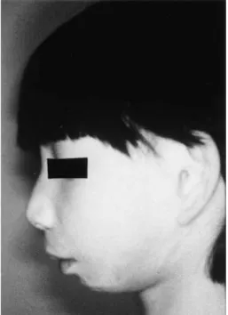
We present a 7-year-old girl with Treacher-Collins syndrome who underwent distraction of the mandi-ble. The patient had marked micrognathia (Figure 1). We simulated the mandible distraction using a
Fig. 1. Preoperative profile of a patient with Treacher-Collins syndrome who has micrognathia.
K. Matsumoto et al. Craniofacial distraction osteogenesis
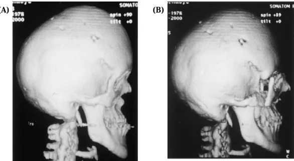
three-dimensional model of the patient’s head build from the preoperative computed tomography data (Figure 2). She was eventually able to take foods that were too hard to eat prior to treatment, and her profile was markedly improved (Figure 3).
MIDFACE
A standard midface advancement consists of si-multaneous fixation following Le Fort osteotomy, but gradual distraction of the midface has been used increasingly for midfacial advancement (23). Soft tissues resistance to advancement of the osteotomized fragment is an issue in conventional osteotomy and rigid fixation for advancements greater than 10 mm, but distraction procedures can solve the problem of resistance, which leads to postoperative relapse. Among the types of Le Fort osteotomy preceding distraction, type I osteotomy has been the most
frequent and has been applied to adult patients with severe midfacial hypoplasia secondary to cleft lip and palate (17). Type III osteotomy has been performed mainly on patients with syndromic craniosynostosis disorders such as Apert, Crouzon, and Pfeiffer syndrome. The Le Fort III osteotomy is justifiably indicated during early childhood for psychological and physiologic reasons (24). The distraction pro-cedure can make the propro-cedures less invasive and allows midface advancement at almost any age, including early childhood (13). However, several craniofacial surgeons have recommended waiting until after 4 years of age due to the fragility of the bone (25).
Polley and Figueroa used an external, adjustable, rigid distraction device for midface advancement of patients with cleft lip and palate (14). The device, which was mounted as a halo onto the temporal and frontal portion of the skull, served to distract the midface after osteotomy by anchoring to the max-illary dental arch. Subsequently, an internal device was developed, which had better patient acceptance and ease of integration into an overall treatment plan (26). Moreover, a new system of miniature distraction devices that can be implanted internal-ly and accessed through a small percutaneous screw was also reported (27). Internal distraction devices can achieve only unidirectional distraction after the placement, whereas the advantage of the external devices is the ability to direct the direction of dis-traction during the advancement process.
(A)
(B)
Fig. 2. A three-dimensional model of the patient’s head showing the distraction devices. (A) Frontal view shows that the distraction devices are attached bilaterally and that the axes of the bilateral devices are parallel to each other. (B) Oblique view shows that the activated device produces a space in the distracted gap.
Fig. 3. Postoperative profile shows marked improvement in appearance.
We treated midface hypoplasia in a 20-year-old female with Crouzon syndrome using a rigid exter-nal distraction device. The patient showed severe exophthalmos and maxillary retrusion, although rela-tively good occlusion had been achieved by long-term orthodontic procedures (Figure 4). It was suggest-ed that this patient’s particular condition could not be resolved by the usual Le Fort III osteotomy/
midface distraction procedure, so we devised a seg-mental approach. The midface, mobilized by Le Fort III osteotomy, was divided into two segments by Le Fort I osteotomy ; each fragment was connected to the rigid external distraction device to be distract-ed separately. Distraction at a rate of 1 mm per day was begun 1 day after the osteotomy. The upper and lower segments were distracted over 17 and 12 days, respectively. The patient’s occlusion was fully cor-rected, and her facial contours were significantly improved. After 3 weeks of consolidation, we removed the distraction device (Figure 5). The clinical course was without complication, and no relapse was ob-served on the cephalogram or computed tomographic scan obtained 1 year after the procedure (Figure 6). Our modified technique was helpful in increasing the usefulness of the external distraction system and in refining the midface distraction procedure (28).
Hemifacial microsomia is the second most com-mon craniofacial anomaly after cleft lip and palate. In this group, mandibular hypoplasia may be asso-ciated with limitation of the vertical growth of the maxilla. In children with hemifacial microsomia, the occlusal changes after distraction of the mandible alone can be corrected orthodontically because there is rapid vertical growth of the maxilla when it is re-leased from the constricting effect of the mandible and soft tissues (29). In the older patients, who have slanted occlusal planes but for whom dental
occlu-Fig. 4. Preoperative profile of a patient with Crouzon syndrome who shows severe exophthalmos and maxillary retrusion.
(A) (B)
Fig. 5. (A) Wires are fixed to the rigid external distraction device during the active lengthening and consolidation period. (B) Postoperative profile shows that the facial contour has been significantly improved.
K. Matsumoto et al. Craniofacial distraction osteogenesis
sion is efficient and stable before the treatment, mandibular distraction alone alters dental occlusion severely and requires unacceptably prolonged ortho-dontic treatment. Therefore, simultaneous distrac-tion of the mandible and maxilla is indicated to cor-rect the facial asymmetry without disturbing the pre-existing dental occlusion (30, 31).
CRANIUM
Craniosynostosis is the term that designates pre-mature fusion of one or more sutures in either the cranial vault or cranial base. The goals of surgery for the newborn with a craniosynostosis are two-fold. One is decompression of the intracranial space to reduce intracranial pressure, prevent visual prob-lems, and permit normal mental development. The other is achievement of satisfactory craniofacial form. Recently, surgical treatment has been performed within the first year of life, and the early effort has resulted in the children looking as good as possible at as early an age as possible to spare the psycho-logical and social trauma associated with craniofacial disfigurement. The surgical treatment includes strip craniectomies, frontal bone advancement, cranial vault remodeling, monobloc or craniofacial advance-ment, and shunt surgery for hydrocephalus (32).
Gradual distraction was used in fronto-orbital ad-vancement and cranial vault remodeling (33-35). The major advantages of gradual distraction were reported to be the elimination of postoperative extradural dead space, preservation of the blood
supply of the bone flap by not detaching the under-lying bone from the dura, and the concomitant ex-pansion of the scalp enabling easy closure of the wound (33). However, the benefits of application of distraction for purely cranial reshaping, which includes fronto-orbital advancement and correction of the plagiocephaly, are ambiguous at present (34, 36). However, distraction for scaphocephaly is indicated (34, 37).
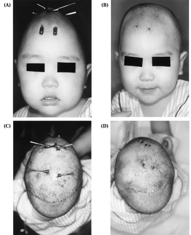
We reported a case of a 1-year-old boy who under-went correction of a scaphocephalic deformity using distraction devices (Figure 7). Double pi-shaped osteotomies were cut, bilateral temporal bone flaps were gradually expanded, and the frontal bone was pulled back simultaneously (Figure 8). The patient showed a good skull contour 2 years postoperatively (Figure 9). Although previous distractions were aimed only at expansion of the cranium, we used the dis-traction technique to successfully shorten the cra-nial vault in the anteroposterior direction. This case demonstrates that the osteotomized bone flaps can be moved in various directions. This feature of the distraction technique may contribute greatly toward meticulous reconstruction of the cranial vault (37).
RECENT ADVANCES
Distraction procedures in craniofacial surgery have some disadvantages, including that the devices may produce scars, a second operation to remove the devices is necessary, the treatment period is pro-longed, infection may occur near the devices, and
(A) (B)
Fig. 6. (A) Preoperative computed tomography scan showing severe maxillary retrusion. (B) Postoperative computed tomogra-phy scan showing that the upper segment is substantially advanced.
minute correction is impossible.
Biodegradable devices made of polylactic acid have been developed to abridge the second operation for removal and stabilization (38, 39). The relative-ly rapid resorption of the implanted components should not interfere with facial growth or provide a nidus for chronic infection. Use of the resorbable devices is a step forward in the evolution of dis-traction osteogenesis (39).
Various agents have been theoretically and experi-mentally implicated as mediators of distraction
osteogenesis. To lessen the time the distraction device is in place on the patient and to accelerate the entire distraction process, a cannulated pin that can provide a route for administering the agents to the precise anatomic point at the optimal time has been developed (40). In addition, the effects of hyaluronic acid, calcium sulfate, and chitosan on early bony consolidation in distraction osteogenesis have been studied in a canine model (41). Calcium sulfate and its combined materials were found to be relatively effective in early bony consolidation.
(A) (B)
(C) (D)
Fig. 7. Frontal views before (A) and after (B) distraction. Top views before (C) and after (D) distraction. K. Matsumoto et al. Craniofacial distraction osteogenesis
The other materials such as several kinds of growth factors may apply to the distraction site in the fu-ture.
Treatment protocols for craniofacial distraction osteogenesis have not yet been established, although there is some degree of consensus (17). Among disorders indicated for distraction, there are sever-al syndromic craniosynostoses, which have
muta-tions in the fibroblast growth factor receptor (FGFR) 2 gene (42-45). Although the relation between the FGFR 2 gene and osteogenesis has not been clarified, some studies have suggested that the FGFR 2 mu-tation accelerates the growth or differentiation ca-pability of osteoblasts (46-48). Clinical studies of the distracted thumb in patients with or without the mutation in the FGFR 2 gene has suggested that osteogenesis at the distraction site may be quicker in Apert syndrome patients with the mutation than in polydactyly patients without the mutation (49). Thus, the efficacy and appropriateness of the dis-traction protocol must be assessed for each indi-vidual disorder.
Distraction osteogenesis in craniofacial surgery has been developed, however, its procedures have not been fully established. The indications for dis-traction must be carefully considered, and the risks of the procedures thoroughly discussed when ob-taining preoperative informed consent from pro-spective patients
REFERENCES
1. Codivilla A : On the means of lengthening in the lower limbs, the muscles and tissues which are shortened through deformity. Am J Orthop Surg 2 : 353-357, 1905
2. Karp NS, Thorne CHM, MaCarthy JG, Sissons HA : Bone lengthening in the craniofacial skel-eton. Ann Plast Surg 24 : 231-237, 1990
Fig. 8. Operative photograph. The patient is in the supine po-sition. Six distraction devices are fixed with microscrews. The activating rods have not yet been inserted into the sheaths of the devices.
(A) (B)
Fig. 9. Frontal view of three-dimensional computed tomography scans before treatment (A) and 2 years after surgery (B). The Journal of Medical Investigation Vol. 50 2003 123
3. Ilizarov GA : The principles of the Ilizarov meth-od. Bull Hosp J Dis Orthop Inst 48 : 1-11, 1988 4. Ilizarov GA : The tension-stress effect on the genesis and growth of tissues : Part I. The in-fluence of stability of fixation and soft-tissue preservation. Clin Orthop 238 : 249-281, 1989 5. Ilizarov GA : The tension-stress effect on the
genesis and growth of tissues : Part II. The in-fluence of the rate and frequency of distrac-tion. Clin Orthp 239 : 263-285, 1989
6. De Bastiani G, Aldegheri R, Renzi-Brivio L, Trivella G : Limb lengthening by callus distrac-tion (callotasis). J Pediatr Orthop 7 : 129-134, 1987
7. Dal Monte A, Donzelli O : Tibial lengthening according to Ilizarov in congenital hypoplasia of the leg. J Pediatr Orthop 7 : 135-138, 1987 8. Martini Z, Castaman E : Tissue regeneration in
the reconstruction of lost bone and soft tissue in the lower limbs : a preliminary report. Br J Plast Surg 40 : 142-148, 1987
9. Kojimoto H, Yasui N, Goto T, Matsuda S, Shimomura Y : Bone lengthening in rabbits by callus dis-traction. The role of periosteum and endosteum. J Bone Joint Surg [Br] 70 : 543-549, 1988 10. Steen H, Fjeld TO, Bjerkreim I, Tevik A, Aldegheri
R, Trivella G : Limb lengthening by diaphyseal corticotomy, callus distraction, and dynamic ax-ial fixation. An experimental study in the ovine femur. J Orthop Res 6 : 730-735, 1988
¨
11. Honig JF, Grohmann UA, Merten HA : Facial bone distraction osteogenesis for correction of malocclusion : a more than 70-year-old concept in craniofacial surgery. Plast Reconstr Surg 109 : 41-44, 2002
12. McCarthy JG, Schreiber J, Karp N, Thorne CH, Grayson BH : Lengthening the human man-dible by gradual distraction. Plast Reconstr Surg 89 : 1-10, 1992
13. Polley JW, Fifueroa AA, Charbel FT, Berkowitz R, Reisberg D, Cohen M : Monobloc craniomaxillofacial distraction osteogenesis in a newborn with se-vere craniofacial synostosis : a preliminary re-port. J Craniofac Surg 6 : 421-423, 1995 14. Polley JW, Figueroa AA : Management of severe
maxillary deficiency in childhood and adolescence through distraction osteogenesis with an external, adjustable, rigid distraction device. J Craniofac Surg 8 : 181-185, 1997
15. Hirabayashi S, Sugawara Y, Sakurai A, Harii K, Park S : Frontoorbital advancement by grad-ual distraction. J Nurosurg 89 : 1058 -1061, 1998
16. Sugawara Y, Hirabayashi S, Sakurai A, Harii K : Gradual cranial vault expansion for the treatment of craniofacial synostosis : a prelimi-nary report. Ann Plast Surg 40 : 554-565, 1998 17. Swennen G, Schliephake H, Dempf R, Schierle H, Malevez C : Craniofacial distraction osteogenesis : a review of the literature. Part 1 : clinical studies. Int J Oral Maxillofac Surg 30 : 89-103, 2001 18. Tomaski SM, Zalzal GH, Saal HM : Airway
ob-struction in the Pierre Robin sequence. Laryn-goscope 105 : 111-114, 1995
19. Benjamin B, Walker P : Management of airway obstruction in the Pierre Robin suquence. Int J Pediatr Otolaryngol 22 : 29-37, 1991
20. Judge B, Hamlar D, Rimell FL : Mandibular distraction osteogenesis in a neonate. Arch Otolaryngo Head Neck Surg 125 : 1029-1032, 1999
21. MaCarthy JG, Williams JK, Grayson BH, Crombie JS : Controlled multiplanar distraction of the mandible : device development and clinical ap-plication. J Craniofac Surg 9 : 322 -329, 1998 22. Diner PA, Kollar E, Martine H, Vazquez MP :
Submerged intraoral device for mandibular lengthening. J Cranio-Maxfac Surg 25 : 116-123, 1997
23. Gosain AK, The Plastic Surgery Educational Foundation DATA Committee : Distraction osteogenesis of the craniofacial skeleton. Plast Reconstr Surg 107 : 278-280, 2001
24. MaCarthy JG, LaTrenta GS, Breitbart AS, Grayson BH, Bookstein FL : The Le Fort III advance-ment osteotomy in the child under 7 years of age. Plast Reconstr Surg 86 : 633-646, 1990 25. Hollier LH, Gosain A, Longaker M, MaCarthy
JG : Craniofacial distraction osteogenesis. J Craniofac Surg 10 : 268-282, 1999
26. Chin M, Toth BA : Le Fort III advancement with gradual distraction using internal devices. Plast Reconstr Surg 100 : 819-830, 1997
27. Cohen SR, Boydston W, Burstein FD, Hudgins R : Monobloc distraction osteogenesis during infancy : report of a case and presentation of a new device. Plast Reconstr Surg 101 : 1919-1924, 1998
28. Matsumoto K, Nakanishi H, Koizumi Y, Seike T, Okazaki M, Yokozeki M, Moriyama K : Seg-mental distraction of the midface in a patient with Crouzon syndrome. J Craniofac Surg 13 : 273-278, 2002
29. Molina F, Ortiz Monasterio F : Mandibular elon-gation and remodeling by distraction : a
fare-K. Matsumoto et al. Craniofacial distraction osteogenesis
well to major osteotomies. Plast Reconstr Surg 96 : 825-840, 1995
30. Ortiz Monasterio F, Molia F, Andrade L, Rodriguez C, Arregui JS : Simultaneous mandibular and maxillary distraction in hemifacial microsomia in adults : avoiding occlusal disasters. Plast Reconstr Surg 100 : 852-861, 1997
31. Cho BC, Shin DP, Park JW, Baik BS : Bimaxillary osteodistraction for the treatment of facial asym-metry in adults. Br J Plast Surg 54 : 491-498, 2001
32. McCarthy JG, Epstein FJ, Wood-Smith D : Craniosynostosis. In : Mccarthy JG, eds. Plas-tic Surgery. WB Saunders Company, Philadel-phia, 1990, pp. 3013-3053.
33. Hirabayashi S, Sugawara Y, Sakurai A, Harii K, Park S : Frontoorbital advancement by grad-ual distraction. J Neurosurg 89 : 1058-1061, 1998 34. Sugawara Y, Hirabayashi S, Sakurai A, Harii K : Gradual cranial vault expansion for the treat-ment of craniofacial synostosis : a preliminary report. Ann Plast Surg 40 : 554-565, 1998 35. Kobayashi S, Honda T, Saitoh A, Kashiwa K :
Unilateral coronal synostosis treated by internal forehead distraction. J Craniofac Surg 10 : 467-471, 1999
36. Polley JW : Commentary on unilateral coronal synostosis treated by internal forehead distrac-tion. J Craniofac Surg 10 : 472, 1999
37. Matsumoto K, Nakanishi H, Seike T, Shinno K, Hirabayashi S : Application of the distrac-tion technique to scaphocephaly. J Craniofac Surg 11 : 172-176, 2000
38. Cohen SR, Holmes RE, Amis P, Fichtner H : In-ternal craniofacial distraction with biodegradable devices : early stabilization and protected bone regeneration. J Craniofac Surg 11 : 354-366, 2000 39. Burstein FD, Williams JK, Hudgins R, Graham L, Teague G, Paschal M, Simms C : Single-stage craniofacial distraction using resorbable devices. J Craniofac Surg 13 : 776 -782, 2002
40. Grayson BH, Rowe NM, Hollier JrLH, Williams JK, McCormick S, Longaker MT, McCarthy JG : Development of a device for the delivery of agents to bone during distraction osteogenesis. J Craniofac Surg 12 : 19-25, 2001
41. Cho BC, Park JW, Baik BS, Kwon IC, Kim IS : The role of hyaluronic acid, chitosan, and cal-cium sulfate and their combined effect on ear-ly bony consolidation in distraction osteogenesis
of a canine model. J Craniofac Surg 13 : 783-793, 2002
42. Reardon W, Winter RM, Rutland P, Pulley LJ, Jones BM, Malcolm S : Mutations in the fibroblast growth factor receptor 2 gene cause Crouzon syndrome. Nature Genet 8 : 98-103, 1994 43. Wilkie AOM, Slaney SF, Oldridge M, Poole MD,
Ashworth GJ, Hockley AD, Hayward RD, David DJ, Pulleyn LJ, Rutland P, Malcolm S, Winter RM, Reardon W : Apert syndrome resultsfrom localized mutations of FGFR2 and is alleic with Crouzon syndrome. Nature Genet 9 : 165-172, 1995
44. Matsumoto K, Urano Y, Kubo Y, Nakanishi H, Arase S : Mutation of the fibroblast growth fac-tor recepfac-tor 2 gene in Japanese patients with Apert syndrome. Plast Reconstr Surg 101 : 307-311, 1998
45. Matsumoto K, Urano Y, Kubo Y, Hirabayashi S, Sakurai A, Nakanishi H : Mutations of the fibroblast growth factor receptor 2 gene in ori-ental patients with Apert syndrome. J Jpn PR S (in Japanese) 18 : 583-586, 1998
46. Fragale A, Tartaglia M, Bernardini S, Di Stasi AMM, Di Rocco C, Velardi F, Teti A, Battaglia PA, Migliaccio S : Decreased proliferation and altered differentiation in osteoblasts from ge-netically and clinically distinct craniosynostotic disorders. Am J Pathol 154 : 1465-1477, 1999 47. Lomri A, Lemonnier J, Hott M, de Parseval N,
Lajeunie E, Munnich A, Renier D, Marie PJ : Increased calvaria cell differentiation and bone matrix formation induced by fibroblast growth factor receptor 2 mutation in Apert syndrome. J Clin Invest 101 : 1310-1317, 1998
48. Yokozeki M, Tanimoto Y, Matsumoto K, Nakanishi H, Hiura K, Moriyama K : Craniofacial and den-tal manifestations in an Apert syndrome patient : participation of osteoblastic cells in phenotypic expression of craniosynostosis syndrome. In : Takada K, Proffit WR, eds. Orthodontics in the 21th Century. Proceedings of an Interna-tional Symposium Orthodontics 2001. Osaka University Press, Osaka, 2003, pp. 151-155 49. Matsumoto K, Nakanishi H, Koizumi Y, Seike
T, Tanimoto Y, Yokozeki M, Hiura K, Moriyama K, Minami M, Urano Y, Hirabayashi S : Cor-rection of a deformed thumb by distraction of the phalanx. Scand J Plast Reconstr Surg Hand Surg 36 : 368-372, 2002