IRUCAA@TDC : Effects of Deuterium Oxide on Streptococcus mutans and Pseudomonas aeruginosa
10
0
0
全文
(2) 175. Bull Tokyo Dent Coll (2010) 51(4): 175–183. Original Article. Effects of Deuterium Oxide on Streptococcus mutans and Pseudomonas aeruginosa Kaname Hirai, Mihoko Tomida*, Yuichiro Kikuchi, Ohmi Ueda, Hiroshi Ando* and Naokazu Asanuma* Department of Microbiology, Matsumoto Dental University, 1780 Hirooka Gobara, Shiojiri, Nagano 399-0781, Japan * Department of Oral Physiology, Matsumoto Dental University, 1780 Hirooka Gobara, Shiojiri, Nagano 399-0781, Japan. Received 25 December, 2009/Accepted for publication 21 May, 2010. Abstract A complex aggregation of microorganisms growing on a solid substrate is termed a biofilm and is considered to be an etiological agents. Pseudomonas aeruginosa and Streptococcus mutans are representative bacteria in such biofilms. It is well known that deuterium oxide (D2O) causes toxic effects on a number of biological systems. We investigated the effects of D2O on growth and biofilm formation of P. aeruginosa and S. mutans. These bacteria were incubated in medium containing D2O (100%, 75% or 0%) at 37°C for 24hr, 48 hr or 72hr. Growth of P. aeruginosa was inhibited by D2O within the first 48hr. However, after 72 hr, growth rate was seen to increase in the D2O-containing medium compared with in medium without D2O. In contrast, the growth of S. mutans in the D2O medium was inhibited within 72hr. The biofilm formation of P. aeruginosa was increased in the D2O medium. Biofilm formation of S. mutans in the D2O medium increased compared with in the medium without D2O, but this increase was only temporary in the case of P. aeruginosa. Compared to biofilm formation in 0% D2O medium marked as 100%, the biofilm formation rate of S. mutans in 75% D2O medium was 143% at 24hr, 146% at 48 hr and 130% at 72 hr. In other D2O concentration media biofilm formation was lower. In 100% D2O medium, biofilm formation rate decreased from 114% at 24hr to 56% at 72hr. The biofilm formation rate of P. aeruginosa in 100% D2O medium was 172% at 24hr, but decreased to 88% at 72hr. Biofilm formation of P. aeruginosa in 75% and 0% D2O media showed no significant difference. We consider that these results were due to stress or alteration in bacterial metabolisms. Key words:. Deuterium oxide —Streptococcus mutans— Pseudomonas aeruginosa— Biofilm. Introduction Accumulated data show two major effects of deuterium oxide (D2O) on the living. system: the “solvent isotope effect (SIE)” and the “deuterium isotope effect (DIE)”16). The SIE is due to the properties of D2O itself, i.e., its high density (p⳱1.11) compared to. 175.
(3) 176. Hirai K et al.. H2O (p⳱0.99) at 20°C and its high viscosity (p⳱1.25) compared to H2O (p⳱1.01) at 20°C9). The SIE involves the structure of water and macromolecules. On the other hand, the DIE involves the ability of D2O to replace H with D in biological molecules: the stretching frequency of the O-D bond is approximately half that of the O-H bond, thus making C-D, O-D, N-D and S-D bonds stronger than their protonated forms (C-H, O-H, etc.)8,32). Although it is not easy to distinguish between the SIE and DIE, long-term effects are probably due to the DIE and short-term effects to the SIE. The DIE involves the metabolic reaction through cleavage of C-H bonds, and if growth and cell division take place in the presence of D2O, some deuterated molecules will form during over periods of days, weeks, or longer. In contrast, the SIE involves isolated cells or enzymes over a much shorter period. The toxic effects of D2O on the nervous system, liver and formation of different blood cells have been studied since the 1960s29). At the cellular level, D2O affects mitosis and membrane function. Mice and rats which were forced to drink D2O instead of H2O died 7 days later12,13) and higher concentrations (ca. 90%) of D2O rapidly killed fish, tadpoles and drosophila20). On the other hand, microorganisms are more tolerant to D2O than multicellular systems30). Algae and bacteria can grow in 100% D2O and utilize a large number of deuterated molecules14). Recently, bacterial biofilms have been considered a form of microbial ecology. Biofilms are packed communities of microbial cells which grow on surfaces and surround themselves with secreted polymers. Biofilms allow microorganisms to survive in hostile conditions, thus their character is significantly different from their planktonic counterparts. Differences are noticed mainly in their growth rate, biochemical composition, virulence and increased resistance to chemical antimicrobials2,5,17,24). Bacterial biofilms are well-organized structured communities. Some investigators describe biofilm as a thin basal layer on the subtrium, with columnar, mushroom-shaped. multibacterial extensions into the lumen of the solution, separated by regions called channels seemingly empty or filled with extracellular polysaccharide3,28). These channels are considered to be at work as a selective transporter of low molecular materials such as water. Biofilm formation is related to the virulence of bacteria24). Streptococcus mutans is a facultatively anaerobic Gram-positive bacterium known to be a cariogenic, and possesses many etiological agents, including the ability to form biofilm. Fermentation is the only metabolic reaction required for this bacterium to manufacture ATP. The matrix of biofilm by S. mutans is considered to be alpha-1-3-glucan. Pseudomonas aeruginosa is well known as the pathogen of hospital infection. P. aeruginosa is respiratory gram negative bacteria. The matrix of biofilm formed by P. aeruginosa is considered to be alginate. These two bacteria form different types of biofilm, and their respective pathogenicities are linked to that ability. In this study, we investigated the effects of pure D2O on the growth and biofilm formation of two types of biofilm-producing bacteria, S. mutans and P. aeruginosa.. Materials and Methods 1. Bacterial strain S. mutans Ingbritt and P. aeruginosa GTC2 strains were used in this study. Bacteria were grown on Trypticase soy agar (Becton, Dickinson and Company, Maryland, USA) at 37°C in an aerobic atmosphere for 48hr. 2. Media and growth conditions As a base medium Trypticase soy broth (Becton, Dickinson and Company) was dissolved in each solvent at a concentration of D2O 100%, 75% or 0%. Density was adjusted to 3%, and the medium supplemented with 5% sucrose for S. mutans and 1% glucose for P. aeruginosa. Under the same conditions, basal medium without glucose or sucrose was also used. These media were sterilized by filtration. Bacterial colonies were picked up from.
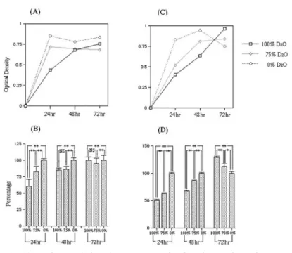
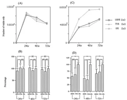
(4) 177. Effects of Deuterium Oxide on Bacteria. Trypticase soy agar (Becton, Dickinson and Company) and suspended in each broth. The suspension was adjusted to OD 0.02 at 655 nm. One hundred l suspension was then dispensed in 96-well polystyrene plates. Three plates of each type of bacteria were prepared and the plates incubated aerobically at 37°C for 24hr, 48hr or 72hr. 3. Bacterial growth Bacteria floating in the medium or adherent to the plastic wells were all considered growing bacteria. Optical density, which is related to bacterial growth, was measured at 655 nm using the Microplate reader (Model 680; Bio-Rad Laboratories, Richmond, CA, USA). The number of viable bacteria was determined by resazurin reduction using Alamar blue (AbD Serotec, Oxford, UK). One hundred l Almar blue, was added to each well and incubated at 37°C for 30 min. The optical density of the samples was measured at 540 nm using the Microplate reader31). 4. Semiquantification of biofilm Bacteria adherent to the plastic wells were considered to be forming a biofilm4). The semiquantification of biofilm formation was carried out using toluidine blue by the method of Tanaka with some modifications27). After measuring bacterial growth, the supernatant was decanted and wells washed twice with distilled water. Each well of the tissue culture plate was incubated at room temperature for 30 min with 100 l of 0.1% toluidine blue to stain the biofilm. The excess stain was removed by washing twice with distilled water. One hundred l ethanol was added to the well and left for 30 min to extract the stain. The optical density of samples was measured at 595 nm with the Microplate reader. 5. Statistical analysis The relationship between growth and biofilm formation rate at each D2O concentration for 24hr, 48hr or 72hr was analyzed using a one-factor ANOVA. Pairwise comparisons among the three concentrations of D2O were made using the Tukey-Kramer test.. Results 1. Bacterial growth Figures 1 and 2 show the results of optical density. Figures 3 and 4 show the viable number of bacteria. Figures 1 and 3 show the progress of P. aeruginosa growth with time. Delayed growth was observed in both basal media and glucose media including D2O. P. aeruginosa was pigmented green in 72hr (data not shown). P. aeruginosa was pigmented particularly strongly in the glucose-supplemented medium. This pigmentation was considered to influence optical density. Optical density of the media with glucose decreased at 72hr (Fig. 1C). This may have been due to the influence of pigmentation. While the number of viable P. aeruginosa in the H2O medium with glucose showed peak growth at 24hr, the number of viable P. aeruginosa in the D2O media with glucose showed peak growth at 48hr (Fig. 3C). At 48hr, D2O medium showed a higher rate of activity of resazurin reduction than H2O medium. Figures 2 and 4 show S. mutans growth. The growth of S. mutans in D2O media was not delayed, but always lower than that in H2O. The number of viable cells was quite different in both the D2O and H2O media when supplemented with sucrose (Fig. 4C). In the basal medium, rates of activity of resazurin reduction of S. mutans showed no significant difference between various concentrations of D2O at 72hr (Fig. 4B). Figures 1B, D, 2B and D show the bacterial growth rates for each experimental condition compared to the growth in 0% D2O medium (marked as 100%). Within the first 48hr, the growth of P. aeruginosa was inhibited by D2O. However, after 72hr, the growth rates reached almost 130% in 100% D2O medium and 110% in 75% D2O medium (Fig. 1D). Growth of P. aeruginosa in 0% D2O medium showed a decline. The growth of S. mutans in 100% and 75% D2O medium was inhibited by D2O within 72hr (Figs. 2C, D). 2. Biofilm formation Figures 5A and C show amounts of biofilm.
(5) 178. Hirai K et al.. Fig. 1 (A) Medium turbidity of P. aeruginosa in basal medium without glucose. (B) Ratio of medium turbidity of P. aeruginosa in basal medium without glucose. (C) Medium turbidity of P. aeruginosa in basal medium with glucose. (D) Ratio of medium turbidity of P. aeruginosa in basal medium with glucose. Bar indicates standard error obtained from three separate assays. **: p⬍0.01, *: p⬍0.05 (Tukey-Kramer).. Fig. 2 (A) Medium turbidity of S. mutans in basal medium without sucrose. (B) Ratio of medium turbidity of S. mutans in basal medium without sucrose. (C) Medium turbidity of S. mutans in basal medium with sucrose. (D) Ratio of medium turbidity of S. mutans in basal medium with sucrose. Bar indicates standard error obtained from three separate assays. **: p⬍0.01..
(6) Effects of Deuterium Oxide on Bacteria. Fig. 3 (A) Number of viable cells of P. aeruginosa in basal medium without glucose. (B) Ratio of activity of resazurin reduction of P. aeruginosa in basal medium without glucose. (C) Number of viable cells of P. aeruginosa in basal medium with glucose. (D) Ratio of activity of resazurin reduction of P. aeruginosa in basal medium with glucose. Bar indicates standard error obtained from three separate assays. **: p⬍0.01 (Tukey-Kramer).. Fig. 4 (A) Number of viable cells of S. mutans in basal medium without sucrose. (B) Ratio of activity of resazurin reduction of S. mutans in basal medium without sucrose. (C) Number of viable cells S. mutans in basal medium with sucrose. (D) Ratio of activity of resazurin reduction of S. mutans in basal medium with sucrose. Bar indicates standard error obtained from three separate assays. **: p⬍0.01 (Tukey-Kramer).. 179.
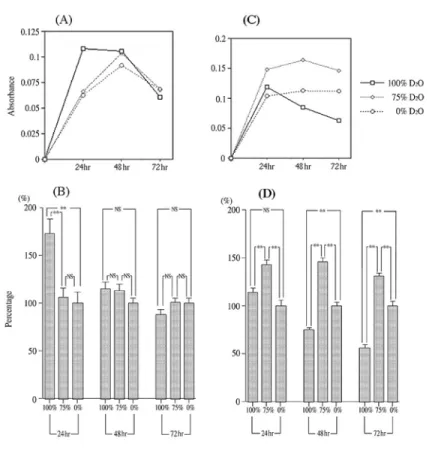
(7) 180. Hirai K et al.. Fig. 5 (A) Amount of biofilm formation of P. aeruginosa in basal medium with glucose. (B) Biofilm formation rates of P. aeruginosa in basal medium with glucose. (C) Amount of biofilm formation of S. mutans in basal medium with sucrose. (D) Biofilm formation rates of S. mutans in basal medium with sucrose. Bar indicates standard error obtained from three separate assays. **: p⬍0.01 (Tukey-Kramer).. measured by staining. P. aeruginosa formed much larger amounts of biofilm in the 100% D2O medium than in the H2O media at 24hr, although this evened out by 48hr. By 72 hr, the amount of biofilm in both mediums began to decline at the same rate. A comparison with S. mutans in Figs. 5A and C revealed a similar pattern in terms of decline in amount of biofilm formation. Figure 5B, however, revealed no remarkable increase in amount of biofilm with culture in 100% D2O medium. Figures 5B and D show biofilm formation rates compared to that in 0% D2O medium (marked as 100% biofilm formation). The biofilm formation rate of P. aeruginosa in 100% D2O medium was 172% at 24hr, but. decreased to 88% at 72hr. Biofilm formation in 75% and 0% D2O medium was not significantly different (Fig. 5B). The biofilm formation rate of S. mutans was higher in 75% D2O medium than in other D2O concentration media: 143% at 24hr, 146% at 48hr and 130% at 72hr. In 100% D2O medium, biofilm formation rate decreased from 114% at 24hr to 56% at 72hr (Fig. 5D). In 100% D2O medium, the biofilm formation of both bacteria decreased gradually.. Discussion Deuterium oxide has been considered harmless to bacteria because no influence.
(8) 181. Effects of Deuterium Oxide on Bacteria. on bacterial growth was observed without any extended lag phase14). Recent reports suggest that D2O affects bacterial metabolism11,19). Escherichia coli revealed D2O at log-phase, with 53% of intracellular water being replaced by D2O15). Intracellular D2O affects the increase of the anaplerotic supply of tricarboxylic acid cycle11). In Rodobacter sphaeroides, the cytochrome C oxidase catalystic cycle was slowed down by deuterium isotope addition19). Cytochrome C oxidase is the terminal enzyme of the respiratory chain, and various aerobic bacteria, including P. aeruginosa, also possess this enzyme22). In this study, D2O (100%, 75%) inhibited growth of P. aeruginosa by approximately 50% compared to 0% D2O for the first 24hr. However, the growth rates in 100% and 75% D2O gradually recovered at 48hr, finally exceeding growth in 0% D2O. These results possibly indicate that D2O does not absolutely inhibit the respiration of P. aeruginosa. However, inhibition was limited until 48hr. At 72hr, media containing D2O did not inhibit, but rather facilitated the bacterial growth. This may be due to the change in respiration metabolism of P. aeruginosa. The growth of S. mutans was inhibited during all test periods by D2O (Figs. 2C, D). The growth pattern differed from that of P. aeruginosa. This may be related to the lack of the TCA cycle and respiration in S. mutans. It seems that D2O does not completely terminate bacterial respiration. Biofilm formation increases under some types of stress. Biofilm formation by microorganisms of the genus Pseudomonas is associated with various stress factors such as oxidative stress18), high temperature25), hydrodynamic stress23) and small amounts of ethanol or antibiotic6,7,10). The biofilm formation of S. mutans also increases by various stress factors such as oxidative stress, nutritional stress and acid circumstance21). In many cases, stress from the environment induces bacteria to form much larger amounts of biofilm. Bioflim may play a role in defending bacteria against stress. In this study, D2O inhibited bacterial growth for a short term, suggesting. that it acted as a stressor on the bacteria, thus inducing biofilm formation. Biofilm formation is also led by the quorum sensing (QS) system. The QS system depends on bacterial cell density. When the population reaches a critical threshold density, bacteria communicate with one another and secrete specific signaling molecules. The signaling molecules in QS systems are, in many cases, acylated homoserine lactones (AHL) in Gram-negative bacteria, small peptides in Gram-positive bacteria or autoinducer-2 (AI-2), which is used both in Gram-negative and Gram-positive bacteria as a universal bacterial language33). Deuterium isotope affects the metabolism of serine1). However, it is not clear how the production of homoserine lactone is related to the metabolism of serine altered by D2O. The QS signal of S. mutans is a 21-amino acid peptide phenomenon, a competent stimulating peptide21,34). Although this peptide includes 4 serines, it is quite different from homoserine lactone, and the sensor of histidine kinase is associated with this peptide26,34). It seems that D2O is not involved in the metabolism of competent stimulating peptides. Both of the two hypotheses regarding various types of stress and QS proposed above seem to indicate that the both are able to explain our results in this study. They both indicate that the D2O effects on the bacteria observed in the present study was due to SIE, as SIE changed little and in a dose-dependent manner. Since we do not yet have clear evidence, further study is needed to clarify the stress effects of D2O on bacteria.. Acknowledgements We would like to thank Professor David M. Carlson, Matsumoto Dental University, for his assistance with the English of this manuscript.. References 1) Born TL, Blanchard JS (1999) Enzyme-catalyzed.
(9) 182. 2) 3) 4) 5) 6). 7). 8) 9) 10). 11). 12). 13). 14) 15). Hirai K et al.. acylation of homoserine: mechanistic characterization of the Escherichia coli metA-encoded homoserine transsuccinylase. Biochemistry 38: 14416–14423. Clote TE (2003) Resistance mechanisms of bacteria to antimicrobial compounds. Int Biodeterior Biodegradation 51:277–282. Costerton JW, Lewandowski Z, Caldwell DE, Korber DR, Lappin-Scott HM (1995) Microbial biofilms. Annu Rev Microbiol 49:711–745. Costerton JW, Stewart PS, Greenberg EP (1999) Bacterial biofilms: a common cause of persistent infections. Science 284:1318–1322. Davie D (2003) Understanding biofilm resistance to antibacterial agents. Nat Rev Drug Discov 2:114–122. Dietrich LEP, Teal TK, Price-Whelan A, Newman DK (2008) Redox-active antibiotics control gene expression and community behavior in divergent bacteria. Science 321: 1203–1206. Edwards K J, Saunders NA (2001) Real-time PCR used to measure stress induced changes in the alginate pathway of Pseudomonas aeruginosa. J Appl Microbiol 91:29–37. Fiher S J, Helliwell JR (2008) An investigation into structural changes due to deuteration. Acta Crystallogr A 64:359–367. Flaumenhaft E, Bose S, Crespi HL, Katz J J (1965) Deuterium isotope effects in cytology. Int Rev Cytol 18:313–361. Fonseca AP, Sousa JC (2007) Effect of shear stress on growth, adhesion and biofilm formation of Pseudomonas aeruginosa with antibiotic-induced morphological changes. Int J Antimicrob Agents 30:236–241. Hochuli M, Szyperski T, Wuthrich K (2000) Deuterium isotope effects on the central carbon metabolism of Escherichia coli grown on a D2O-containing minimal medium. J Biomol NMR 17:33–42. Ikeda M, Hirono M, Kishino M, Matsuura J, Sakakibara M, Yoshioka T (2000) Examination of microgravity effects on spontaneous Ca2Ⳮ oscillations in AtT20 pituitary cells using heavy water. J Gravit Physiol 7:63–64. Ikeda M, Suzuki S, Kishio M, Hirono M, Sugiyama T, Matsuura J, Suzuki T, Sota T, Allen CN, Konishi S, Yoshioka T (2004) Hydrogendeuterium exchange effects on beta-endorphin release from AtT20 murine pituitary tumor cells. Biophys J 86:565–575. Johnstone DB (1962) Growth of Azobacter in deuterium oxide. J Bacteriol 83:867–870. Kreuzer-Martin HW, Lott MJ, Ehleringer JR, Hegg EL (2006) Metabolic processes account for the majority of intracellular water in logphase Escherichia coli cells as revealed by hydro-. gen isotopes. Biochemistry 45:13622–13630. 16) Kushner DJ, Baker A, Dunstall TG (1999) Pharmacological uses and perspectives of heavy water and deuterated compounds. Can J Physiol Pharmacol 77:79–88. 17) Lewis K (2007) Persister cells, dormancy and infectious disease. Nat Rev Microbiol 5:48–56. 18) Rodriguez-Rojas A, Blazquez J (2009) The Pseudomonas aeruginosa pfpI gene plays an antimutator role and provides general stress protection. J Bacteriol 191:844–850. 19) Salomonsson L, Branden G, Brizezinski P (2008) Deuterium isotope effect of proton pumping in cytoclome c oxidase. Biochemi Biophys Acta 1777:343–350. 20) Samis HV, Baird MB, Massie HR (1974) Deuterium oxide effect on temperature-dependent survival in populations of Drosophila melanogaster. Science 183:427–428. 21) Senadheera D, Cvitkovitch DG (2008) Quorum sensing and biofilm formation by Streptococcus mutans, Bacterial Signal Transduction and Drug Targets (Adv EXP Med Biol 631), Utsumi R ed., pp.178–188, Kluwer Academic/ Plenum Publishers, New York. 22) Silvestrini MC, Falcinelli S, Ciabatti I, Cutruzzolà F, Brunori M (1994) Pseudomonas aeruginosa nitrite reductase (or cytochrome oxidase): an overview. Biochimie 76:641–654. 23) Simoes M, Simoes LC, Vieira MJ (2008) Physiology and behavior of Pseudomonas fluorescens single and dual strain biofilms under hydrodynamics stresses. Int J Food Microbiol 128: 309–316. 24) Smith RS, Iglewski BH (2003) P. aeruginosa quorum-sensing system and virulence. Curr Opin Microbiol 6:56–60. 25) Srivastava S, Yadav A, Seem K, Mihara S, Chaudhary V, Nautiyal CS (2008) Effect of high temperature on Pseudomonas putida NBRI0987 biofilm formation and expression of stress sigma factor RpoS. Curr Microbiol 56:453–457. 26) Suntharalingam P, Cvitkovitch DG (2005) Quorum sensing in streptococcal biofilm formation. Trends Microbiol 13:3–6. 27) Tanaka G, Shigeta M, Komatsuzawa H, Sugai M, Suginaka H, Usui T (2000) Effect of clarithromycin on Pseudomonas aeruginosa biofilm. Chemotherapy 46:36–42. 28) Ten Cate JM (2006) Biofilms, a new approach to the microbiology of dental plaque. Odontol 94:1–9. 29) Thomson AE, Brarnsley EA, Young L (1963) Biochemical studies of toxic agents. 14. The biosynthensis of ethylmercapturic acid. Biochem J 86:145–152. 30) Vengrus TV, Zatsepina GN, Kramskoi MN,.
(10) Effects of Deuterium Oxide on Bacteria. Goriunov NN (1994) Various changes in the average membrane potential and number of T-lymphocytes in Balb/c mice at different physiological state in environments with various levels of D2O. Biofizika 39:116–122. 31) Vidal-Aroca F, Meng A, Minz T, Page MGP, Dreier J (2009) Use of resazurin to detect mefloquine as an efflux-pump inhibitor in Pseudomonas aeruginosa and Escherichia coli. J Microbiol Methods 79:232–237. 32) Wade D (1999) Deuterium isotope effects on noncovalent interactions between molecules. Chem Biol Interact 117:191–217. 33) Winans SC (2002) Bacterial esperanto. Nat Struct Biol 9:83–84.. 183. 34) Zhang K, Ou M, Wang W, Ling J (2009) Effects of quorum sensing on cell viability in Streptococcus mutans biofilm formation. Biochem Biophys Res Commun 379:933–938. Reprint requests to : Dr. Kaname Hirai Department of Microbiology, Matsumoto Dental University, 1780 Hirooka Gobara, Shiojiri, Nagano 399-0781, Japan Tel & Fax: +81-263-51-2084 E-mail: kaname8bac@po.mdu.ac.jp.
(11)
図
関連したドキュメント
Based on the models of urban density, two kinds of fractal dimensions of urban form can be evaluated with the scaling relations between the wave number and the spectral density.. One
Solutions of integral equa- tions are expressed by the inverse operators of auxiliary exterior and interior boundary value problems, i.e., theorems on the solvability of
We present sufficient conditions for the existence of solutions to Neu- mann and periodic boundary-value problems for some class of quasilinear ordinary differential equations.. We
Abstract The representation theory (idempotents, quivers, Cartan invariants, and Loewy series) of the higher-order unital peak algebras is investigated.. On the way, we obtain
Then it follows immediately from a suitable version of “Hensel’s Lemma” [cf., e.g., the argument of [4], Lemma 2.1] that S may be obtained, as the notation suggests, as the m A
Similarly, an important result of Garsia and Reutenauer characterizes which elements of the group algebra k S n belong to the descent algebra Sol( A n−1 ) in terms of their action
I ) basic cellular material which is used by living cells as raw material for mitosis; 2) a generic non-utilisable material which may inhibit mitosis. Numerical
Motivated by ongoing work on related monoids associated to Coxeter systems, and building on well-known results in the semi-group community (such as the description of the simple
