Posted at the Institutional Resources for Unique Collection and Academic Archives at Tokyo Dental College,
Title
Immunohistochemical expression of Ki67 and p53 by
cellsin odontogenic epithelial islands in the walls
of dentigerouscysts and keratocystic odontogenic
tumors
Author(s)
Matsuzaka, K; Okudaira, S; Inoue, K; Hashimoto, K;
noue, T
Journal
日本口腔検査学会雑誌, 7(1): 10-15
URL
http://hdl.handle.net/10130/3654
Immunohistochemical expression of Ki67 and p53 by cells
in odontogenic epithelial islands in the walls of dentigerous
cysts and keratocystic odontogenic tumors
Matsuzaka K
! *, Okudaira S
!, Inoue K, Hashimoto K, Inoue T
Department of Clinical Pathophysiology, Tokyo Dental College, Tokyo, Japan !: These authors contributed equally to this work. *:2-9-18, Misakicho, Chiyoda-ku, Tokyo, 101-0061 JAPAN Tel: +81-3-6380-9252 Fax: +81-3-6380-9606 e-mail: matsuzak@tdc.ac.jp Abstract Backgroud: This study investigated the proliferation activity using Ki67- and p53-positive ratios of cells in the odontogenic epithelium in cyst walls of dentigerous cysts (DCs) and keratocystic odontogenic tumors (KCOTs). Methods: Specimens of KCOTs (73 cases) and DCs (457 cases) without inflammatory cell infiltrations were used in this study. Specimens fixed with 20% buffered formalin were embedded in paraffin, sectioned and stained with hematoxylin and eosin. Immunohistochemical staining was carried out using antibodies to Ki67 and p53. The odontogenic epithelial islands in the cyst walls were observed with a microscope, and the mean number of odontogenic epithelial islands, the mean number of cells per odontogenic epithelial island, the ratio of Ki67-positive cells per odontogenic epithelial island and the ratio of p53-positive cells per odontogenic epithelial island were calculated. Results: The ratios of DCs and KCOTs with odontogenic islands in cyst walls were 21.4% (98 cases) and 35.6% (26 cases), respectively. Immunohistochemically, some cells were positive for Ki67 in the epithelial islands. The mean number of odontogenic epithelial islands in KCOTs was larger than in DCs, and the number of cells in epithelial islands in KCOTs was larger number than in DCs. The ratios of Ki67 and p53-positive cells in KCOTs was higher than in DCs. Conclusion: When odontogenic epithelial islands, which are characterized by cell proliferation activity and neoplasia, are observed in cyst walls, considerable attention must be given to the differential diagnosis because residual odontogenic epithelial islands might be recurrences not only in KCOTs but also in DCs. Key words: dentigerous cyst, keratocystic odontogenic tumor, epithelial islands, Ki67, p53 received:Oct 17th 2014 accepted:Nov 28th 2014JJ S E D P Vol. 7 No. 1: , 2015 Introduction Odontogenic epithelial islands in cyst walls can often be observed in radiolucent lesions of the mandibles and maxillae 1). Keratocystic odontogenic tumors (KCOTs) have the potential for aggressive behavior, recurrence and genetic abnormalities 2). A unilocular radiolucency associated with the crown of a tooth is a classic diff erential diagnosis in which a dentigerous cyst (DC) is the most likely entity. DCs arise from the dental follicle of unerupted teeth. The other specific diseases on the differential list have more troubling biologies and include KCOTs. The walls of DCs frequently have nests of odontogenic epithelium which may be numerous 3).
KCOTs are benign uni- or multi- cystic, intraosseous tumors of odontogenic origin, with a characteristic lining of parakeratinized stratified squamous epithelium (WHO). KCOTs are lined by a regular parakeratinized stratified squamous epithelium, usually consisting of about 5 to 8 cell layers. There is a well-defi ned often palisaded, basal layer of columnar basal cells that tend to tbe oriented away from the basement membrane and are often intensely basophilic 2).
It is known that odontogenic epithelial islands are present in the walls of odontogenic cystic lesions, KCOTs and DCs as well. In KCOT walls, some daughter cysts which have proliferated
from odontogenic epithelial islands were thought to be infiltrations of tumor cells. Investigations of proliferative activity and tumor inhibitor gene expression of odontogenic epithelial island cells play important roles to determine the prognosis and to decide on courses of treatment, such as only to enucleate or to enucleate with scraping around the bone. Ki67 and proliferating cell nuclear antigen (PCNA) is known the marker of cell proliferation activity. Seyedmajidi et al. reported p53 and PCNA expression in the lining epithelium of KCOTs, radicular cysts, calcifying cystic odontogenic tumors and DCs 4). There are some immunohistochemical
reports detecting proliferating cells 4) 5), but those
reports demonstrated the cell proliferation activity in the lining epithelium. In the cyst wall, however, various sizes of odontogenic epithelial islands are often observed, which are also related to the recurrence of the lesions.
So, the purpose of this study was to investigate the cell proliferation activity using Ki67-positive ratio and p53-positive ratio of cells in the odontogenic epithelium in cyst walls of DCs and KCOTs. Material and methods The ratio of DCs and KCOTs was determined for all intraosseous lesions submitted by the Chiba Hospital dentigerous cyst others ameloblastoma dental sac/follicle cystic lesion calcifying cystic odontogenic tumor radicular cyst/granuloma keratocystic odontogenic tumor 37.7 % 6.0 % 47.1 % 0.2 % 3.6 % 1.5 % 2.1 % 1.8 % Fig. 1 Graph of the ratios for radiolucent lesions of maxillae and mandibles The ratio of DCs is 37.7% and of KCOTs is 6.0%. DCs are the second most common type of lesion. 10 - 15
and the Suidobashi Hospital, Tokyo Dental College, from April 2011 to March 2013. The mean numbers of odontogenic epithelial islands of DCs and KCOTs were counted. The mean numbers of cells composing the odontogenic epithelial islands of DCs and KCOTs were counted. While all Specimens of KCOTs (73 cases) and DCs (457 cases), 20 cases of KCOTs and DCs with odontogenic epithelial islands without inflammatory cell infiltrates were used for immunohistochemical analysis. Specimens were routinely fixed with 20% buffered formalin and were embedded in paraffin. Paraffin sections approximately 4 µm were used in this study. Specimens were routinely Hematoxylin and eosin. Morphological characteristics of odontogenic epithelial islands were observed. Immunohistochemical staining was carried out using antibodies to Ki67 (MIB-1; 1:100, DAKO, Glostrup, Denmark) and to p53 (1:100, DAKO, Glostrup, Denmark). Briefly, after deparaffinization, the sections were microwaved for 30 minutes at 60ºC for antigen retrieval. Endogenous peroxidase activity was blocked by incubating the sections with 3% H2O2 in methanol for 30 minutes. To
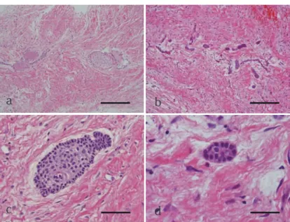
prevent non-specific reactions, sections were incubated with 3% bovine serum albumin for 30 minutes in a humidity chamber. After washing in PBS, sections were incubated with the primary antibody to Ki67 or p53 for 2 hours. The sections were then incubated with horseradish-peroxidase-conjugated secondary antibody (Histofine, MAX-PO multi, NICHIREI, Tokyo, Japan) for 30 minutes in the humidity chamber. Finally, bound antibody was visualized using 0.01% 3,3’-diaminobenzidine, and sections were counterstained with hematoxylin. The odontogenic epithelial islands in the cyst walls were observed with a microscope, and the mean number of odontogenic epithelial islands, the mean number of cells per odontogenic epithelial island, the ratio of Ki67-positive cells per odontogenic epithelial island and the ratio of p53-positive cells per odontogenic epithelial island were calculated. Meanwhile, all cells of odontogenic epithelial islands were intended for the ratios of Ki67 and p53-positive cells. Results Of all 3,360 specimens collected from 2011 to 2013 in the Clinical Laboratory at the Chiba Hospital and the Suidobashi Hospital, Tokyo Dental College, 457 cases (13.6%) were DCs and 73 cases (2.2%) were KCOTs. The ratios of DCs and KCOTs of the maxillary and mandibular radiolucent lesions, which included DCs, KCOTs, radicular cysts/granulomas, calcifying odontogenic tumors, cystic lesions, dental sacs/ follicles, ameloblastomas and others, were 37.7% and 6.0%, respectively (Fig. 1). The ratios of DCs and KCOTs with odontogenic islands in the cyst walls were 21.4% (98 cases) and 35.6% (26 cases), respectively (Fig. 2). Histologically, two types of epithelial islands were observed, one arranged in a cord type and the other in an alveolar configuration type (Fig. 3). Most of the odontogenic epithelial
DCs KCOTs
mean number of odontogenic islands / slide 9.1 islands 24.3 islands
mean number of cells / odontogenic epithelial island 29.1 cells 47.3 cells
ratio of Ki67-positive cells / odontogenic epithelial island 2.92 ± 0.01 % 6.57 ± 0.03 % ratio of p53-positive cells / odontogenic epithelial island 0.41 ± 0.31 % 0.80 ± 0.83 % Table 1 Statistical analyses of odontogenic epithelial islands in DC and KCOT 500 400 300 200 100 0 cases dentigerous cyst keratocystic odontogenic tumor presence of odontogenic island absence of odontogenic island
Fig. 2 Graph of the numbers of DCs and KCOTs, and the numbers and ratios of odontogenic epithelial islands in the cyst wall 98 cases (21.4%) of the total number of 457 DC cases included odontogenic epithelial islands, and 26 cases (35.6%) of the total number of 73 KCOTs included odontogenic epithelial islands. 21.4 % 78.6 % 64.4 % 35.6 %
JJ S E D P Vol. 7 No. 1: , 2015 Fig. 3 Histochemical staining of DCs and KCOTs a, b: DCs, c, d:KCOTs. Cord type (a, c) and alveolar configuration type (b, c) can be observed. Bar: 100 μm Fig. 4 Immunohistochemical staining for Ki67 a, b: DCs, c, d:KCOTs. Some cells are positive for Ki67 in a, c, d, not in b. Bar: 100 μm Fig. 5 Immunohistochemical staining for p53 a, b: DCs, c, d:KCOTs. Some cells in c are positive for p53, but only few cells are
a
b
c
d
a
b
c
d
a
b
c
d
10 - 15islands in DCs were arranged in the cord like structure (Fig. 3a,b), and only a few islands were of the alveolar configuration structure (Fig. 3c,d). On the other hand, most odontogenic epithelial islands in the KCOTs were in an alveolar configuration structure. Immunohistochemically, some cells reacted with Ki67 in the epithelial islands. Specifically, there was a tendency for large numbers of Ki67-positive cells in odontogenic epithelial islands in an alveolar configuration structure (Fig. 4). p53-positive cells could be observed in odontogenic epithelial islands of KCOTs, especially in the alveolar configuration type, but only few p53-positive cells were observed in the cord type of odontogenic epithelial islands in KCOTs, and both the cord and the alveolar configuration types of DCs (Fig. 5). Table 1 shows the mean numbers of odontogenic epithelial islands, the numbers of cells in an epithelial island, and the ratio of Ki67-positive cells. The mean number of odontogenic epithelial islands in KCOTs was larger than in DCs, and also the number of cells in islands in KCOTs was larger than the number in DCs. The ratio of Ki67-positive cells in DCs was 2.92 ± 0.01% and in KCOTs was 6.57 ± 0.03%. The ratio of p53-positive cells in DCs was 0.41 ± 0.31% and in KCOTs was 0.80 ± 0.83%. Discussion This study characterized the proliferative activity and tumor inhibitor gene expression of odontogenic epithelial islands in DCs and in KCOTs. DCs are known to be developmental cysts, and KCOTs are known to be developmental cysts, but the WHO working group recommended the term KCOT as it better reflects its neoplastic nature in 2005. KCOTs are potentially aggressive lesions. Surgeons and dentists should carefully follow up after treatment because of the common presence of daughter cysts and a tendency for multiplicity 2). DCs are the second most common type of odontogenic cyst after radicular cysts 3). The ratio of DCs in this study was 37.7% which is the second most common type of lesion in radiolucent lesions in the jaw after radicular cysts / granulomas. Further, Manor et al. reported that the ratio of radicular cysts was 48% 6). Otherwise, the ratio of KCOTs was 6.0%
in this study. Manor et al. reported about cystic lesions of the jaw, and found that 23 cases of the 322 cystic lesions of the jaw (7%) were KCOTs (6). This study about the ratio of KCOTs was similar to the previous report. Ki67 expression in the superbasal layer and p53 expression in the basal and superbasal layers are generally observed. Odontogenic epithelial islands are composed of several kinds of cells, such as basal cells and/or suprabasal cells. Ki67 is an important marker for the detection of the cell proliferation index and is a useful indicator as a neoplastic marker 5) - 9). In this study, the ratio of Ki67-positive cells in odontogenic islands of DCs was 2.92% and of KCOTs was 6.57%. The ratio of Ki67-positive cells in odontogenic epithelial islands of KCOTs was higher than DCs. Some studies have reported that proliferating cells in the lining epithelium of KCOTs are significantly higher than in DCs 4)8)10). Although
this study investigated odontogenic epithelial islands, the ratio of proliferating cells in odontogenic epithelial islands of KCOTs was higher than in DCs. This suggests that odontogenic epithelial islands of KCOTs are proliferating neoplastically. Superficially, odontogenic epithelial islands of DCs seem to be in a resting environment, but their activity of proliferation has been presented. Once the DNA is repaired, the cell cycle arrest ends, but if DNA is not successfully repaired, p53 induces apoptosis. Mutations in the p53 gene can inhibit the repair of DNA damage and lead to neoplastic proliferation of damaged cells 11) 12). p53 can occur in wild-type and mutant
forms. p53 has an important role in controlling the expression of cellular proliferation inhibiting genes, and mutation of the p53 gene can prevent its inhibitory role, resulting in oncogenic activities and neoplastic changes. Wild-type p53 is expressed in small amounts, has a short half-life and cannot be detected using immunohistochemistry. Its detection can be possible in conditions where the protein is
JJ S E D P Vol. 7 No. 1: , 2015
expressed in large amounts or accumulates in mutant cells 4)13). In this study, the p53-positive ratio of
KCOTs was higher than DCs. This phenomenon also reveals that odontogenic epithelial islands of KCOTs have a neoplastic nature, but in DCs it has less of an effect on neoplasticity. In conclusion, when odontogenic epithelial islands are characterized for cell proliferation activity and neoplastic nature in the cyst wall, patients have to be followed up because of possibilities of recurrence not only in KCOTs but also in DCs. References 1) Lin HP, Wang YP, Chen HM, Cheng SJ, Sun A, Chiang CP: A clinicopathological study of 338 dentigerous cysts, J Oral Pathol Med, 42:462-467, 2013 2) Barnes L, Eveson JW, Reichart P, Sidransky D (eds): World Health Organization Classification of Tumours, Pathology and Genetics of Head and Neck Tumours, IARC POress: Lyon, 2005
3) Marx ER, Stern D. Dentigerous cyst In; Oral and Maxillofacial Pathology A rationale for diagnosis and treatment, pp. 579-583, 2003 4) Seyedmajidi M, Nafarzadeh S, Siadati S, Shafaee S, Bijani A, Keshmiri N: p53 and PCNA expression in keratocystic odontogenic tumors compared with selected odonotogenic cysts, Int J Mol Cell Med Autumn, 2:185-193, 2013 5) de Oliveira RG, Costa A, Meurer MI, Vieira DS,
Rivero ER: Immunohistochemical analysis of matrix metalloproteinases (1, 2, and 9), Ki67, and myofibroblasts in keratocystic odontogenic tumors and pericoronal follicles, J Oral Pathol Med, 43:282-288, 2014. 6) Manor E, Kachko L, Puterman MB, Szabo G, Bodner L: Cystic lesions of the jaws - a clinicopathological study of 322 cases and review of the literature, Int J Med Sci, 9: 20-26, 2012. 7) de Vicente JC, Torre-Iturraspe A, Guiterrez AM, Lequerica-Fernandez P: Immunohistochemical comparative study of the odontogenic keratocysts and other odontogenic lesions, Med Oral Pathol Oral Cir Bucal, 15:709-715, 2010
8) Li TJ, Browne RM, Matthews JB: Epithelial cell proliferation in odontogenic keratocysts: a comparative immunocytochemical study of Ki67 in simple, recurrent and basal cell naevus syndrome (BCNS)-associated lesions, J Oral Pathol Med, 24:221-226, 1995
9) de Oliveira MG, Lauxen Ida S, Chaves AC, Rados PV, Sant'Ana Filho M: Odontogenic epithelium: immunolabeling of Ki-67, EGFR and survivin in pericoronal follicles, dentigerous cysts and keratocystic odontogenic tumors, Head Neck Pathol, 5:1-7, 2011 10)Piattelli A, Fioroni M, Santinelli A, Rubini C: P53 protein expression in odontogenic cysts, J Endod, 27:459-461, 2001 11)Levine AJ: p53 the cellular gatekeeper for growth and division, Cell, 88:323-331, 1997 12)Nylander K, Dabelsteen E, Hall PA: The p53 molecule and its prognostic role in squamous cell carcinomas of the head and neck, J Oral Pathol Med, 29:413-425, 2000. 13)de Oliveira MG, Lauxen Ida S, Chaves AC. Rados PV,
Sant’Ana Filho M: Immunohistochemical analysis of the patterns of p53 and PCNA expression in odontogenic cystic lesions, Med Oral Pathol Oral Cir Bucal, 13: E275-280, 2008


