IRUCAA@TDC : Clinical validity of the interactive and low-dose three-dimensional dento-alveolar imaging system, tuned-aperture computed tomography
10
0
0
全文
(2) Bull. Tokyo dent. Coll., Vol. 44, No. 3, pp. 159⬃167, August 2003. 159. Clinical Report. CLINICAL VALIDITY OF THE INTERACTIVE AND LOW-DOSE THREE-DIMENSIONAL DENTO-ALVEOLAR IMAGING SYSTEM, TUNED-APERTURE COMPUTED TOMOGRAPHY YOSHIHIKO HAYAKAWA*, KAZUHIRO YAMAMOTO*, YUJI KOUSUGE*, NORIO KOBAYASHI*, MAMORU WAKOH*, HIROSHI SEKIGUCHI**, MASASHI YAKUSHIJI** and ALLAN G. FARMAN*** * Department of Oral and Maxillofacial Radiology, Tokyo Dental College, 1-2-2 Masago, Mihama-ku, Chiba 261-8502, Japan ** Department of Pediatric Dentistry, Tokyo Dental College, 1-2-2 Masago, Mihama-ku, Chiba 261-8502, Japan *** Division of Radiology and Imaging Sciences, Department of Surgical and Hospital Dentistry, School of Dentistry, The University of Louisville, Louisville, Ky 40292 USA. Received 7 April, 2003/Accepted for Publication 12 May, 2003. Abstract Based on technical studies and clinical examinations, the clinical value of a new three-dimensional dento-alveolar imaging system, Tuned-Aperture Computed Tomography (TACT®), were examined for dental applications. TACT is a conventional and low-cost tomosynthesis method in which the benefit of digitization is fully utilized. The clinical information yield of TACT has been examined for the detection of dental caries, periodontal defects and radicular fractures, and also for the pre-surgical assessment of implant placement and impacted teeth. In this article, we introduce basic TACT technology, review the literature pertaining to in vitro and in vivo studies, and describe the outline of our study of TACT to determine its clinical value in the assessment of impacted maxillary teeth. Key words:. Tuned-aperture computed tomography—Intraoral radiography — Tomosynthesis— Impactions — Flat panel detector. INTRODUCTION Because newly developed, small, flat-panel detectors with high sensitivity to X-ray and comparatively low-noise characteristics have. been utilized for intraoral radiography, tomosynthesis has become practical for dentoalveolar imaging. TACT®, Tuned-Aperture Computed Tomography, is an interactive, low-dose, low-cost, three-dimensional (3-D). 159.
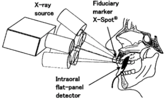
(3) 160. Y. HAYAKAWA et al.. tomosynthesis system in which the benefit of digitization is fully utilized29,30). A 3-D imageforming algorithm, TACT, which can be implemented with virtually any projectionbased imaging system capable of digitized output, produces true 3D data from any number of arbitrarily oriented two-dimensional projections. In the middle of the 1990’s, the principal theory of TACT was described by Webber (Wake Forest University, School of Medicine, Winston-Salem, North Carolina, USA)29,30). The outline of TACT theory will be described in this article. We also introduce basic TACT technology, review the literature pertaining to TACT, and outline our study of TACT to determine its clinical value in assessment of impacted teeth in the maxillary incisor region.. THEORY TACT is a technique for 3-D diagnostic imaging which can be carried out with low dosage. Application of the algorithm is not only applied to dento-alveolar imaging but also to digital mammography24,32) and scintimammography8). TACT technology is now licensed to Instrumentarium Imaging (Tuusula, Finland). As shown in Fig. 1, intraoral radiographs are made in different directions while a small flat-panel detector is held in the oral cavity. The images are obtained by “component projections” and are called TACT basis images. The projections are made at with different angles and repeated four to six times. The TACT workbench (Verify Software, WinstonSalem, North Carolina, USA) stacks the images, inputs locations of fiduciary markers for each basis image, and reconstructs a series of arbitrary multi-planar cross-sectional images. Such reconstruction of a series of arbitrary section images can produce pseudo-holograms. Iterative restoration to remove blurring is also available after three-dimensional models are built. TACT is based on tomosynthesis and aperture theory29,30).. Fig. 1 Schematic presentation of component projections for obtaining TACT base images. A fiduciary marker is attached at the buccal surface of the oral mucosa. Four to six exposures are made from different directions while the small flatpanel detector is held in the oral cavity.. LITERATURE REVIEW Dental Caries: Abreu et al. studied TACT in dental caries detection1–4,25). Effects in caries detectability were examined under various acquisition parameters of basis images, presence or absence of iterative restorations, and the choice of reconstruction algorithms. Their 1999 report4) indicated that TACT images did not improve caries detection over that using film or digital radiography, but they nevertheless used their research data to calculate the optimum parameter settings of TACT for caries detection. In 1997, Tyndall et al. had previously explained that the relatively poorer performance yielded by the digital control images suggested that increased information capacity associated with more modern CCD detectors might improve the diagnostic performance of TACT displays25). In contrast, in 1998, Nair et al. showed that TACT images held the promise of a more effective imaging modality than film or individual digital images for the detection of recurrent caries22). Periodontal status: Chai-U-Dom et al. examined TACT for the in vitro detection of pericrestal and periodontal bone gain6,7). They indicated that TACT associated with digital subtraction radiography provided greater sensitivity while the technical flexibility remained. Nair et al. concluded that TACT was a more effective and accurate imaging modality for.
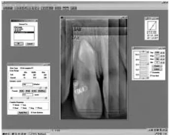
(4) CLINICAL VALIDITY OF TACT. 161. Fig. 2 A selected screen of the TACT Workbench. The tool box, “Clinical TACT slice control”, enables the operator to control the position and the interval of the reconstructed sectional images.. non-destructive quantification of osseous changes within healing bony defects20,21). Ramesh et al. conducted research on the detection of simulated periodontal defects and concluded that TACT images processed by weak iterative restoration provided the best performance23). Nair et al. also reported that TACT performed significantly better than planar images for the detectability of traumainduced radicular fractures, vertical root fractures, and mandibular fractures in their in vitro studies18,19). Implants: The clinical value of TACT for presurgical implant imaging was examined by Liang et al., who found that TACT may provide an alternative to conventional tomography for dental implant imaging14,15). Morant et al. reported that TACT was significantly more accurate than standard application of the buccal object rule for assessing the relationship between tooth roots and the inferior alveolar canal17). Since 1998, we have also reported several basic studies of TACT, including collaborative work with the inventor, Webber9,34,35,39). We analyzed the accuracy of depth discrimination affected by projection geometry and the number of projections in our studies.. CLINICAL APPLICATION: IMPACTED TOOTH ASSESSMENT As a further step in our research, we performed a clinical study on the diagnostic information yield of TACT in the assessment of impacted teeth in comparison with conventional dentoalveolar imaging11,36–38). 1. Procedures for TACT A small flat-panel detector system, CompuRay® (Yoshida Dental Trading, Tokyo, Japan) was employed as the intraoral X-ray detector. This is known as the RVG-ui sensor (Trophy Radiology, Marne-la-Vallée, France) in the rest of the world. Basis images are collected and stacked. The input of each projection position using the fiduciary marker on individual images is carried out, and a reconstruction algorithm is applied. Stability for maintaining continuity in geometric projection conditions is required for the generation of 3D images, such as CT, at the time of collection of image data. However, TACT enables an arbitrary setup of projection directions, because the position of the fiducial marker is always available. The operation for cross-sectional reconstruction can be carried out interactively. The.
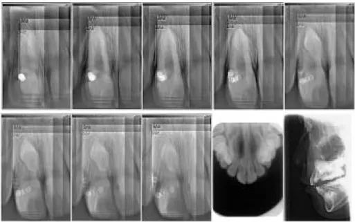
(5) 162. Y. HAYAKAWA et al.. Fig. 3 Patient is a 9-year-old female. An impacted tooth is located in the maxillary incisor region. Based on the six component projections, TACT images were constructed. Eight sectional images are arranged in order from the buccal side (upper left) to the lingual side (lower center). These TACT images were selected at the same intervals. The occlusal and lateral radiographs are presented at the lower right.. reconstruction of a series of dento-alveolar depths is easily realized in compliance with diagnostic tasks. A selected screen of the TACT Workbench is shown in Fig. 2. In accordance with the results of previous in vitro studies34,35,39), in which influences of the number of and the angle of component projections to the accuracy of depth discrimination were reported, five or six component projections with conical beam-projection array per reconstruction, and approximately 20 degrees of angular disparity were chosen. The fiduciary marker we used was a 1.5 mm diameter X-Spot® lead pellet (Beekley, Bristol, Connecticut, USA). The X-Spot was packed under an adhesive tab used to help keep track of sensor positioning. The coordinates on each image were inputted into a reconstruction algorithm as reference points. 2. Case presentation The case presented in Fig. 3 is an examination of an impacted tooth in the anterior region of the maxilla. Impacted teeth do often occur in the anterior maxilla, and their occur-. rence is significant in early mixed dentition in view of both esthetics and occlusion. TACT was applied for the radiological diagnosis of the status of the impacted tooth and its positional relationship with surrounding tissue and adjacent teeth. Various radiographic techniques have been reported for recognizing the position of an impacted (clinically “missing”) tooth. Radiological diagnosis is necessary for clarifying the morphologic status of impacted teeth and their relationship with the surrounding tissues. Traditionally, the intraoral radiograph with an orthogonal projection has been used with an additional mesial, distal, inferior, or superior projection. With such parallax methods, the position of the impacted tooth in relation to its surrounding tissues can be estimated, but with limited accuracy. Given these limitations, it is possible that TACT might be used to study the morphology of impacted teeth in the incisor and canine regions of the maxilla. As indicated in Fig. 3, TACT revealed the morphological relationship between the impacted tooth and surrounding tissues,.
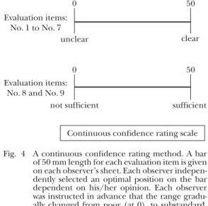
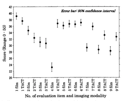
(6) 163. CLINICAL VALIDITY OF TACT. Table 1 Items used in the clinical evaluation Anatomical structure depiction (unclear — clear) 1. An outer contour of the impacted tooth: dental crown 2. An outer contour of the impacted tooth: dental root 3. Developmental status of the dental root of the impacted tooth 4. Buccal/palatal position of the impacted tooth 5. Upper/lower position of the impacted tooth 6. Mesial/distal position of the impacted tooth 7. Relationship between the impacted tooth (permanent tooth germ in particular) and adjacent teeth Subjective assessments (not sufficient — sufficient) 8. Estimate how useful you consider the existing radiographic information to be when considered only with regard to its potential for altering clinical treatment options. 9. Estimate the extent to which you feel that you have all the information you need to be confident in your clinical assessment of the patient’s surgical prognosis.. including the regular teeth. Both the impacted tooth and the roots of adjacent teeth are sharply depicted in different sectional images with different depths in the buccolingual direction. All the TACT images were processed by the installed iterative restoration to remove blurring. 3. Statistical analysis Previously, TACT has been compared variously with conventional intraoral, occlusal, rotational panoramic, and lateral cephalographic examinations of impacted maxillary incisors. Since the results are already published37,38), only a brief outline is given in this report. TACT was used to examine 20 dental impactions in 20 patients. Sixteen dentists trained to read TACT image reconstructions independently carried out the following tasks: (1) clarity of depiction of the impacted tooth and its surrounding tissues, (2) estimation of confidence in clinical assessments of the patient by using the provided images, and (3) estimation of the diagnostic potential of TACT for altering the selection of treatment options. Table 1 lists the seven objective criteria for the evaluation of image clarity and an additional two subjective assessments. The continuous confidence-rating method was employed for each evaluation (Fig. 4). Mean. 0 Evaluation items: No. 1 to No. 7 unclear. 0 Evaluation items: No. 8 and No. 9 not sufficient. 50. clear. 50. sufficient. Continuous confidence rating scale Fig. 4 A continuous confidence rating method. A bar of 50 mm length for each evaluation item is given on each observer’s sheet. Each observer independently selected an optimal position on the bar dependent on his/her opinion. Each observer was instructed in advance that the range gradually changed from poor (at 0), to substandard, acceptable, good, and finally excellent (at 50) in sequence.. scores compare the observers’ evaluations for structural clarity, observers’ perceptions of the potential for TACT altering patient’s treatment options, and observers’ confidence in the reading of the position of the impacted tooth. Collected data were analyzed by means of t-tests performed with StatView (Abacus Concepts, Berkeley, Calif. USA). The a priori was set at 0.05. Mean scores and 95% confidence intervals.
(7) 164. Y. HAYAKAWA et al.. Fig. 5 Chart comparing observers’ evaluation of depiction of anatomic structures (items 1 to 7) and observers’ subjective assessments (items 8 and 9). Each error bar expresses the 95% confidence interval.. are indicated in Fig. 5. The clarity of the outer contour (items 1 and 2) was rated relatively low with TACT (p⬍0.05). In some cases, the impacted teeth and surrounding tissues were not completely depicted by TACT because of the small imaging area of the CCD sensor. Scores for accuracy in determination of the “buccal/palatal position of the impacted tooth,” and the “relationship between the impacted tooth (permanent tooth germ in particular) and adjacent teeth” and subjective assessments (task 2 and 3) were high for TACT compared with those of conventional radiographs. TACT was perceived to improve depiction of the buccal/palatal position of the impacted tooth and its relationship with adjacent teeth (p⬍0.0001). TACT outperformed conventional images in terms of both subjective assessments for clinical decision making. The added diagnostic value with TACT varied from case to case.. DISCUSSION Our research on TACT imaging, which was recently published, is the second in vivo. study38). The first such study was reported by the inventor, Webber and Messura31). His group concluded that TACT displays were more diagnostically informative and had more impact on potential treatment options than did conventional radiographs. Some in vitro studies, which were mentioned in the short review section, indicated that TACT is sensitive or compatible in comparison with conventional dento-alveolar radiography examinations to detect dental caries, periodontal bone dynamics and other diagnostic tasks. Since the image detector of intraoral radiography is changed from film to flat-panel detector (CCD or C-MOS sensor), TACT becomes convenient as such digital procedures are utilized. The digital revolution of the recent decade has put interactive and low-dose three-dimensional imaging systems, such as TACT tomosynthesis, into practice26). In a recent study, TACT projection data was utilized for rendering 3-D models for quantitative analysis of image subtraction methods13). In addition to research for the optimization of TACT procedures and algorithms16,27,28), Webber recently used a hand-held X-ray system33), in an attempt to increase the user-.
(8) CLINICAL VALIDITY OF TACT. friendliness of TACT for the use in dental offices. For the last decade, researchers have carried out 3-D dento-alveolar imaging by using singleor multi-row spiral CT5) and cone-beam CT images10,12,40). In comparison with such CT systems, TACT is characterized by a low-cost and low-dose 3-D image-forming system, which has been demanded by the medical and dental community in recent years. In this article, we introduced the basic TACT technology, reviewed the literature, and described the outline of our study. Our in vivo study shows the validity of using interactive 3-D TACT to improve the benefits of a digital flat-panel detector employed for intraoral radiography in the assessment of dental impactions.. REFERENCES 1) Abreu, M., Jr., Tyndall, D.A. and Ludlow, J.B. (2001). Generation of TACT image slices using different reconstruction algorithms: Effects on natural caries detection. Oral Surg Oral Med Oral Pathol Oral Radiol Endod 92, 576– 581. 2) Abreu, M., Jr., Tyndall, D.A., Ludlow, J.B. and Nortje, C.J. (2001). The effect of the number of iterative restorations on tuned aperture computed tomography for approximal caries detection. Dentomaxillofac Radiol 30, 325–329. 3) Abreu, M., Jr., Tyndall, D.A., Ludlow, J.B. and Nortje, C.J. (2002). Influence of the number of basis images and projection array on caries detection using tuned aperture computed tomography (TACT). Dentomaxillofac Radiol 31, 24–31. 4) Abreu, M., Jr., Tyndall, D.A., Platin, E., Ludlow, J.B. and Philips, C. (1999). Two- and threedimensional imaging modalities for detection of caries: A comparison between film, digital radiography and tuned aperture computed tomography (TACT). Dentomaxillofac Radiol 28, 152–157. 5) Cavalcanti, M.G., Ruprecht, A. and Vannier, M.W. (2002). 3D volume rendering using multislice CT for dental implants. Dentomaxillofac Radiol 31, 218–223. 6) Chai-U-Dom, O., Ludlow, J.B., Tyndall, D.A. and Webber, R.L. (2001). Detection of simulated periodontal bone gain by digital subtraction radiography with tuned-aperture computed. 7). 8). 9). 10). 11). 12). 13). 14). 15). 165. tomography. The effect of angular disparity. Dentomaxillofac Radiol 30, 92–97. Chai-U-Dom, O., Ludlow, J.B., Tyndall, D.A. and Webber, R.L. (2002). Comparison of conventional and TACT (Tuned Aperture Computed Tomography) digital subtraction radiography in detection of pericrestal bonegain. J Periodontal Res 37, 147–153. Fahey, F.H., Grow, K.L., Webber, R.L., Harkness, B.A., Bayram, E. and Hemler, P.F. (2001). Emission tuned-aperture computed tomography: A novel approach to scintimammography. J Nucl Med 42, 1121–1127. Farman, A.G., Scheetz, J.P., Eleazer, P.D., Edge, M.J., Morant, R.D., Limrachtamorn, S. and Yamamoto, K. (2001). Tuned-aperture computed tomography accuracy in tomosynthetic assessment for dental procedures. In The Proceedings of the 15th International Congress of Computer Assisted Radiology and Surgery. (H.U., Lemke, M.W., Vannier, K., Inamura, A.G., Farman and K. Doi eds.) pp.654–658, Springer, Berlin. Hashimoto, K., Arai, Y., Iwai, K., Araki, M., Kawashima, S. and Terakado, M. (2003). Comparison of a new limited cone beam computed tomography machine for dental use with a multidetector row helical CT machine. Oral Surg Oral Med Oral Pathol Oral Radiol Endod 95, 371–377. Hayakawa, Y., Yamamoto, K., Kobayashi, N., Kousuge, Y., Wakoh, M. and Farman, A.G. (2002). Clinical trial of small flat-panel detector in dento-alveolar imaging. Innervision 18(4), 88–91. (in Japanese) Heiland, M., Schulze, D., Adam, G. and Schmelzle, R. (2003). 3D-imaging of the facial skeleton with an isocentric mobile C-arm system (Siremobil Iso-C3D). Dentomaxillofac Radiol 32, 21–25. Lehmann, T.M., Hemler, P.F. and Webber, R.L. (2002). Virtual radiographs computed from TACT volume data as a gold standard for image registration prior to subtraction. Dentomaxillofac Radiol 31, 187–192. Liang, H., Tyndall, D.A., Ludlow, J.B. and Lang, L.A. (1999). Cross-sectional presurgical implant imaging using tuned aperture computed tomography (TACT). Dentomaxillofac Radiol 28, 232–237. Liang, H., Tyndall, D.A., Ludlow, J.B., Lang, L.A. and Nunn, M.E. (2001). Accuracy of mandibular cross-sectional imaging with tunedaperture computed tomography (TACT), iteratively reconstructed TACT, and multidirectional, linear, and transverse panoramic tomography. Oral Surg Oral Med Oral Pathol Oral Radiol Endod 91, 594–602..
(9) 166. Y. HAYAKAWA et al.. 16) Linnenbrugger, N.I., Webber, R.L. and Lehmann, T.M. (2002). Implementation of a generalized TACT algorithm for arbitrary source-object distances. Dentomaxillofac Radiol 31, 249–256. 17) Morant, R.D., Eleazer, P.D., Scheetz, J.P. and Farman, A.G. (2001). Array-projection geometry and depth discrimination with Tuned-Aperture Computed Tomography for assessing the relationship between tooth roots and the inferior alveolar canal. Oral Surg Oral Med Oral Pathol Oral Radiol Endod 91, 252–259. 18) Nair, M.K., Nair, U.P., Grondahl, H.G. and Webber, R.L. (2002). Accuracy of tuned aperture computed tomography in the diagnosis of radicular fractures in non-restored maxillary anterior teeth— An in vitro study. Dentomaxillofac Radiol 31, 299–304. 19) Nair, M.K., Nair, U.D.P., Grondahl, H.G., Webber, R.L. and Wallace, J.A. (2001). Detection of artificially induced vertical radicular fractures using tuned aperture computed tomography. Eur J Oral Sci 109, 375–379. 20) Nair, M.K., Seyedain, A., Agarwal, S., Webber, R.L., Nair, U.P., Piesco, N.P., Mooney, M.P. and Grondahl, H.G. (2001). Tuned aperture computed tomography to evaluate osseous healing. J Dent Res 80, 1621–1624. 21) Nair, M.K., Seyedain, A., Webber, R.L., Nair, U.P., Piesco, N.P., Agarwal, S., Mooney, M.P. and Grondahl, H.G. (2001). Fractal analyses of osseous healing using tuned aperture computed tomography images. Eur Radiol 11, 1510–1515. 22) Nair, M.K., Tyndall, D.A., Ludlow, J.B. and May, K. (1998). Tuned aperture computed tomography and detection of recurrent caries. Caries Res 32, 23–30. 23) Ramesh, A., Ludlow, J.B., Webber, R.L., Tyndall, D.A. and Paquette, D. (2002). Evaluation of tuned-aperture computed tomography in the detection of simulated periodontal defects. Oral Surg Oral Med Oral Pathol Oral Radiol Endod 93, 341–349. 24) Suryanarayanan, S., Karellas, A., Vedantham, S., Glick, S.J., D’Orsi, C.J., Baker, S.P. and Webber, R.L. (2000). Comparison of tomosynthesis methods used with digital mammography. Acad Radiol 7, 1085–1097. 25) Tyndall, D.A., Clifton, T.L., Webber, R.L., Ludlow, J.B. and Horton, R.A. (1997). TACT imaging of primary caries. Oral Surg Oral Med Oral Pathol Oral Radiol Endod 84, 214–225. 26) Webber, R.L. (1999). The future of dental imaging. Where do we go from here? Dentomaxillofac Radiol 28, 62–65. 27) Webber, R.L. and Bettermann, W. (1999). A method for correcting for errors produced by. 28). 29). 30). 31). 32). 33). 34). 35). 36). variable magnification in three-dimensional tuned-aperture computed tomography. Dentomaxillofac Radiol 28, 305–310. Webber, R.L. and Hendrickson, J.L. (2000). A comparison of the effect of task-specific with fixed beam alignment in tuned-aperture computed tomography. Dentomaxillofac Radiol 29, 223–229. Webber, R.L., Horton, R.A., Tyndall, D.A. and Ludlow, J.B. (1997). Tuned-aperture computed tomography (TACT): Theory and application for three-dimensional dento-alveolar imaging. Dentomaxillofac Radiol 26, 53–62. Webber, R.L., Horton, R.A., Underhill, T.E., Ludlow, J.B. and Tyndall, D.A. (1996). Comparison of film, direct digital, and tunedaperture computed tomography images to identify the location of crestal defects around endosseous titanium implants. Oral Surg Oral Med Oral Pathol Oral Radiol Endod 81, 480–490. Webber, R.L. and Messura, J.K. (1999). An in vivo comparison of diagnostic information obtained from tuned-aperture computed tomography and conventional dental radiographic imaging modalities. Oral Surg Oral Med Oral Pathol Oral Radiol Endod 88, 239–247. Webber, R.L., Underhill, H.R. and Freimanis, R.I. (2000). A controlled evaluation of tunedaperture computed tomography applied to digital spot mammography. J Digit Imaging 13, 90–97. Webber, R.L., Webber, S.E. and Moore, J. (2002). Hand-held three-dimensional dental X-ray system: Technical description and preliminary results. Dentomaxillofac Radiol 31, 240– 248. Yamamoto, K., Farman, A.G., Webber, R.L., Horton, R.A. and Kuroyanagi, K. (1998). Effects of projection geometry and number of projections on accuracy of depth discrimination using tuned-aperture computed tomography (TACT) in dentistry. Oral Surg Oral Med Oral Pathol Oral Radiol Endod 86, 126–130. Yamamoto, K., Farman, A.G., Webber, R.L., Horton, R.A. and Kuroyanagi, K. (1999). Effects of number of projections on accuracy of depth discrimination using tuned-aperture computed tomography (TACT) for threedimensional dentoalveolar imaging of lowcontrast details. Oral Surg Oral Med Oral Pathol Oral Radiol Endod 88, 100–105. Yamamoto, K., Hayakawa, Y., Kobayashi, N., Kousuge, Y., Wakoh, M., Sekiguchi, H., Yakushiji, M. and Farman, A.G. (2002). TunedAperture Computed Tomography (TACT®) to assess impacted teeth using 2D slices and 3D pseudo-holograms. In The Proceedings of the 16th International Congress of Computer Assisted.
(10) CLINICAL VALIDITY OF TACT. Radiology and Surgery (CARS2002). (H.U., Lemke et al. eds.) p.1117, Springer-Verlag, Berlin. 37) Yamamoto, K., Hayakawa, Y., Kobayashi, N., Kousuge, Y., Wakoh, M., Sekiguchi, H., Yakushiji, M. and Farman, A.G. (2002). Diagnostic value of tuned-aperture computed tomography for three-dimensional dentoalveolar imaging. Eizo Jyoho Medical 34, 1026– 1031. (in Japanese) 38) Yamamoto, K., Hayakawa, Y., Kousuge, Y., Wakoh, M., Sekiguchi, H., Yakushiji, M. and Farman, A.G. (2003). Diagnostic value of tuned-aperture computed tomography versus conventional dentoalveolar imaging in assessment of impacted teeth. Oral Surg Oral Med Oral Pathol Oral Radiol Endod 95, 109–118. 39) Yamamoto, K., Nishikawa, K., Kobayashi, N., Kuroyanagi, K. and Farman, A.G. (2000). Evaluation of tuned-aperture computed tomog-. 167. raphy depth discrimination for image series acquired variously with linear horizontal, linear vertical, and conical beam projection arrays. Oral Surg Oral Med Oral Pathol Oral Radiol Endod 89, 766–770. 40) Ziegler, C.M., Woertche, R., Brief, J. and Hassfeld, S. (2002). Clinical indications for digital volume tomography in oral and maxillofacial surgery. Dentomaxillofac Radiol 31, 126– 130. Reprint requests to: Dr. Yoshihiko Hayakawa Department of Oral and Maxillofacial Radiology, Tokyo Dental College, 1-2-2 Masago, Mihama-ku, Chiba 261-8502, Japan.
(11)
図
+2
関連したドキュメント
In addition, early ischemic change in clinical images with matrix sizes of 256×256 and 128×128 processed using three imaging filters (Gaussian, smoothing, unsharp mask) from
7.A Ogura, K Hayakawa, T Miyati, F Maeda: The effect of susceptibility of gadolinium contrast media on Diffusion-weighted imaging and the apparent Diffusion coefficient. Dose MD,
1) Tamaki N, Cuidlines for clinical use of cardiac nuclear medicine (JSC 2005). Neuronal imaging using SPECT. Eur J Nucl Med Mol Imaging. Role of MIBG myocardial scintigraphy in
[Publications] Taniguchi, K., Yonemura, Y., Nojima, N., Hirono, Y., Fushida, S., Fujimura, T., Miwa, K., Endo, Y., Yamamoto, H., Watanabe, H.: "The relation between the
Nov, this definition includ.ing the fact that new stages on fundamental configuration begin at the rows 23 imply, no matter what the starting configuration is, the new stages
Moreover, to obtain the time-decay rate in L q norm of solutions in Theorem 1.1, we first find the Green’s matrix for the linear system using the Fourier transform and then obtain
We provide an accurate upper bound of the maximum number of limit cycles that this class of systems can have bifurcating from the periodic orbits of the linear center ˙ x = y, y ˙ =
Maria Cecilia Zanardi, São Paulo State University (UNESP), Guaratinguetá, 12516-410 São Paulo,
