Posted at the Institutional Resources for Unique Collection and Academic Archives at Tokyo Dental College,
Title
Analysis of bone marrow stem and SP cells derived
from the femur, humerus and ilium of aged and young
rats
Author(s)
Soejima, Y; Matsuzaka, K; Kokubun, K; Kaketa, A;
Inoue, T
Journal
日本口腔検査学会雑誌, 6(1): 58-65
URL
http://hdl.handle.net/10130/3297
Analysis of bone marrow stem and SP cells derived from
the femur, humerus and ilium of aged and young rats
Soejima Y
1), Matsuzaka K
1), 2), Kokubun K
1), 2), Kaketa A
1), Inoue T
1), 2) *1. Department of Clinical Pathophysiology, Tokyo Dental College 2. Oral Health Science Center, Tokyo Dental College
*:2-9-18 Misaki-cho, Chiyoda-ku, Tokyo, 101-0061 JAPAN TEL: +81-3-6380-9252 FAX : +81-3-6380-9606
e-mail: [email protected] Abstract
The aim of this study was to compare the abundance of bone marrow stem cells and side population (SP) cells derived from two different long bones (femur and humerus) and a flat bone (ilium) of rats and to investigate aging-related changes in stem cells. We performed histological and immunohistochemical analyses of the bone marrow derived from the femur, humerus and ilium of young and aged rats. Histological examination revealed a higher number of adipose cells in the bone marrow. Proliferating cell nuclear antigen (PCNA) -positive cells were predominantly observed peripheral to the endosteum, in the femur and humerus. In the ilium, PCNA-positive cells were observed throughout the bone marrow. There was a similar ratio of CD34 and Stro1-positive cells in all bone types of aged and young rats. Bmi 1-positive cells were tendency abundant in all bone types of young rats, particularly Bmi-1 positive cells in the ilium of young rat was much higher than those of aged rats. Real-time polymerase chain reaction assays indicated that Cbfa1 mRNA expression in the young rats of ilium was significantly higher than that of aged rats of both humerus and femur. In addition, in young rats, Bmi-1 mRNA expression was higher in all bone types, and the number of stem cells was significantly higher. p16 mRNA expression was higher in aged rats for all bone types, and the incidence of cell cycle inhibition increased with age. There were no significant differences in any of the investigated factors in terms of site differences. Fluorescence-activated cell sorting analysis showed that the proportion of SP cells was slightly higher in aged rats than in young rats for all bone types but there were probably no difference. Higher proportion of SP cells were observed in the ilium of both young and aged rats compare to long bones. These results suggest that the qualitative reduction in stem cell function is compensated by an increase in the proportion of stem cells in aged rat.
Key words:aging; bone marrow; side population cells; RT-PCR; FACS Resved:December 20th 2013 accepted:March 20th 2014
JJ S E D P Vol. 6 No. 1: , 201458 - 65
Introduction
Hematopoietic and mesenchymal stem cells are present in the bone marrow and have the ability to self-replicate, differentiate into red blood cells or mesenchyme or to maintain an undifferentiated state1)-5). In addition, the adenosine-5′-triphosphate-binding cassette subfamily G member 2 (ABCG2) transporter, which is an extracellular channel, is expressed on the cell membranes of those cells. Furthermore, the DNA-binding dye Hoechst 33342 is transported into the cytoplasm by these cells owing to the function of ABCG2. Therefore, side population (SP) cellular fractions can be isolated utilizing this property of Hoechst 33342 and the stem cells that are abundantly present in the cell group6).
Aging is associated with a progressive loss of the regulation of cellular, tissue and organ interactions, which ultimately result in senescence. Aging can influence the decline in the regenerative potential of tissue and cellular functions of various organs. Reductions in the numbers and functions of somatic cells suggest that all body tissues undergo changes with age7). In contrast, age-related changes in hematopoietic stem cells (HSCs) and bone marrow SP cells are characterized by an increase in cell number and a reduction in function with age8)9). Clinical trials and animal studies have shown that the regenerative potentials of bone and other tissues declines with age because of decreases in the number or frequency of stem cells present in adult organs10)-13). Therefore, understanding age-related functions and biological changes that occur in stem cells will improve the clinical success of therapeutic applications of stem cells in regenerative medicine. However, few studies have investigated age-related changes in stem cells and SP cells in rat bone marrow (RBM)14).
We aimed to compare the abundance of bone marrow stem cells and SP cells derived from two different long bones (femur and humerus) and a flat bone (ilium) of rats and to investigate aging-related changes in stem cells.
Materials and Methods 1. Animals
All animal studies were conducted in compliance with the Guidelines for the Treatment of Experimental Animals at Tokyo Dental College (Approval Number: A03-0611-1). All samples were obtained from 4-week-old male Sprague–Dawley (SD) rats (n = 10) weighing 120–150 g each as young rats and from 60-week-old male SD rats (n = 10) weighing 800 g each as aged rats (Sankyo Lab Service Co., Tokyo, Japan).
2. Histological and immunohistochemical analyses Young and aged rats were sacrificed by cervical dislocation, and the femur, humerus and ilium were extracted. Bones from each group were fixed with 10% neutral buffered formalin and then were decalcified with 10% EDTA for 14 days at room temperature. After dehydration with ethanol and embedding in paraffin, a total of 30 paraffin sections of 3 µm were prepared for each group and were stained with hematoxylin and eosin. Specimens were observed by light microscopy (Axiophoto 2; Carl Zeiss, Oberkochen, Germany).
For immunohistochemical staining, paraffin sections were deparaffinized with xylol, mycrowaved for 20 min at 65 ℃ for antigen retrieval, and then incubated in 3% hydrogen peroxide with methanol for 13 min at room temperature to block endogenous peroxidase activity. After antigen retrieval, sections were treated with 3% bovine serum albumin or 10% goat serum for 30 min at room temperature. Anti-proliferating cell nuclear antigen (PCNA; 1:200; Dako Cytomation, Denmark), anti-CD34 (1:100, Dako Cytomation), anti-Bmi-1 (1:500; Abcam Inc., Cambridge, MA, USA) and anti-Stro-1 (1:200; R&D Systems, Inc., Minneapolis, MN, USA) were used as primary antibodies. The sections were incubated at room temperature for 60 min and were then incubated with a biotinylated secondary antibody, NICHIREI-Histofine simple-stain MAX-PO® (Nichirei, Tokyo, Japan), for 30 min at room temperature. Thereafter, the sections were rinsed with phosphate-buffered saline, stained with NICHIREI-Histofine simple-stain Diaminobenzidine® (Nichirei) and counterstained with hematoxylin.
Sample preparation for reverse transcription-polymerase chain reaction (RT-PCR) analyses and fluorescence-activated cell sorting (FACS).
RBM cells were isolated from the femur, humerus and ilium of young and aged rats according to the method described by Maniatopoulos et al.15). In brief, the proximal and distal ends of each bone were removed, and RBM cells were obtained from the diaphysis by flushing with the culture medium described below, using a stainless steel #26 hypodermic needle (Terumo Co., Tokyo, Japan) mounted on a 3-mL syringe (Terumo). The extracted material was minced with scissors and incubated in a solution of 3 mg/mL collagenase type-I (Gibco, Carlsbad, CA, USA) and 4 mg/mL dispase (Gibco) for 1 h at 37 °C to prepare single-cell suspensions for RT-PCR analysis and FACS.
3. Real-time RT-PCR analysis
Total RNA was obtained from 1.0 × 10⁵ RBM cells using an RNeasy Plus Micro Kit (Qiagen, G e r m a n t o w n , M D, U S A ) , a c c o r d i n g t o t h e manufacturer’s protocol. The quantity of isolated RNA was 8–12 ng/mL for a single RT-PCR run, as
measured by spectrophotometry (Nano drop® ND-1000; Thermo Fisher Scientific, Wilmington, DE, USA). Total RNA was reverse-transcribed to yield complementary DNA (cDNA) using a QuantiTect Reverse Transcription Kit (Qiagen). RT-PCR products were analyzed by quantitative real-time RT-PCR using a TaqMan Gene Expression Assay (Applied Biosystems, Foster City, CA, USA) for the following target genes: Cbfa1 (Rn01512296_m1), Bmi-1 (Rn0Bmi-1487363_mBmi-1), pBmi-16Ink4a (Rn00589664_ m1) and b-actin (Rn01768120_m1). PCR was performed using the 7500 fast Real-time PCR System (Applied Biosystems). Gene expression was quantified by the TaqMan Gene Expression Assay as the second step in a two-step RT-PCR. Assays were performed in 20-mL singleplex reactions containing TaqMan Fast Universal PCR Master Mix, TaqMan Gene Expression Assays, distilled water and cDNA, according to the manufacturer’s protocol (Applied Biosystems). Reaction conditions consisted of primary denaturation at 95 °C for 20 sec, cycling for 40 cycles of 95 °C for 3 sec and 62 °C for 30 sec. Expression levels of the selected genes were determined by quantitative RT-PCR of RBM cells isolated from young and from aged rats. Relative mRNA expression levels were determined after normalizing the cycle threshold values from each gene with the internal control (b-actin).
4. Cell staining with Hoechst 33342 and FACS analysis
RBM cells were passed through a 40-mm nylon mesh (Cell Strainer; BD Biosciences, San Jose, CA, USA) and resuspended at 1.0 × 10⁶ cells/mL in Hank’ s Balanced Salt Solution (HBSS; Sigma-Aldrich, St. Louis, MO, USA) with 2% fetal calf serum (Sigma-Aldrich), HEPES buffer (Gibco) and 1% penicillin/ streptomycin (Gibco). Cell suspensions were incubated in a staining medium containing 5 mg/ mL Hoechst 33342 (Sigma-Aldrich) at 37 °C for 90 min. Propidium iodide (Sigma-Aldrich) was added at a concentration of 2 mg/mL to exclude non-viable cells. Analysis and cell sorting were performed using FACS (Aria™; Becton-Dickinson, San Jose, CA, USA). Hoechst 33342 was excited at a wavelength of 350
Young Aged
Humerus
Femur
Ilium
Fig. 1: Hematoxylin and eosin staining of the humerus, femur and ilium of young and aged rats. In terms of morphological view, there were no difference in any of the bone, however, more capillaries in young rat and adipose cells in old rat of ilium were observed.
JJ S E D P Vol. 6 No. 1: , 2014
nm with a laser, and fluorescence was measured with 450/50-nm (Hoechst blue) and 530/30-nm band pass (Hoechst red) optical filters. A 505-nm long-pass diachronic mirror was used to separate the emission wavelengths. An inhibitor of the ABC transporter family involved in the dye efflux of Hoechst 33342 was used to ensure the isolation of SP cells. Non-SP cells are considered as main population (MP) cells. 5. Statistical analyses
One-way analysis of variance (ANOVA) and the multiple-comparison Scheffè test were used to analyze the data.
Results
1. Histological observations
Histological examination revealed that there were no difference in any of the bone in terms of cell density, but a higher number of capillaries in the bone marrow of ilium of young rats and adipose cells in the bone marrow of ilium of aged rats (Fig. 1).
2. Immunohistochemical observations
PCNA-positive cells were predominantly observed peripheral to the endosteum in the femur and homerus, particulary in the ilium of both young and aged rats. PCNA-positive cells were observed throughout the bone marrow in ilium of both young and aged rats (Fig. 2). There was a similar ratio of both CD34 and Stro-1 positive cells were observed throughout the bone marrow in aged and young rats for all bone types (Fig. 3, 4). Bmi 1-positive cells in the young rats of humerus was tendency higher than those of any of the aged bone (Fig. 5A).
3. Quantitative RT-PCR
Real-time PCR results indicated that Cbfa1 mRNA expression in the young rats was tendency higher in that of aged rats for any of bone(Fig. 6A). In addition, in young rats, Bmi-1 mRNA expression was higher for all bone types, and the number of stem cells was significantly higher (Fig. 6B). p16 mRNA expression was higher in aged rats for all bone types, and the incidence of cell cycle inhibition increased with age
Fig. 2: Immunohistochemical staining for proliferating cell nuclear antigen (PCNA) in the humerus, femur and ilium of young and aged rats. Positive cells were observed peripheral to the endosteum in any of the bones. PCNA-positive cells were observed throughout the bone marrow in ilium of both young and aged rats
Original Magnification: x66. bar: 200 mm
Young Aged
Humerus
Femur
Ilium
Fig. 3: Immunohistochemical staining of CD34 in the humerus, femur and ilium of young and aged rats. Positive cells were observed throughout the bone marrow.
Original Magnification: x66. bar: 200 mm
Young Aged
Humerus
Femur
Ilium
(Fig. 6C). There were no significant differences in any of the investigated factors in terms of site differences. 4. Proportion of SP cells in RBM
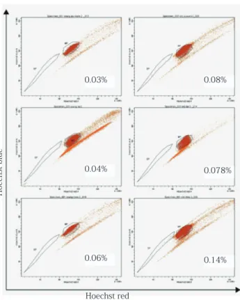
In young rats, the humerus, femur and ilium were composed of approximately 0.03%, 0.04% and 0.06% SP cells, respectively (Fig. 7). In aged rats, the humerus, femur and ilium were composed of approximately 0.08%, 0.078% and 0.14% SP cells, respectively (Fig. 7). In addition, higher proportion of SP cells were observed in the ilium of both young and aged rats compare to long bones. However, there were probably no difference among any of them.
Discussion
Many studies have described biological age-related changes in human and murine organs. In this study, we performed histological and immunohistochemical analyses of bone marrow cells derived from the femur, humerus and ilium of young and aged rats. Immunohistochemical staining was conducted with primary antibodies for PCNA, which is a cell proliferation marker, as well as for Bmi-1 and CD34, which are hematopoietic stem cell markers, and
Stro-1, which is a mesenchymal stem cell marker. 5-Bromo-2-deoxyuridine, which is incorporated into nuclear DNA during the S phase of the cell cycle, was used to label proliferating cells16). PCNA is a 36-kDa acidic nonhistone nuclear protein that functions as an auxiliary protein for DNA delta polymerase and is mandatory for DNA synthesis. 5-Bromo-2-deoxyuridine-positive cells, which may be progenitor cells, were observed in the bone marrows of young and aged rats. In addition, these cells were observed in different areas in the femur, humerus and ilium. Stro-1 is an early marker of different bone marrow mesenchymal stem cell and pre-osteogenic populations, which infers a possible perivascular niche for these cell populations in situ. The expression of Stro-1 has been shown to be progressively lost after cell proliferation and differentiation into mature osteoblasts in vitro17)-19). Bmi-1 and CD34 were used to detect bone marrow hematopoietic stem cells. In this study, Bmi-1 immunostaining indicated higher numbers of HSCs in young rats, and Stro-1 immunostaining showed an increase in the proportion of stem cells with increasing age. Therefore, these findings suggest that the qualitative reduction in stem cell function is compensated by an increase in the proportion of stem cells.
In addition, we conducted real-time PCR with primers specific to the following mRNAs: Cbfa1, which is a transcription factor that promotes osteoblastic differentiation, Bmi-1, which is a hematopoietic stem cell marker, and p16, which is a cell cycle inhibitor. Cbfa1 is activated at the onset of osteogenesis and is considered to be a robust marker of osteogenic commitment20)-22). p16-INK4A, a cell-cycle regulatory protein, interacts with CDK4 and CDK6, inhibits their ability to interact with cyclin D, and inhibits phosphorylation of the retinoblastoma protein by CDK4 or CDK6 and their entry into the S phase of the cell cycle. Expression of p16 increases as cells age, thereby reducing the capacity for self-replication8)23). Our results suggest that changes in Cbfa1 expression induce the active differentiation of stem cells into osteoblastic cells in aged rats. However, p16 and Bmi-1 expression were reciprocal,
Fig. 4: Immunohistochemical staining of Stro-1 in the humerus, femur and ilium of young and aged rats. Positive cells were observed throughout the bone marrow.
Original Magnification: x66. bar: 200 mm
Young Aged
Humerus
Femur
JJ S E D P Vol. 6 No. 1: , 2014
suggesting that Bmi-1 inhibits or delays cell aging and may be involved in mechanisms that maintain stem cell numbers.
Since SP cells from mouse bone marrow were first established by Goodell et al.6), their presence has been reported in many body organs24)-27). However, the proportion of SP cells varies depending on the species, tissue and organ. In this study, each type of bone marrow cell was isolated and SP cells were analyzed by FACS to determine the dye affinity with Hoechst 33342. Furthermore, we investigated the differences in the proportion of SP cells in the bone marrow of each type of bone. Moreover, this study found a difference in the proportion of SP cells between young and aged rats. There was a marked increase in the number of SP cells that were associated with aging, suggesting a quantitative compensation for qualitative reductions that occur with aging. The differences between the bone types, observed as a high number of SP cells in the ilium, is considered to support the widespread use of iliac bone marrow in clinical practice. Nanci et al.28) reported that undifferentiated mesenchymal stem cells decrease in number with age, as observed in this study, and Garvin et al.29) reported a decrease
Young Aged
Humerus
Femur
Ilium A
Fig.5 Immunohistochemical staining of Bmi-1 in the humerus, femur and ilium of young and aged rats.
Original Magnification: x66. bar: 200 mm
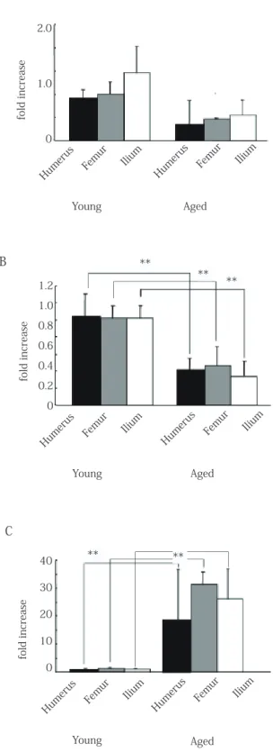
2.0 1.0 0 fold increase Humerus Femur Ilium Humerus Femur Ilium Young Aged ** 1.2 1.0 0.8 0.6 0.4 0.2 0 fold increase Humerus Femur Ilium Humerus Femur Ilium Young Aged ** ** ** 40 30 20 10 0 fold increase Humerus Femur Ilium Humerus Femur Ilium Young Aged **
Fig. 6 Mean mRNA expression levels. The levels of Cbfa1 (A), Bmi-1 (B) and p16 (C) mRNAs were normalized against ß-actin (**, p > 0.01; *, p > 0.05).
6A: Cbfa1 mRNA expression in the young rats was tendency higher in that of aged rats for any of bone.
6B: Bmi-1 mRNA expression in young rats was higher for all bone types, and the number of stem cells was significantly higher.
6C: p16 mRNA expression was higher in aged rats for all bone types, and the incidence of cell cycle inhibition increased with age.
A
B
C
in SP cell numbers in human bone marrow with age. However, Pearce et al.9) reported that numbers of HSCs and SP cells increase with age in mice and suggested that if the functionality of stem cells decreases with age, then the increased number of cells and the maintained quality would compensate for the absence of exclusive function.
These results suggest that the reduction in stem cell function is compensated by an increase in the proportion of stem cells in aged rat. Taken all together, the iliac bone marrow of both young and aged rats is considered appropriate for regeneration therapy.
Acknowledgements
We would like to thank Miss Saori Takano and members of the Department of Clinical Pathophysiology for their technical assistance.
References
1) Izadpanah R, Joswig T, Tsien F, Dufour J, Kirijan JC, Bunnell BA: Characterization of multipotent mesenchymal stem cells from the bone marrow of rhesus macaques, Stem cells and development, 14: 440-451, 2005
2) Majumdar MK, Thiede MA, Mosca JD, Moorman M, Gerson SL: Phenotypic and functional comparison of cultures of marrow-derived mesenchymal stem cells (MSCs) and stromal cells Journal of Cellular Physiology, 176: 57-66, 1998
3) Pittenger MF, Mackay AM, Beck SC, Jaiswal RK, Douglas R, Mosca JD, Moorman MA, Simonetti DW, Craig S, Marshak DR : Multilineage potential of adult human mesenchymal stem cells Science (New York, NY), 284: 143-147, 1999 4) Prockop DJ : Marrow stromal cells as stem cells for
nonhematopoietic tissues Science (New York, NY), 276: 71-74, 1997
5) Woodbury D, Schwarz EJ, Prockop DJ, Black IB : Adult rat and human bone marrow stromal cells differentiate into neurons J Neuroscience Res, 61: 364-370, 2000
6) Goodell MA, Brose K, Paradis G, Conner AS, Mulligan RC: Isolation and functional properties of murine hematopoietic stem cells that are replicating in vivo The J Experimental Medicine, 183: 1797-1806, 1996
7) Sherr CJ, DePinho RA: Cellular senescence: mitotic clock or culture shock? Cell, 102: 407-410, 2000
8) Beausejour CM, Campisi J: Ageing: balancing regeneration and cancer, Nature, 443: 404-405, 2006
9) Pearce DJ, Anjos-Afonso F, Ridler CM, Eddaoudi A, Bonnet D : Age-dependent increase in side population distribution within hematopoiesis: implications for our understanding of the mechanism of aging, Stem cells, 25: 828-835, 2007 10) Conboy IM, Rando TA: Aging, stem cells and tissue
regeneration: lessons from muscle, Cell cycle, 4: 407-410, 2005
11) Meyer KC: The role of immunity in susceptibility to respiratory infection in the aging lung, Respiration Physiology, 128: 23-31, 2001
12) Rando TA: Stem cells, ageing and the quest for immortality, Nature, 441: 1080-1086, 2006
13) Stenderup K, Rosada C, Justesen J, Al-Soubky T, Dagnaes-Hansen F, Kassem M: Aged human bone marrow stromal cells maintaining bone forming capacity in vivo evaluated using an improved method of visualization, Biogerontology, 5: 107-118, 2004
14) Asumda FZ, Chase PB: Age-related changes in rat bone-marrow mesenchymal stem cell plasticity, BMC Cell Biology, 12: 44, 2011
15) Maniatopoulos C, Sodek J, Melcher AH: Bone formation in vitro by stromal cells obtained from bone marrow of young adult rats, Cell Tissue Res, 254: 317-330, 1988 16) Rakic P: Adult neurogenesis in mammals: an identity crisis
The Journal of neuroscience: J Neuroscience, 22: 614-618, 2002
17) Ahdjoudj S, Lasmoles F, Oyajobi BO, Lomri A, Delannoy P, Marie PJ: Reciprocal control of osteoblast/chondroblast and osteoblast/adipocyte differentiation of multipotential clonal human marrow stromal F/STRO-1(+) cells, J Cellular Biochemistry, 81: 23-38, 2001
18) Gronthos S, Zannettino AC, Graves SE, Ohta S, Hay SJ, Simmons PJ: Differential cell surface expression of the Humerus
Femur
Ilium
Hoechst blue
Hoechst red
Fig. 7 Fluorescence-activated cell sorting (FACS) analysis. The rat bone marrow cells were analyzed for Hoechst 33342 efflux by FACS.
In young rats, the humerus, femur and ilium were composed of approximately 0.03%, 0.04% and 0.06% SP cells, respectively. In aged rats, the humerus, femur and ilium were composed of approximately 0.08%, 0.078% and 0.14% SP cells, respectively. Higher proportion of SP cells were observed in the ilium of both young and aged rats compare to long bones.
0.03% 0.08%
0.04% 0.078%
JJ S E D P Vol. 6 No. 1: , 2014 STRO-1 and alkaline phosphatase antigens on discrete
developmental stages in primary cultures of human bone cells Journal of bone and mineral research, J American Soci Bone Mineral Res, 14: 47-56, 1999
19) Stewart K, Walsh S, Screen J, Jefferiss CM, Chainey J, Jordan GR, Beresford JN: Further characterization of cells expressing STRO-1 in cultures of adult human bone marrow stromal cells Journal of bone and mineral research, J American Soci Bone Mineral Res, 14: 1345-1356, 1999
20) Ducy P: Cbfa1: a molecular switch in osteoblast biology Developmental dynamics, an official publication of the American Association of Anatomists, 219: 461-471, 2000 21) Nishimura R, Hata K, Harris SE, Ikeda F, Yoneda T: Core-binding factor alpha 1 (Cbfa1) induces osteoblastic differentiation of C2C12 cells without interactions with Smad1 and Smad5, Bone 31: 303-312, 2002
22) Satomura K, Krebsbach P, Bianco P, Gehron Robey P: Osteogenic imprinting upstream of marrow stromal cell differentiation, J Cellular Biochemistry 78: 391-403, 2000 23) Kim WY, Sharpless NE: The regulation of INK4/ARF in
cancer and aging, Cell, 127: 265-275, 2006
24) Challen GA, Bertoncello I, Deane JA, Ricardo SD, Little MH: Kidney side population reveals multilineage potential and renal functional capacity but also cellular heterogeneity, J American Soc Nephrology JASN, 17: 1896-1912, 2006 25) Uezumi A, Ojima K, Fukada S-i, Ikemoto M, Masuda S,
Miyagoe-Suzuki Y, Takeda Si: Functional heterogeneity of side population cells in skeletal muscle, Biochemical Biophysical Res Commun, 341: 864-873, 2006
26) Yamahara K, Fukushima S, Coppen SR, Felkin LE, Varela-Carver A, Barton PJR, Yacoub MH, Suzuki K: Heterogeneic nature of adult cardiac side population cells, Biochemical Biophysical Res Commun, 371: 615-620, 2008
27) Zhang L, Hu J, Hong T-P, Liu Y-N, Wu Y-H, Li L-S: Monoclonal side population progenitors isolated from human fetal pancreas, Biochemical Biophysical Res Commun, 333: 603-608, 2005
28) Nanci A SA, Simmer J: Dentin-pulp Complex,Ten Cate’s Oral Histology, St Louis: CV Mosby, pp, 191–238, 2008 29) Garvin K, Feschuk C, Sharp JG, Berger A: Does the number
or quality of pluripotent bone marrow stem cells decrease with age? Clinical Orthopaedics and Related Res, 465: 202-207, 2007