近畿大学学術情報リポジトリ
32
0
0
全文
(2) 2. Memoirs of The School of B.O.S.T. of Kinki University No. 5 (1999). studied well. In particular, the effects of hierarchical substructures of tendons and ligaments on the remodeling remain almost unknown. As a step for the understanding of the mechanisms of the remodeling phenomenon, we have been doing a series of microscopic studies on the biomechanical properties and the remodeling of collagen fascicles, which are substructure of tendons and ligaments. This paper summarizes our experimental findings on the biomechanical properties of collagen fascicles obtained from (1) non-treated tendons, (2) stress-shielded tendons, .and (3) frozen and stress-shielded tendons. In addition, the article deals with a review on previously published macroscopic studies related to the biomechanical properties and the remodeling of tendons and ligaments, and comparison the remodeling of microstructure to that of bulk tendons and ligaments. 2. Biomechanical Properties of Collagen Fascicles from Patellar Tendons. Over the past two decades, extensive studies have been performed on the mechanical and structural properties of knee joint tendons and ligaments [2 - 4 J. These tendons and ligaments contain 70 to 80% collagen by dry weight [5 - 8 J; besides collagen, they contain elastin, proteoglycans, glycolipids, cells, and water. Collagen is a basic structural component of soft and hard tissues in animals, and gives mechanical integrity and strength to their bodies with different structures in different tissues and organs. The collagen in tendons and ligaments has a hierarchical substructure composing of collagen fascicles, fibrils, and molecules [8 -10J. Therefore, basic knowledges of the structure and mechanical properties of such substructures as collagen fascicles and fibrils are essential to knee joint biomechanics.. 2.1 Materials and Methods Skeletally matured female Japanese white rabbits aged 6 months were used for the experiments. Collagen fascicles having the diameter and length of approximately 300 f.1 m and 15 mm (Fig. 1), respectively, were very carefully dissected with a surgical knife in parallel to the axis of the patellar tendon. During the resection of patellar tendons and the dissection of collagen fascicles, tendon substance was kept moist with physiological saline solution of room temperature. A newly designed apparatus was used for the measurement of the diameter of collagen fascicles (Fig. 2). The lateral image of a specimen immersed in physiological saline solution of 37°C was enlarged by a low magnification microscope, taken with a CCD camera, and then processed with a video dimension analyzer. The diameters were measured from 36 directions, while the fascicle was intermittently rotated with a stepping motor at the angular interval of 5 degrees. The cross-sectional area was calculated from averaging these diameters, assuming that the cross section is circular..
(3) 3. Fig. 1 A collagen fascicle and its cross-sectional shape.. Stepping motor Video dimension analyzer Fascicle CCO camera. Thermostatic bath \. Microscope. Weight. Fig. 2 Apparatus for the measurement of cross-sectional area. A specially designed micro tensile tester was used to study the mechanical properties of collagen fascicles (Fig. 3). After measuring the cross-sectional area of each fascicle, small acrylic blocks were attached to both ends. One of the blocks was fixed to a load cell, and the other one was attached to the crosshead of a linear stage. Tensile tests were carried out by moving the stage with a microprocessor-controlled stepping motor via a ball screw. For the.
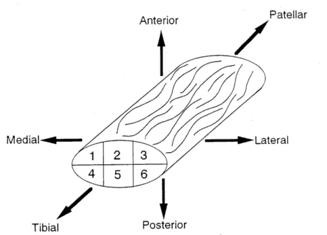
(4) 4. Memoirs of The School of B.O.S.T. of Kinki University No. 5 (1999). measurement of strain, two markers were drawn on a collagen fascicle with a stain (Nigrosine) about 5 mm apart; the strain measured with this method is the strain on specimen surface [2, 11]. The distance between the makers was measured with the above-mentioned video dimension analyzer using tracking function. During tensile testing, specimens were immersed in physiological saline solution of 37°C. Tensile load and the distance between the above-mentioned two markers w'ere recorded on a personal computer. Stress was calculated from dividing tensile load by the initial cross-sectional area of each fascicle; strain was determined from dividing the increment of the distance between the two markers by the initial distance. From' these data, stress-strain curves, tensile strength, and strain at failure were obtained.. X-T recorder. A. I Video dimension analyzer. Program controller. I -. "'-. Linear stagl9 Ball screw. Stepping ~. I. lo:~r 11. I. ~. ceo camera. Pulse generator. ~. Load cell. /. Fascicle. \~f-~\. :=. \. -. \. I. .=] Computer I. " ., J ~ I. 7. ,. l. H. I I. r-... Thermostatic bath. I I. -----. Test bath I. Fig. 3 Micro tensile tester used to determine the mechanical properties of collagen fascicles. Patellar tendons obtained from the left knees were used to study locational dependence of the mechanical properties of collagen fascicles. Each tendon was transversely divided into 6 blocks (Fig. 4). Five collagen fascicles were obtained from each block in the above-mentioned manner. First a preload of 0.01 N was applied to a fascicle, and zero strain was defined at this load. Then, each fascicle was preconditioned with ten cycles of loading and unloading between 0 and 2 % strain at the strain rate of approximately 1.5 % / sec followed by stretch to failure at the same rate. Other patellar tendons obtained from the left knees were used for relaxation tests. -Five collagen fascicles were randomly obtained from the medial and lateral one-thirds of each tendon. Five of the 6 remaining central one-third tendons were used for the relaxation test of bulk patellar tendons which is ·described in the next section. Following the same.
(5) 5. preconditioning as that described above, these fascicle specimens were deformed to approximately 2 % strain (2.1 ± 0.4%) at a rate of approximately 1.5 %/ sec and, then, load was measured for the period of 350 seconds while maintaining the strain. Anterior. ~.Lateral. Medial ....... ~....-. 2. 4. /. Tibial. Patellar. 3. 5. 6. l. Posterior. Fig. 4 Sampling location of collagen fascicles in a whole patellar tendon.. 2 . 2 Results and Discussion To our knowledge, this is the first study that determined the mechanical properties of collagen fascicles obtained from the rabbit patellar tendon. Stressstrain relations of collagen fascicles had initial toe regions, followed by linear portions between 2 and 5 % strain and finally by decreased slopes at higher strain (Fig. 5). There were no statistically significant differences in the tensile properties among 6 locations (Fig. 6). Averaged tangent modulus, tensile strength, and strain at failure of all collagen fascicles (210 specimens) were 216 ± 68 MPa, 17.2 ± 4.1 MPa, and 10.9 ± 1.6%, respectively, where the tangent modulus was defined by the slope of a stress-strain curve between 2 and 5 % strain. Biomechanical studies of non-reconstituted, chemically non-treated raw collagens have been done mostly on those harvested from the rattail tendon. Their tensile strength varied from 10 to 110 MPa, primarily depending on strain rate and animal age [11-14J. Mechanical properties of reconstituted collagen fibers have also been studied rather well, and their tensile strength ranged from 8 to 91 MPa [14-17]. These values are similar to those of non-reconstituted raw collagens obtained from intact rat tail tendons. Kato et al. [14J studied the tensile properties of rat tail tendon fibers of 50-100 f1 m in diameter, and reported that their tangent modulus, tensile strength, and strain at failure determined at 100 %/ min strain rate were 478 ± 130 MPa, 32.6 ± 12.0 MPa, and 6.7 ± 3.1 %, respectively. These tangent modulus and tensile strength were much larger than those obtained from the present study. These differences may.
(6) Memoirs of The School of B.O.S.T. of Kinki University No. 5 (1999-). be attributable to the differences of animal species (rat vs. rabbit) and tissues (tail tendon vs. patellar tendon) because of different body weight and activities in different animals, different load applied to different tissues, and so on. In addition, different methods for the measurement of cross-sectional area (calibrated eyepiece of microscope vs. VDA) may have caused the different results. In contrast to many studies of mechanical properties of collagen fibers obtained from rat tail tendons, there have been no studies on the micromechanical properties of stress-bearing tendons and ligaments, except for the work done by Wilmink et al. [18J on the collagen fibers obtained from the equine flexor tendon. They reported that the elastic moduli of the collagen fibers of 100 to 200 Jl m in diameter obtained from the superficial digital flexor tendon were 135 ± 18 and 139 ± 30 MPa for young and old groups, respectively. These moduli were smaller than the tangent modulus obtained from the present study. This difference may be also attributable to the differences of animal species (horse vs. rabbit) and tissues (flexor tendon vs. patellar tendon).. 30. -+--. 1. --0-. 2 3 Location number --{J4 --A--- 5 --IS- 6. -.....-. .CO D.... ~. 20. (n=7, Mean ± S.D.). o U) C/). ~. Cf). 10. +. Breaking point. o~-~-~~-~~~~--~~~~~-~~~. o. 5. 10. 15. Strain E (%) Fig. 5 Stress-strain relations of collagen fascicles obtained from different locations. The relations were essentially similar among 6 locations..
(7) 7. 400 (n = 7, Mean ± S.D.). as 0... e 300 I-. LU (f). -§. 200. "C. o. E. ~C) 100 c: CI1. Io~~~~~~~~--~--~. 2. 3. 456. Location number. 30 (n = 7,Mean ± S.D.). :5 C) c:. ~. ~ .Ci.j. 10. c:. ~. 2. 3. 456. Location number. 20 (n. =7, Mean ± S.D.). c:. ·cu 5. ~. o~~~~~~~~--~--~. 2. 3. 456. Location number. Fig. 6 Tensile properties of collagen fascicles obtained from each location. One-way ANOVA indicated no significant differences in each parameter among locations. Mechanical properties of the collagen fascicles obtained from patellar tendons were greatly different from those of bulk tendons (Fig. 7) ; for example, their tensile strength was approximately 42 % of that of the tendons. This difference is possibly attributable to such interactions as frictional force between collagen fascicles as well as between collagen fascicles and ground substance. To study the effects of these factors, we carried out tensile tests of the patellar tendon.
(8) 8. Memoirs of The School of B.O.S.T. of Kinki University No. 5 (1999). specimens which were split into collagen fascicles. Their tensile strength was approximately 67 % of that of the normal bulk tendons, which implies a contribution of the above--mentioned interactions to the strength of bulk tendons. However, the strength and mechanical behavior of split tendons were not the same as to those of isolated fascicles. This difference may be ascribed to the difference in the diameter of collagen fasciCles between both cases and, therefore, to that in the total surface area of separated fascicles. We could not split tendons into fascicles of 300 f1 m in diameter, preserving bone attachments with no damage. The strain to failure of the isolated collagen fascicles was 178% of that of the bulk tendons. In addition to the effects of the interaction between collagen fascicles and ground substance, the multiformity of the crimp morphology of collagen fibrils in bulk tendons, which is defined by non-uniform crimp interval and amplitude, may be a reason for the difference. Fibrils in collagen fascicles and bulk tendons both have undulating crimp patterns. However, crimp interval and amplitude are different between them, which may have resulted in different strain to failure.. 60. -. 50 .. (Mean ± S.D.) Patellar tendon (n = 7). ct1. a.. ~. 40 Split patellar tendon (n. = 6). b 30 en en Q). ~. +oJ. Cf). 20 10. 5. 10. 15. Strain E (%) Fig. 7 Stress-strain curves of bulk patellar tendons, patellar tendons with split into collagen fascicles, and collagen fascicles. There were no statistically significant differences in the tensile properties of collagen fascicles among 6 locations (Figs. 5 and 6). No study has been done on the locational dependence of the mechanical properties of collagen fascicles which have the diameter of about 300 f1 m. However, several investigators [3, 19-21J have tested multiple tendons and ligaments divided into several· fascicles. For example, Butler et al. [19J reported that there were no significant differences.
(9) 9. in the mechanical properties between the central one-third and the medial onethird of the human patellar tendon. This result is similar to the result obtained from the present experiment. Yamamoto et al. [2 ] showed in the rabbit patellar tendon that the mechanical properties of the medial one-third were essentially similar to those of the central one-third, although the lateral one-third had 16% higher tensile strength than the other portions. Chun et at. [22J "reported that the tensile strength of the most medial one-sixth fascicle .. bone unit cut out from a human patellar tendon was significantly lower than those of the central and the lateral one-sixth units. These results are somewhat different from the present results. Collagen fascicles are relatively thin, and collagen fibrils in the fascicles are straighter and are aligned in more parallel than those in tendons. In addition, effects of the bone insertion on the orientation of collagen fibrils should disappear in collagen fascicles. Therefore, the anatomical orientation of collagen fascicles, which was functionally designed, has been changed in isolated collagen fascicles. For these reasons, the locational dependence of the mechanical properties may have been different between collagen fascicles and divided tendons with bone ends. It is also presumed that the interaction between fascicles is different at different locations of the patellar tendon, and this difference may have induced the locational dependence of the mechanical properties of the tendon. Stress relaxation was initially rapid but became gradual with time in both collagen fascicles and patellar tendons. Stress relaxation was smaller in collagen fascicles than in patellar tendons; stress decreased to approximately 70% and 50 % of the initial stress at 300 seconds in collagen fascicles and bulk tendons, resp ectively (Fig. 8), and the difference was statistically significant. Viscoelastic properties of tendons and ligaments have been studied by many investigators [23-26J. Johnson et al. [26J reported that the reduction of stress in human patellar tendons was about 40% at 300 seconds. This result is fairly similar to the present result on the rabbit patellar tendon. There have been no studies on the relaxation and viscoelastic characteristics of collagen fascicles and fibrils. Such a larger stress relaxation in bulk tendons than in collagen fascicles might be attributable to the contribution of ground substances between fascicles as well as to the wavy structure of fibrils in tendons. Furthermore, it may be ascribed to difference in swelling in saline solution and different rates of change in tissue water content caused by different specimen geometry like the ratio of surface to volume. The initial stresses applied to collagen fascicles (2.3 ± 0.5 MPa) and bulk tendon (6.7 ± 2.9 MPa) were approximately 13.2 and 11.8% of their tensile strength, respectively. Because the difference between these percentages was very small, the difference of initial stress should have no influence on the difference in stress relaxation..
(10) 10. Memoirs of The School of B.O.S.T. of Kinki University No. 5 (1999). - 100. ~ c:. 0 ctS >< ctS. "..j:j. CD "en en ()) "-. +-'. '2-Q"2-§-o.o-ON Collagen fascicle (n =6). 80. ~ITI. 60. 40. Patellar tendon (n. en. '1:J. ()) (.). 20. = 5). (Mean ± S.D.). ~. '1:J. ()). a:. 0. 0. 100. 200 Time T. 300. 400. (sec). Fig. 8 Relaxation curves of collagen fascicles and patellar tendons. Initial strains were 2.1 ± 0.4 and 1.9 ± 0.5% for collagen fascicles and patellar tendons, respectively. Patellar tendons developed significantly larger stress relaxation than collagen fascicles. 3. Mechanical Properties of Collagen Fascicles from Stress-Shielded Patellar Tendons In response to applied mechanical stress, living tissues and organs change their dimensions and properties. This phenomenon is called remodeling or functional adaptation. Several recent studies have shown that fibrous connective tissues such as tendons and ligaments also have this ability, and the effects of stress deprivation and stress enhancement on the biomechanical properties of tendons and ligaments have been studied extensively. For example, the effects of stress deprivation have been experimentally studied in immobilized animal knees [27-30]. Woo et al. [30J reported that knee immobilization decreased the strength of the medial collateral ligament in the rabbit. On the other hand, several studies have shown that exercise and training increased the strength of knee joint tendons and ligaments [31-33 J. Hayashi and his colleagues [34 J developed novel experimental techniques for changing stress more quantitatively than immobilization and exercise. They have applied the techniques to a series of studies on the biomechanical, morphometrical, and histological responses of knee joint tendons and ligalnents to stress [34-36 J, and have documented the phenomena of the remodeling of knee joint tendons and ligaments. However, the mechanisms have not been studied well. Collagen is a major protein of tendons and ligaments, and comprises 70 to 80 % of their dry weight [6 - 8 J. Tendons and ligaments have a hierarchical structure composing of collagen fascicles, fibrils, and molecule [9 J. Besides collagen and water, they contain cells, proteoglycans, fibronectin, elastin, actin,.
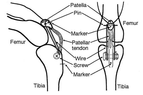
(11) 11. and a few other glycoproteins. The effects of each structural component on the remodeling of tendons and ligaments remain almost unknown; few biomechanical studies have been done even on collagen fascicles and fibrils, in spite of the fact that basic knowledge of their mechanical properties is essential not only to tendon and ligament biomechanics but also to the development of reconstruction methods for damaged tendons and ligaments using autografts.. 3.1 Materials and Methods Animal experimentation was conducted under the Guidelines of the Committee for Animal Care and Use of Osaka University. Mature female Japanese white rabbits were used for the experiments of 1, 2, and 3 week stress shielding. Surgery was applied to the right knee under intravenous pentobarbital anesthesia in a fashion similar to that done by Yamamoto et al [37J After a stainless steel pin (1 mm diameter) and a stainless steel screw (3 mm diameter) were inserted into the patella and the tibial tubercle, respectively, two stainless steel pin markers (0.7 mm diameter) were embedded in the patella and the tibial tubercle, and the distance between them was measured using a caliper (Fig. 9). Then, a flexible stainless steel wire (0.97 mm diameter) was hooked between the pin and the screw, and the patellar tendon was slackened by pulling the patella toward the tibial tubercle using the wire. When the distance between the pin markers was shortened by approximately 6 mm from the original distance, the wire was fixed. We confirmed from visual inspection that the patellar tendon was loosened at all knee flexion angles. Postoperatively, no immobilization treatment was applied, and the animals were allowed daily activities in cages for 1, 2, or 3 weeks until sacrifice. During the period, the rabbits did not favor their right legs except for one day after surgery.. Tibia Fig. 9 Method for stress shielding.. Tibia.
(12) 12. Memoirs of The School of B.O.S.T. of Kinki University Nn 5 (1999). Each animal was killed by an overdose injection of thiamylal sodium under pentobarbitone anesthesia. After sacrifice, the patellar tendon with the entire patella' and tibia (patellar tendon-bone complex) was cut 'out from each limb, and the surrounding tissue was removed carefully and thoroughly. The cross~ sectional area of each patellar tendon (Aw) was measured using an area micrometer under a constant pressure of' 0.12 MPa. The device design and measurement procedure were reported previously [2 J. The length of the patellar tendon (La) was defined as the distance between the distal end of the patellar attachment of the patellar tendon and the proximal end of the tibial attachment on the dorsicentrai tendon surface. It was measured with a caliper while a constant tensile load of 0.5 N was applied to the patellar tendon in the axial direction by suspending a weight with thread.' After the tibia was attached to a holder and the patella was picked up with a forceps, the patellar tendon was pulled lightly by moving the forceps. Each patellar tendon was divided into 6 blocks (Fig. 3). Collagen fascicles having the diameter and length of approximately 300 f.1 m and 15 mm, respectively, were carefully dissected from each block by moving a surgical knife in parallel to the axis of. the patellar tendon. The cross-sectional area was calculated from averaging the diameters of each collagen fascicles, assuming that the cross section was circular. The details of apparatus and procedures for the determination of cross-sectional area have been described previously [38J. The mechanical and viscoelastic properties of the fascicles were determined a micro tensile tester specially designed for collagen fascicles [38l Stress was calculated from dividing tensile load by the initial cross-sectional area of each fascicle; strain was obtained from the distance between the above-mentioned two markers and the initial distance. From each patellar tendon-bone complex prepared for histological observation, approximately 3 mm long patellar tendon substance was sampled for the observation of the transverse cross section. These histological specimens were stained with hematoxylin and eosin after fixed in a buffered 10 % formalin solution, decalcified, and cast in paraffin blocks for sectioning. The numbers of fibroblasts per 1 mm 2 in different 5 areas in each location shown in Fig. 3 were determined using an image analyzer, and averaged.. 3.2 Results and Discussion Stress shielding significantly increased the cross-sectional area of patellar tendons (Table 1). The length of 2 - and 3 -week stress-shielded tendons were significantly shorter than that of the control tendon, although there was no significant difference between the control and the 1-week stress-shielded tendons. The dimensions and lengths of the stress-shielded patellar tendons were very similar to the results obtained by Yamamoto et a1. [37J which indicates that the experimental model is of high reproducibility..
(13) 13. Table 1 Dimensions of the control and the stress-shielded patellar tendons, expressed as mean (SD). Groups Stress-shiel ded Dimension. Control. 1 wk. 2wks. 3 wks. Whole cross-sectional area Aw (mm2). 14.1 (1.7). 18.9 (0.9)*. 32.8 (3.4)*. 31.7 (3.5)*. Length Lo (mm). 19.2 (0.2). 19.4 (0.9). 18.4 (0.6)*. 15.2 (1.4)*. * Significantly different from Control group (p <: 0.05).. The stress-strain relations of collagen fascicles were essentially similar among 6 locations either in the control or in the stress-shielded patellar tendons (Fig. 10). No significant effects of fascicular locations on the tensile strength (Fig. 11), tangent modulus, and strain at failure were observed by one-way ANOV A at any period, where the tangent modulus was defined by the slope of a stressstrain curve between 2 and 5 % strain in the control, and 1 - and 2 -week stress-shielded collagen fascicles, and between 1, and 2.5% strain for the 3 -week stress-shielded ones. Therefore, these mechanical parameters were averaged for all the locations in each tendon. There have been many studies on the change of the mechanical properties of tendons and ligaments which occurs in response to immobilization and exercise. However, few studies have been done on the biomechanical changes of substructural components of tendons and ligaments. In the present study, we studied the mechanical properties of collagen fascicles which were obtained from stress-shielded rabbit patellar tendons. The tangent modulus progressively and markedly decreased with increase in the period of stress shielding, and statistically significant differences between the control and the stress-shielded collagen fascicles were observed at 2 and 3 weeks (Fig. 12). The tensile strength of collagen fascicles decreased more rapidly and markedly than the tangent modulus: it decreased to approximately 74%, 44%, and 19% at 1, 2, and 3 weeks, respectively, compared to the control fascicles. The decrease of strain at failure was less marked than those of tangent modulus and tensile strength; however, there were also statistically significant differences in the strain at failure between the control and the 2 - and 3 -week stress-shielded fascicles. Yamamoto et al. [37J studied the effects of stress shielding on the mechanical properties of bulk patellar tendons in the rabbit, and found that stress shielding progressively and markedly decreased the tangent modulus and tensile strength. These decreases were very much different from those in collagen fascicles observed in the present study; for example, the tensile strength of collagen fascicles decreased to 74%, 44%, and 19% of the control value at 1, 2, and 3 weeks, respectively, while that of bulk tendons decreased to 50%, 13%, and 9 %, respectively. The tangent modulus also showed differences essentially similar to the tensile strength..
(14) 14. Memoirs of The School of B.O.S.T. of Kinki University No. 5 (1999). 25. --+- 1 --0-. 2. -~ 3 Location number. -. 4 -....- 5 --I::r-- 6. --0-. 20. CCS. a. ~15 c. ~ 10 ~. +-'. (J). 5. o. o. 5 Strain. c. 15. 10 (%). Fig. 10 Stress-strain relations to failure of the control and the stressshielded collagen fascicles obtained from different locations. There were no significant differences in the relations among 6 locations. To avoid complication, only mean values are shown in this figure.. o. -m. 30. a.. ~. Control (n = 7). ~ Stress-shielded for 1 wk (n = 6). Stress-shielded for 2 wks (n =6) ~ Stress-shielded for 3 wks (n = 6) Mean:t S.D.. §. t;D. o. ..c: +-'. 20. 0'). C. Q). ~. +-'. en Q). ·00. 10. c. Q). I-. 1. 2. 3. 4. 5. 6. Location number Fig. 11 Tensile strength of collagen fascicles obtained from each location. One-way ANOVA indicated no significant difference among locations..
(15) 15. o. Control (n. =7). r.;a Stress-shielded for 1 wk (n = 6) EJ Stress-shielded for 2 wks (n = 6) lm Stress-shielded for 3 wks (n = 6). -. 25. OJ. a.. ~. I-. W200 ::J ::J. E 100. +-'. C. b ....-. ~ (J). C. OJ. CD. '-. ::J. -ca 'ffi. 10. CD. c 5 'ffi. CD. (f). '00 5 C. CD. 0). I-. CD. C. o. --. 15. W 10. ID. 0>. "0. Mean:t S.D.. ~ 0. 20. ..c:: 15. (J). * P < 0.05, vs Control. '-. +-'. I-. o. 0. 0. Fig. 12 Mechanical properties of the control and the stress-shielded collagen fascicles (Averaged for all locations). Our previous study on the mechanical properties of collagen fascicles obtained from non-treated, normal patellar tendons [38J suggested that the strength of bulk patellar tendons is possibly attributable not only to the strength of collagen fascicles but also to the mechanical properties of ground substances and mechanical interaction between fascicles. The larger response of bulk tendons to stress shielding compared to collagen fascicles may indicate that the degenerative changes of patellar tendons caused by stress shielding are ascribed to those of collagen fascicles as well as of interaction between fascicles. It has been demonstrated that immobilization induces substructural changes of periarticular fibrous tissue, including reduction of water and glycosaminoglycans [39J, and increase in the number of reducible cross-links [40J, No differences in collagen type [41J nor collagen mass between immobilized and control fibrous tissues have been detected [42J, These results also indicate that changes in ground substance and interaction between collagen fibrils may play an important role in the remodeling of tendons and ligaments. Biochemical and microstructual studies on the effects of stress shielding on tendons and ligaments are suggested. The number of fibroblasts increased markedly with the time of stress shielding; dense and plump fibroblasts were observed in the stress-shielded patellar tendons (Table 2 and Fig, 13). However, there were almost no locational dependences not only in the control patellar tendons but also in the stress-shielded ones at each period. These results are similar to those obtained from the longitudinal sections of stress-shielded patellar tendons [37J. The increase of fibroblasts is possibly attributable to the proliferation of intrinsic fibroblasts and the invasion of fibroblasts from surrounding tissues, most probably from infrapatellar fat pad [43 J. It was expected that non-uniform surrounding tissues induced.
(16) 16. Memoirs of The School of B.O.S.T. of Kinki University. No. 5 (1999). locational differences of fibroblast number and mechanical properties of collagen fascicles. However, there were no remarkable locational differences in the fibroblast number on the transverse cross sections of stress-shielded patellar tendons (Table 2), which was consistent with the result that the mechanical properties of collagen fascicles were independent of the cross-sectional locations. Location number 2. 3. 5. 4. 6. Control. Stress-shielded (1 wk). Stress-shielded (2 wks). Stress-shielded (3 wks). 100 pm. Fig. 13 Microphotograph of each location of the control and the stressshielded patellar tendons (stained by hematoxylin and eosin).. Table 2 Number of fibroblasts per mm 2 in each location of the control and the stress-shielded patellar tendons. Mean (SD) values for 5 areas are shown. Groups Stress-shielded Location number. Control. 1 wk. 2 wks. 3 wks 1756 (371). 1. 275 (50). 382 (178). 2122 (610). 2. 283 (110). 382 (95). 1977 (765). 1884 (545). 3. 198 (55). 641 (190). 1366 (456). 2456 (575). 4. 275 (121). 412 (145). 1534 (318). 2366 (611). 5. 229 (50). 366 (139). 1420 (404). 2122 (616). 6. 252 (95). 443 (85). 2061 (659). 2114 (411). Stress relaxation was initially rapid but became gradual with time, and was larger in the stress-shielded collagen fascicles than in the control fascicles (Fig. 14). The control fascicles showed 38.8 ± 7.9% decrease in stress at 600 seconds, whereas the fascicles stress-shielded for 1 and 3 weeks exhibited mean 49.3 ± 2.1% and mean 52.0 ± 7.9% decrease, respectively. The differences between the control group and stress-shielded groups were statistically significant; however,.
(17) 17. there were no significant differences among the' stress-shielded groups. In constant to many studies of the viscoelastic properties of normal tendons and ligaments [24, 26J, there have been few studies on remodeled tissues. In the present study, the stress relaxation of stress-shielded collagen fascicles was larger than that of control fascicles. Ground substances such as proteoglycan are considered to be closely related to the viscoelastic behavior of tendons and ligaments [44J. The result from the present relaxation tests implies that the characteristics and mass of ground substances were changed by stress shielding. ----- Control Stress-shielded for 1 wk - - 0 - Stress-shielded for 2 wks --t.r- Stress-shielded for 3 wks. 100 .. --. 80. 0. 60. ~ 0. c:. ~. ctS X ctS. CD. --0-. 40. (n = 6, Mean± S.D.). ex: 20. o~~~~~~~~~--~~--~--~--~~. o. 100. 200. 300. 400. 500. 600. 700. Time T (sec) Fig. 14 Relaxation curves of the control and the stress-shielded collagen fascicles. Initial strains were 2.5 ± 0.2, 2.2 ± 0.5, 2.4 ± 0.3, and 2.1± 0.5% (Mean ± S.D.) for the control, 1 -, 2 -, and 3 -week stress-shielded collagen fascicles , respectively. Patellar tendon grafts have commonly been used for the reconstruction of damaged anterior cruciate ligaments. A number of investigators have reported that the strength of the' grafts decreases soon after reconstruction and then gradually increases with time [45-47]. The early reduction of graft strength is a serious problem, because it leads to failure of the grafts during postoperative rehabilitation. To solve this problem, synthetic augmentation devices have been developed and used clinically [48-51]. However, many studies have shown that the augmentation devices shield the stress to be exerted on the grafts, which leads to weakening and resorption of the grafts [48, 51]. Although stress shielding is one of the most important problems in the augmentative reconstruction of the anterior cruciate ligament, its effects on graft strength have not been fully documented, and the mechanisms have not been understood well. The results obtained from the present study would be useful as basic knowledge for the reconstruction of damaged anterior cruciate ligaments using autografts..
(18) 18.. Memoirs of The School of B.O.S.T. of Kinki University No. 5 (1999). 4. Mechanical Properties of Collagen Fascicles from In-Situ Frozen and Stress-Shielded Pa.tellar Tendons. One of the standard methods for the reconstruction of injured anterior cruciate ligaments (ACLs) is the replacement with autogenous grafts; the patellar tendon, iliotibial band, hamstring muscle, and semitendinous tendon have been often used for this purpose. After transplantation into the intraarticular environment, the grafts manifest a variety of changes. One of the earliest changes is the ischemic necrosis of fibroblasts [52-55J. This occurs within 2 to 4 weeks after surgery, followed by revascularization and cellular proliferation in 4 to 20 weeks [52, 53, 55J. Eventually, graft strength decreases soon after reconstruction and, then, gradually increases with time [51, 56, 57J. However, the strength recovers to only 50 % or less than 50 % of that of the control ACL even 2 years after reconstruction [56-58J. The early reduction of graft strength is a serious problem, because excessive stress leads to the failure of grafts during postoperative rehabilitation. To overcorne this problem, augmentation devices are commonly used, and many experimental studies have been done to evaluate their feasibility [49-51, 59, 60J. Some of those studies have shown that augmentation is effective for the reconstruction of the ACL [49, 50J, while others have indicated that it does not work [51, 59J. With this technique, mechanical stress applied to the grafts is reduced or completely removed during recovery process, which strongly influences the results of the treatment. However, the effects of stress shielding on the mechanical strength of autogenous grafts have not been studied well. To know the effects of stress shielding, Yamamoto et al. [37J and Ohno et al. [61] have developed a unique experimental technique, and have applied it to an autograft model using the rabbit patellar tendon [61-64J. In these experiments, the patellar tendon was frozen in situ to kill fibroblasts without surgically disturbing the anatomical orientation, physiological tension, and bone attachment of the tendon, considering that fibroblast necrosis occurs in autografts transplanted for the reconstruction of the ACL. Then, tension in the patellar tendon was removed or reduced by pulling a metallic wire or a textile strand hooked between the patella and the tibial tubercle. The results have indicated that the tangent modulus and tensile strength of the patellar tendon were significantly decreased by complete stress shielding even if there existed no fibroblasts. However, the mechanisms of the decrease in strength have not been studied yet. Tendons and ligaments have hierarchical substructures composing of closely packed collagen fascicles, fibrils, and molecules, which are oriented in their axial direction so as to resist tensile load. They also contain elastin, proteoglycans, glycolipids, cells (fibroblasts), and water. In these components, fibroblasts are thought to play an important role in the response of tendons and ligaments to stress, because they synthesize and digest collagen and their metabolism changes depending on environmental stimuli and functional needs, which alters the ultrastructure, chemistry, and mechanical properties of tendons and ligaments [36J. If there are no fibroblasts, the properties of collagen fascicles and fibrils should not change in response to stress. Therefore, we hypothesized that the mechanical properties of collagen fascicles in autografts is.
(19) 19. not changed by stress shielding. To examine the hypothesis, we carried out tensile tests of the collagen fascicles obtained from in situ frozen and stressshielded patellar tendons.. 4.1 Materials and Methods Skeletally mature female Japanese white rabbits were used for the experiment. They were divided into two groups: frozen (Fr) and frozen/stress-shielded (Fr /SS) groups. Surgical treatments were applied to their right patellar tendons, while non-treated left patellar tendons were used to obtain control data. Rabbits from each group were killed at 2, 3, and 6 weeks, respectively, after surgery; animals were used for biomechanical testing and histological observation. Surgery was performed under intravenous pentobarbital anesthesia. The anterior part of the right knee was exposed through a midline longitudinal skin incision, and two stainless steel pins (0.7 mm diameter) were embedded in the patella and in the tibial tubercle as markers (Fig. 15). The subcutaneous retinacula were incised longitudinally along the side edges of the patellar tendon and, then, the posterior surface of the tendon was separated from the infrapatellar fat pad. After these preparations, the patellar tendon was frozen in situ by the procedure reported elsewhere [61, 65J. Briefly, a patch of silicone rubber sheet was inserted between the patellar tendon and the fat pad to make a bath; the distal half of the patella and the whole tendon substance except for the tibial attachment were included inside the bath (Fig. 15). A wood piece was placed between the silicone sheet and the fat pad to prevent the fat pad from freezing. The silicone sheet bath was quickly filled with liquid nitrogen, and the patellar tendon was immersed in the solution for 1 minute. Then, physiological saline solution was poured in the bath to thaw the frozen tendon. After both side edges of the patellar tendon were sutured to the retinacula with 4 - 0 nylon thread, the distance between the two markers (initial distance) was measured with a caliper. For the Fr group, the skin was closed with a standard technique, followed by the application of temporary dressing. Patellar tendon. Liquid nitrogen. ....- - Marker. Femur Fig. 15 Schematic diagram of in situ freezing technique..
(20) 20. Memoirs of The School of B.O.S.T. of Kinki University No. 5 (1999). After the freezing procedure, tension in the patellar tendon of the Fr /SS group was completely released in the same fashion as that reported previously [37J. Briefly, a stainless steel wire was installed between the patella and the tibial tubercle and, then pulled to completely release tension in the patellar tendon (Fig. 9). No immobilization treatment was applied, and all animals were allowed regular activities in cages. Each animal was killed by an overdose injection of thiamylal sodium under pentobarbital anesthesia. The patellar tendon with the entire patella and tibia (patellar tendon-bone complex) was cut out from each limb, and the surrounding tissue was removed carefully and thoroughly. After the tibia was attached to a holder and the patella was picked up with a forceps, the patellar tendon was pulled lightly by moving the forceps. Each tendon was divided into anterior and posterior portions. Approximately 10 collagen fascicles each having the diameter and the length of approximately 300 J1 m and 15 mm, respectively, were carefully dissected from each portion with a surgical knife in parallel to the axis of the tendon. To measure the cross-sectional area of each fascicle, we used a specially designed a ppara tus [ 38 J. After measuring the cross-sectional area of each fascicle, their mechanical properties were determined using a micro tensile tester specially designed for collagen fascicles. [38 J. Patella-patellar tendon-tibia complexes for histological observation were removed from the knees immediately after sacrifice, and the surrounding tissue was carefully removed. Approximately 1 mm long patellar tendon substance was sampled for histological observation of the transverse cross section. Each specimen was stained with hematoxylin and eosin. Fibroblast density, defined as the number of fibroblasts per 1 mm 2 area, was determined using an image analyzer (PIAS-III, PlAS, Osaka, Japan). Its distribution on each entire transverse cross section was determined, and fibroblast densities in all image areas (1 mm 2 for each) were averaged for each anterior and posterior portion, the average was used to represent the fibroblast density in each portion.. 4.2 Results and Discussion The stress-strain curves of the collagen fascicles of the Fr, Fr/SS, and control groups were linear between 2 and 5 % strain, with toe region under 2 % strain (Fig. 16). There were no noticeable differences in the shape of stress-strain curves between the fascicular locations. The tangent modulus, which was defined by the slope of a stress--strain curve between 2 and 5 % strain, and the tensile strength of the Fr/SS group were lower than those of the control group (Fig. 17) ; for example, there were statistically significant differences in the tensile strength in each portion between the control and Fr /SS groups at 3 and 6 weeks (Fr/SS 3 wand Fr/SS 6 w). As compared with the Fr/SS group, changes in the tangent modulus and tensile strength in the Fr group were smaller at every period, and there were no significant differences between the control and Fr groups except for the tensile strength of the posterior portion at 6 weeks (Fr 6 w). Significant differences between the Fr and Fr/SS groups were observed in the tensile strength of the posterior portion at 2 weeks (Fr 2 wand Fr /SS 2 w) and that of the both portions at 3 (Fr 3 wand Fr/SS 3 w) and 6 weeks (Fr 6 w.
(21) 21. and Fr /SS 6 w), and also in the tangent modulus of the anterior portion at 6 weeks. Only in the Fr /SS group at 3 weeks (Fr / SS 3 w), there were significant differences in the tangent modulus and tensile strength between the two portions; the posterior portion had smaller modulus and strength than the anterior portion. There were no significant differences in the strain at failure among all the experimental groups and locations. Previously, Ohno et al. [61] applied the above-mentioned surgical treatments to the rabbit patellar tendon, and studied the mechanical properties and histology of the bulk tendon. The study showed that the tangent modulus and tensile strength of the bulk tendon progressively and markedly decreased in the Fr /SS groups until 6 weeks after surgery. For example, the tensile strength in the Fr /SS group decreased to 32 and 23 % of those of the non-treated, control tendon at 2 and 3 weeks, respectively. Although there was no significant difference in the tensile strength between the Fr and the control groups at 2 weeks, the strength decreased to 78% of the control tendon at 3 weeks. The differences of the tensile strength between the Fr/SS and control groups at 2 and 3 weeks and between the Fr and control groups at 3 weeks were statistically significant; in particular, it was noteworthy that there was a significant difference between the Fr /SS and control groups at 2 weeks when almost no fibroblasts were observed in the tendon. This result suggested that non-cellular mechanisms worked for the remodeling of the tendon.. - - 0 - Con!. (ant.) - + - Cont. (pos.) - i r - Fr2w (ant.) - - . - Fr2w (pos.) - - 0 - Fr3w (ant.). 30. ----. C?. - - 0 - Con!. (ant.) - + - Con!. (pos.) - i r - Fr/SS2w (ant.) - - . - Fr/SS2w (pos.) - - 0 - Fr/SS3w (an!.) - - - - Fr/SS3w (pos.) ---3V- Fr/SS6w (an!.) - . . - Fr/SS6w (pos.). 30. Fr3w (pos.). ---3V- Fr6w (an!.). a. 20. -..-. -. Fr6w (pos.). ~. b. b. en en. en en. ~ 10. ~ 10. (f). (f). Mean ± S.D.. t o. 10. Mean ± S.D.. t. Breaking point. 20. Strain E (%). 30. o. 10. Breaking point. 20. Strain E (%). Fig. 16 Stress-strain relations of collagen fascicles obtained from the anterior and posterior portions in the patellar tendons of the control (left and right), Fr (left), and Fr/SS (right) groups at each week.. 30.
(22) 22. Memoirs of The School of B.O.S. T. of Kinki University No. 5 (1999) 400. co a.. ~. o Anterior part. * p < 0.05. •. Posterior part. (n. =5, Mean ± S.D.). -300. # P < 0.05, vs. Control. f-. LU. CJ). ::l. :s 200 "C. T. o. T. T. E c:. f. (].) 100 0'). c:. ~ o 40. Control. Fr2w. Fr/SS2w. Fr3w. o Anterior part •. Fr/SS3W. Fr6w. * p < 0.05. Posterior part. # P < 0.05,. VS.. Fr/SS6~. Control. (n = 5, Mean ± S.D.). * T. * * *. T. #. # T. o. Control. Fr2w. i. Fr/SS2:-'Fr3W. ~~S3W. T. i Fr6w. Fr/SS6w. 30. o ;?. •. ~. (n. Anterior part Posterior part. =5, Mean ± S.D.). III. W 20 (].) '-. .2. '(ij. o. T. T. Control. Fr2w. Tl. Fr/SS2w. Fr3w. 1. Fr/SS3w. T. Fr6w. T. Fr/SS6~. Fig. 17 Mechanical properties of collagen fascicles obtained from the anterior and posterior portions in the patellar tendons of the control, Fr, and Fr/SS groups at each week..
(23) 23. At 2 weeks, almost no fibroblasts were observed except for a few in the peripheral part of the patellar tendon both in the Fr and Fr /SS groups (Figs. 18 and 19). The number of fibroblasts increased at 3 and 6 weeks in both groups. At 3 weeks, fibroblasts proliferated in the posterior portion much more than in the anterior portion; however, the difference disappeared at 6 weeks. The fibroblast density in the Fr /SS group was higher than that in the control group at 6 weeks, although there was no difference between the Fr and the control groups. The normal patellar tendon mainly consists of collagen fibrils, fibroblasts, and ground substance. To know the mechanisms of its response to stress shielding, it is prerequisite to investigate the role of each structural component. Fibroblasts metabolize collagen and change the ultrastructure, chemistry, and mechanical properties of tendons and ligaments, depending upon environmental stimuli and functional needs [36]. Therefore, under acellular condition, the properties of collagen fascicles which are bundles of collagen fibrils should not be affected by stress. To confirm this, the mechanical properties of collagen fascicles were determined in the present study. However, the experiments showed that the tensile strength and tangent modulus of collagen fascicles were lower in the Fr/SS group than in the Fr group at 2 weeks, while there existed few fibroblasts in both groups (Figs. 18 and 19); the difference was significant in the tensile strength in the posterior portion (Fig. 17). That is, the tensile properties of collagen fascicles were changed by stress shielding even in the absence of fibroblasts. The above-mentioned result was also observed in bulk patellar tendons; however, the difference in the tensile strength between the Fr/SS and Fr groups was much less in collagen fascicles than in bulk tendons. At 2 weeks, the difference of the tensile strength of collagen fascicles between the two groups was only 10% (Fig. 17), while the difference in bulk tendons was 26% [61]. These results suggest that interfascicular networks and/ or ground substances have some role in the response of tendon to stress shielding.. -E. E 1200. o Anterior part. ID. • Posterior part. -E. 1000. ::::3. ~. 800. ~. .~. 600. Q) ~. Ci5. 400. ctS. ::c. e. 200. u:. o. ..0. Control. Fr2w Fr/SS2w Fr3w Fr/SS3w Fr6w Fr/SS6w. Fig. 18 Fibroblast density in the transverse cross sections of the anterior and posterior portions in the patellar tendons of the control, Fr, and Fr/SS groups at each week..
(24) 24. Memoirs of The School of B.O.S. T. of Kinki University. Anterior part. No. 5 (1999). Fr2w. Fr6w. FrISS2w. FrlSS6w ~.. .. .:. •. !. •. • t. ... .!. ----L.........:...:.._........... ---l. 10Dpm Control. Fr2w. Fr6w. '.. . .... .. .~ ". ~e;-~i*;;;:~~. Posterior part. .<';:;'~~~"J .. l. i ~;~~~i~i~·~~S4.,~~,J. Fig. 19 Microphotograph of the anterior and posterior portions in the patellar tendons of the control, Fr, and Fr / SS groups at each week. After 2 weeks, the tangent modulus and the tensile strength of collagen fascicles also showed a tendency of gradually decreasing with time, with larger decreases in the Fr / SS group than in the Fr group and in the posterior portion than in the anterior portion. However, these changes in the mechanical properties of collagen fascicles were much smaller than those of bulk tendons. In addition, the difference in the tensile strength between the Fr a nd Fr / SS groups at each period was much less in collagen fascicles than in bulk tendons. The smaller differences in the mechanical properties between the two groups in collagen fascicles than in bulk tendons observed at 3 and 6 weeks, when there appeared many fibroblasts, also support the speculation that interfascicular networks and / or ground substances may contribute to the response of tendon to stress shielding. Tsuchida et al. [64J have studied the microstructure and ultrastructure of the patellar tendons from the two groups. Microstructurally, the occupation ratio of collagen fiber bundles in the Fr group was 78 and 95% at 3 and 6 weeks, respectively, while that in the Fr / SS group was 56 and 72%, respectively, In the.
(25) 25. Fr/SS group, numerous large extracellular spaces were observed in the patellar tendon at both 3 and 6 weeks. Ultrastructurally, the area occupied by collagen fibrils in the Fr group was 67 and 61 % at 3 and 6 weeks, respectively, while that of the Fr / SS group was 60 and 48 %, respectively. The existence of extracellular spaces only in the Fr /SS group and the differences in the reduction of net volume of collagen bundles and fibrils between the Fr and the Fr /SS groups also imply that the response of frozen tendons to stress shielding is related to ground substances and mechanical interaction between collagen fascicles. Although the tensile strength of collagen fascicles in the posterior portion was significantly lower in the Fr /SS group than in the Fr group at 2 weeks, the differences of the strength between the two groups were only 10 and 13% in the anterior and posterior portions, respectively, at this period. There were few fibroblasts at 2 weeks in the both portions in both groups (Figs. 18 and 19). At 6 weeks, fibroblast density in the Fr group was similar to that in the control tendon in both portions; however, there were much more fibroblasts in the Fr/SS group than in the Fr and control groups. At this period, the tensile strength and tangent modulus of collagen fascicles in the Fr /SS group were significantly smaller than those in the Fr group except for the tangent modulus in the posterior portion (Fig. 17). That is, the reduction of strength and modulus caused by stress shielding was much larger at 6 weeks (hyper cellular) than at 2 weeks (acellular), and the stress shielding condition 'proliferated fibroblasts at 6 weeks. These results indicate that fibroblasts also serve for the remodeling of tendons and ligaments as widely known [66, 67J. There were significant differences in the tangent modulus and tensile strength between the anterior and posterior portions in the 3 week Fr /SS group (Fr / SS 3 w, Fig. 17), which corresponded well to the higher proliferation of fibroblasts in the posterior portion than in the anterior portion (Fig. 18). This result also implies a relationship between fibroblasts and tendon remodeling. The higher fibroblast density in the posterior portion than in the anterior portion is attributable to faster and greater invasion of fibroblasts into the posterior part of the patellar tendon from the infrapatellar fat pad. In the stress-strain curve of the bulk patellar tendon from the 3 week Fr /SS group, stress reached a maximum value at about 5 % strain, followed by a slight decrease, and then it increased again until failure [61J. This bimodal stress-strain curve of the bulk tendon observed only in this group may be ascribed to the non-uniform distribution of tensile properties of collagen fascicles in the tendon. Previously, the authors determined the mechanical properties of collagen fascicles obtained from stress-shielded, but non-frozen rabbit patellar tendons [68J. The tangent modulus and tensile strength of the fascicles were also progressively decr~ased by stress shielding. However, these decreases were much faster and more marked compared with the fascicles obtained from the frozen and stress-shielded patellar tendon in the present study. For example, the tensile strength of the collagen fascicles obtained from the stress-shielded tendon decreased to approximately 40% of the control value at 2 weeks, while that from the frozen and stress-shielded tendon decreased to only 90% of the control.
(26) 26. Memoirs of The School of B.O.S.T. of Kinki University No. 5 (1999). value. In constant to many fibroblasts observed in the stress-shielded, but nonfrozen tendon [68J, there were few fibroblasts in the frozen and stress-shielded tendon (Fr /SS group) at 3 weeks. These results again indicate a strong relationship between fibroblasts and the change in the mechanical properties of tendons, that is, fibroblasts also have a very important role in the remodeling of tendons and ligaments.. 5. Conclusions The results obtained from our three experiments done on the collagen fascicles are summarized in Fig. 20 together with the results from our previous studies on the bulk patellar tendons. Stress shielding significantly changed the mechanical and viscoelastic properties of the collagen fascicles. However, these changes were much smaller than those observed in bulk tendons. These results may indicate that the interaction between collagen fascicles, contribution of ground substances, and crimp structure of collagen fibrils have important roles in the remodeling of tendons and ligaments. In addition, the mechanical properties of collagen fascicles in in situ frozen tendons (an autograft model) are affected by stress shielding even under acellular condition, although the decrease of strength was smaller than that observed in bulk tendons. These results also support the speculation that interfascicular networks and / or ground substances may contribute to response of tendon to stress shielding. Studies of more microscopic level, i.e. the level of collagen fibrils and molecules are suggested. ---0- Collagen fascicle (55) ________ Bulk tendon (55). ..-..... gc 120. ---0-. Collagen fascicle from anterior part (Fr/5S). --0-. Collagen fascicle from posterior part (Fr/SS). -I),-. Collagen fascicle from anterior part (Fr). u. ----t:r-. Collagen fascicle from posterior part (Fr). o. _____ Bulk tendon (Fr/55). '0100. --6-- Bulk tendon (Fr). Q) 0). «'. +-'. a5. 80. ~. Q). e:-. 60. en. 0. .c +-'. 40. 0'). C Q) '--. +-'. en. 20. ~. ·00 c Q). I-. 0. 0. 2. 4. 6. 8. Time T (weeks). Fig. 20 Summary of the experimental results on the effects of stress shielding (S8) , freezing (Fr) , and stress shielding after freezing (Fr /S8) upon the tensile strength of collagen fascicles and bulk pa tellar tendons..
(27) 27. Acknowledgments. All experimental work was done at Biomechanics Laboratory, Division of Mechanical Science, Department of Systems and Human Science, Graduate of School of Engineering Science, Osaka University. The author appreciates his collaborates, Drs Kozaburo Hayashi, Noritaka Yamamoto, and Mr. Susumu Tokura. This research work was financially supported in part by the Grat-in-Aid for Scientific Research on Priority Areas [Biomechanics J (K. Hayashi, no. 04237102; N. Yamamoto, nos. 04237201, 05221201, and 06213222), and the Grant-inAid for Developmental Scientific Research (B) (2) (K. Hayashi, no. 07558124) from the Ministry of Education, Science and Culture, Japan. References. [ 1 J Fung, Y. C., 1990, "Biomechanics-Motion, Flow, Stress, and Growth," Springer, -Verlag, pp. 499-546. [ 2 J Yamamoto, N., Hayashi, K., Kuriyama, H., Ohno, K., Yasuda, K., and Kaneda, K., 1992, "Mechanical Properties of the Rabbit Patellar Tendon," Trans. ASME, J. Biomech. Eng., Vol. 114, pp. 332-337. [3 J Butler, D. L., Kay, M. D., and Stouffer, D. C., 1986, "Comparison of Material Properties in Fascicle-Bone Units from Human Patellar Tendon and Knee Ligaments, J. Biomech., Vol. 19, pp. 425-432. [4 J Woo, S. L-Y., Gomez, M. A., Seguchi, Y., Endo, C. M., and Akeson, W. H., 1983, "Measurement of Mechanical Properties of Ligament Substance From a Bone-Ligament-Bone Preparation," J. Orthop. Res., Vol. 1, pp.22-29. [5 J Neuman, R. E., and Logan, M. A., 1950, "The Determination of Collagen and Elastin in Tissues," J. BioI. Chern., Vol. 186, pp. 549-556. [ 6 J Frank, C., Amiel, D., and Akeson, W. H., 1983, "Healing of the Medial Collateral Ligament of the Knee: A Morphological and Biochemical Assessment in Rabbits," Acta Orthop. Scand., Vol. 54, pp. 917-923. [7 J Frank, C., Woo, S. L-Y., Amiel, D., Harwood, F. L., Gomez, M. A., and Akeson, W. H., 1983, "Medial Collateral Ligament Healing: A Multidisciplinary Assessment in Rabbits," Am. J. Sports Med., Vol. 11, pp. 379-389. [ 8 J Amiel, D., Frank, C., Harwood, F. L., Fronek, J., and Akeson, W. H., 1984, "Tendons and Ligaments: A Morphological and Biochemical Comparison," J. Orthop. Res., Vol. 1, pp. 257-265. [ 9 J Kastelic, J., and Galeski, A., and Baer, E., 1978, "The Multicomposite Structure of Tendon," Connect. Tissue Res., Vol. 6, pp. 11-23. [10J Clark, J. M., and Sidles, J. A., 1990, "The Interrelation of Fiber Bundles in the Anterior Cruciate Ligament," J. Orthop. Res., Vol. 8, pp. 180-188. [11] Haut, R. C., 1983, "Age-Dependent Influence of Strain Rate on the Tensile Failure of Rat-Tail Tendon," Trans. ASME, J. Biomech. Eng., Vol. 105, pp. 296-299. [12J Haut, R. C., 1986, "The Influence of Specimen Length on the Tensile Failure Properties of Tendon Collagen," J. Biomech., Vol. 19, pp. 951-955. [13J Morein, G., Goldgefter, L., Kobyliansky, E., Goldschmidt-Nathan, M., and Nathan, H., 1978, "Changes in the Mechanical Properties of Rat Tail Tendon.
(28) 28. Memoirs of The School of B.O.S.T. of Kinki University No. 5 (1999). During Postnatal Ontogenesis," Anat. Embryol., Vol. 154, pp. 121-124. [14J Kato, Y. P., Christiansen, D. L., Hahn, R. A., Shieh, S-J., Goldstein, J. D., and Silver, F. H., 1989, "Mechanical Properties of Collagen Fibers: A Comparison of Reconstituted and Rat Tail Tendon Fibers," Biomaterials, Vol. 10, pp. 38-42. [15J Law, J. K., Parsons, J. R., Silver, F. H., and Weiss, A. B., 1989, "An Evaluation of Purified Reconstituted Type I Collagen Fibers," J. Biomed. Mat. Res., Vol. 23, pp. 961-977. [16J Dunn, M. G., Avasarala, P. N., and Zawadsky, J. P., 1993, "Optimization of Extruded Collagen Fibers for ACL Reconstruction," J. Biomed. Mat. Res., Vol. 27, pp. 1545-1552. [17J Wang, M-C., Pins, G. D., and Silver, F. H., 1994, "Collagen Fibres with Improved Strength f.or the Repair of Soft Tissue Injures," Biomaterials, Vol. 15, pp. 507-512. [18J Wilmink, J., Wilson, A. M., and Goodship, A. E., 1992, "Functional Significance of the Morphology and Micromechanics of Collagen Fibres in Relation to Partial Rupture of the Superficial Digital Flexor Tendon in Racehorses," Res. in Veterinary Science, Vol. 53, pp. 354-359. [19J Butler, D. L., Grood, E. S., Noyes, F. R., Zernicke, R. F., and Brackett, K., 1984, "Effects of Structure and Strain Measurement Technique on the Material Properties of Young Human Tendons and Fascia," J. Biomech., Vol. 17, pp. 579-596. [20J Butler, D. L., Guan, Y., Kay, M. D., Cummings, J. F., Feder, S. M., and Levy, M. S., 1992, "Location-Dependent Variations in the Material Properties of the Anterior Cruciate Ligament," J. Biomech., Vol. 25, pp. 511-518. [21] Hollis, J. M., Marcin, J. P., Horibe, S., and Woo, S. L-Y., 1988, "Load Determination in ACL Fiber Bundles Under Knee Loading," Trans. Orthop. Res. Soc., Vol. 13, pp. 58. [22J Chun, K. J., Butler, D. L., Bukovec, D. B., Gibbons, M. J., and Stouffer, D. C., 1989, "Spatial Variation in Material Properties in Fascicle-Bone Units from Human Patellar Tendon," Trans. Orthop. Res. Society, Vol.14, pp. 214. [23J Pradas, M. M., and Calleja, R. D., 1990, "Nonlinear Viscoelastic Behavior of the Flexor Tendon of the Human Hand," J. Biomech., Vol. 23, pp. 773-781. [24J Kwan; M. K., Lin, T. H-C., and Woo, S. L-Y., 1993, "On the Viscoelastic Properties of the Anteromedial Bundle of the Anterior Cruciate Ligament," J. Biomech., Vol. 26, pp. 447-452. [25J Lam, T. C., Frank, C. B., and Shrive, N. G., 1993, "Changes in the Cyclic and Static Relaxations of the Rabbit Medial Collateral Ligament Complex During Maturation," J. Biomech., Vol. 26, pp. 9-17. [26J Johnson, G. A., Tramaglini, D. M., Levine, R. E., Ohno, K., Choi, N..,Y., and Woo, S. L-Y., 1994, "Tensile and Viscoelastic Properties of Human Patellar Tendon," J. Biomech., Vol. 12, pp. 796-803. [27J Amiel, D., Woo, S. L-Y., Harwood, F. L., and Akeson, W. H., 1982, "The Effect of Immobilization on Collagen Turnover in Connective Tissue: A Biochemical-Biomechanical Correlation," Acta Orthop. Scand., Vol. 53, pp. 325-332..
(29) 29. [28J Muneta, T., Yamamoto, H., Takakuda, K., Sakai, H., and Furuya, K., 1993, "Effects of Postoperative Immobilization on the Reconstructed Anterior Cruciate Ligament," Am. J. Sports Med., Vol. 21, pp. 305-313. [29J Noyes, F. R., 1977, "Functional Properties of Knee Ligaments and Alterations Induced by Immobilization: A Correlative Biomechanical and Histological Study in Primates," Clin. Orthop., Vol. 123, pp. 210-242. [30J Woo, S. L-Y., Gomez, M. A., Sites, T. J., Newton,. P.O., Orlando, C. A., and Akeson, W. H., 1987, "The Biomechanical and Morphological Changes in the Medial Collateral Ligament of the Rabbit After Immobilization and Remobilization," J. Bone Joint Surg., Vol. 69A, pp. 1200-1211. [31] Laros, G. S., Tipton, C. M., and Cooper, R. R., 1971, "Influence of Physical Activity on Ligament Insertions in the Knees of Dogs," J. Bone Joint Surg., Vol. 53A, pp. 275-286. [32J Tipton, C. M., James, S. L., Mergner, W., and Tcheng, T-K., 1970, "Influence of Exercise on Strength of Medial Collateral Knee Ligaments of Dogs," Am. J. Physiol., Vol. 218, pp. 894- 902. [33J Woo, s. L-Y., Ritter, M. A., Amiel, D., Sanders, T. M., Gomez, M. A., Kuei, S. C., Garfin, S. R., and Akeson, W. H., 1980, "The Biomechanical and Biochemical Properties of Swine Tendons - Long Term Effects of Exercise on the Digital Extensors," Connect. Tissue Res., Vol. 7, pp. 177-183. [34J Hayashi, K., Yamamoto, N., and Yasuda, K., 1996, "Response of Knee Joint Tendons and Ligaments to Mechanical Stress," Hayashi, K., Kamiya, A., and Ono, K., editors, Biomechanics - Functional Adaptation and Remodeling, Springer-Verlag, pp. 185-212. [35J Hayashi, K., 1996, "Biomechanical Studies of the Remodeling of Knee Joint Tendons and Ligaments," J. Biomech., Vol. 29, pp. 707-716. [36J Yasuda, K., and Hayashi, K., 1996, "Remodeling of Tendon Autograft in Ligament Reconstruction," Hayashi, K., Kamiya, A., and Ono, K., editors. Biomechanics - Functional Adaptation and Remodeling, Springer-Verlag, pp. 213-250. [37J Yamamoto, N., Ohno, K., Hayashi, K., Kuriyama, H., Yasuda, K., and Kaneda, K., 1993, "Effects of Stress Shielding on the Mechanical Properties of Rabbit Patellar Tendon," Trans. ASME, J Biomech. Eng., Vol. 115, pp. 23-28. [38J Yamamoto, E., Hayashi, K., and Yamamoto, N., 1999, "Mechanical Properties of Collagen Fascicles from the Rabbit Patellar Tendon," Trans. ASME, J. Biomech. Eng., Vol. 121, pp. 124-131. [39J Akeson, W. H., Woo, S. L-Y., Amiel, D., Coutts, R. D., and Daniel, D., 1973, "The Connective Tissue Response to Immobility: Biochemical Changes in Periarticular Connective Tissue of the Immobilized Rabbit Knee," Clin. Orthop., Vol. 93, pp. 356-362. [40J Akeson, W. H., Amiel, D., Mechanic, G.L., Woo, S. L-Y., Harwood, F. L., and Hamer,. M. L., 1977, "Collagen Cross-Linking Alterations in Joint Contractures: Changes in the Reducible Cross-Links in Periarticular Connective Tissue Collagen After Nine Weeks of Immobilization," Connect. Tissue Res., Vol. 5, pp. 15-19..
(30) 30. Memoirs of The School of B.O.S.T. of Kinki University No. 5 (1999). [41J Amiel, D., Akeson, VV. H, Harwood, F. L., and Mechanic, G. L., 1980, "The Effect of Immobilization on the Types of Collagen Synthesized in Periarticular Connective Tissue," Connect. Tissue Res., Vol. 8, pp. 27-32. [42J Akeson, W. H., Amiel, D., La Violette, and D., Secrist, D., 1968, "The Connective Tissue Response to Immobility: An Accelerated Aging Response?," Exp. Geront., Vol. 3, pp. 289-30l. [43J Yamamoto, N., and Hayashi, K., 1995, "Effects of Stress Shielding on the Mechanical Properties of Rabbit Patellar Tendon: Effects of Inhibiting the Invasion of Fibroblasts," J. Jap. Soc. Clin. Biomech., Vol. 16, pp. 119-122. [44J Frank, C., and Shrive, N. G., 1994, "Ligament," Nigg, B. M, and Herzog, W., editors. Biomechanics of the Musculo-Skeletal System, John Wiley & Sons Ltd, pp. 106-130. [45J Ballock, R. T., Woo, S. L-Y., Lyon, R.M., Hollis, J. M., and Akeson, W. H., 1989, "Use of Patellar Tendon Autograft for Anterior Cruciate Ligament Reconstruction in the Rabbit: A Long-Term Histologic and Biomechanical Study," J. Orthop. Hes., Vol. 8, pp. 474-485. [46J Butler, D. L., Grood, E. S., Noyes, F. R., Olmstead, M. L., Hohn, R. B., Arnoczky, S. P., and Siegel, M. G., 1989, "Mechanical Properties of Primate Vascularized vs. Nonvascularized Patellar Tendon Grafts: Changes Over Time," J. Orthop. Res., Vol. 7, pp. 68-79. [47J Clancy, W. G., Narechania, R. G., Rosenberg, T. D., Gmeiner, J. G., Wisnefske, D. D., Lange, T. A., 1981, "Anterior and Posterior Cruciate Ligament Reconstruction in Rhesus Monkeys: A Histological, Microangiographic, and Biomechanical Analysis," J. Bone Joint Surg., Vol. 63A, pp. 1270-1284. [48J Andrish, J. T., and Woods, L. D., 1984, "Dacron Augmentation in Anterior Cruciate Ligament Reconstruction in Dogs," Clin. Orthop., Vol. 183, pp. 298302. [49J Jackson, W. D., Grood, E. S., Arnoczky, S. P., Butler, D. L., and Simon, T. M., "Cruciate Reconstruction Using Freeze Dried Anterior Cruciate Ligament Allograft and a Ligament Augmentation Device (LAD)," Am. J. Sports Med., Vol. 15, pp. 528-538. [50J Kennedy, J. C., Roth, J. H., Mendenhall, H. V., and Sanford, J. B., 1980, "Intraarticular Replacement in the Anterior Cruciate Ligament-Deficient Knee," Am. J. Sports Med., Vol. 8, pp. 1 - 8 . [51J Yoshiya, S., Andrish, J. T., Manley, M. T., and Kurosaka, M., 1986, "Augmentation of Anterior Cruciate Ligament Reconstruction in Dogs with Prostheses of Different Stiffnesses," J. Orthop. Res., Vol. 4, pp. 475-485. [52J Amiel, D., Kleiner, (J. B., and Akeson, W. H., 1986, "The Natural History of the Anterior Cruciate Ligament Autograft of Patellar Tendon Origin," Am. J. Sports Med., Vol. 14, pp. 446-462. [53J Arnoczky, S. P., Werren, R. F., and Ashlock, M. A., 1986, "Replacement of the Anterior Cruciate Ligament Using a Patellar Tendon Allograft," J. Bone & Joint Surg., Vol. 68A, pp. 376..:385. [54J Butler, D. L., Grood, E. S., Noyes, F. R., Olmstead, M. L., Hohn, R. B., Arnoczky, S. P., and Siegel, M. G., 1989, "Mechanical Properties of Primate Vascularized Versus Nonvascularized Patellar Tendon Grafts: Change Over.
(31) 31. [55J. [56J. [57J. [58J. [59J. [60J. [61]. [62J. [63J. [64J. [65J. [66J. [67J. Time," J. Orthop. Res., Vol. 7, pp. 68-79. Clancy, W. G., Narechania, R. G., Rosenberg, T. D., Gmeiner, J. G., Wisnefske, D. D., and Lange, T. A., 1981, "Anterior and Posterior Cruciate Ligament Reconstruction in Rhesus Monkey," J. Bone & Joint Surg., Vol. 63A, pp. 1270-1284. Ballock, R. T., Woo, S. L-Y., Lyon, R. M., Hollis, J. M., and Akeson, W. H., 1989, "Use of Patellar Tendon Autograft for Anterior Cruciate Ligament Reconstruction in the Rabbit: A Long-Term Histologic and Biomechanical Study," J. Orthop. Res., Vol. 7, pp. 474-485. Cabaud, H. E., Feagin, J. A., and Rodkey, W. G., 1980, "Acute Anterior Cruciate Ligament Injury and Augmented Repair: Experimental Studies," Am. J. Sports Med., Vol. 8, pp. 395-40l. O'Donoghue, D. H., Frank, G. R., Jeter, G. L., Johnson, W., Zeiders, J. W., and Kenyon, R., 1971, "Repair and Reconstruction of the Anterior Cruciate Ligament in Dog: Factors Influencing Long-Term Results," J. Bone & Joint Surg., Vol. 63A, pp. 710-718. Andrish, J. T., and Woods, L. D., 1984, "Dacron Augmentation in Anterior Cruciate Ligament Reconstruction in Dogs," Clin. Orthop., VoL 183, pp. 298302. MaCarthy, J. A., Steadman, J. R., Dunlap, J., Shively, R., and Stonebrook, S., 1990, "A Nonparallel, Nonisometric Synthetic Graft Augmentation of a Patellar Tendon Anterior Cruciate Ligament Reconstruction: A Model for Assessment of Stress Shielding," Am. J. Sports Med., Vol. 18, pp. 43-49. Ohno, K., Yasuda, K., Yamamoto, N., Kaneda, K., and Hayashi, K., 1993, "Effects of Complete Stress-Shielding on the Mechanical Properties and Histology of in Situ Frozen Patellar Tendon," J. Orthop. Res., Vol. 11, pp. 592-602. Ishida, H., Yasuda, K., Hayashi, K., Yamamoto, N., and Kaneda, K., 1996, "Effects of Resumption of Loading on Stress-:-Shielded Autografts After Augmentation Procedures," Am. J. Sports Med., Vol. 24, pp. 510-517. Majima, T., Yasuda, K., Yamamoto, N., Kaneda, K., and Hayashi, K., 1994, "Deterioration of Mechanical Properties of the Autograft in Controlled Stress-Shielded Augmentation Procedures: An Experimental Study with Rabbit Patellar Tendon," Am. J. Sports Med., Vol. 22, pp. 821-829. Tsuchida, T., Yasuda, K., Kaneda, K., Hayashi, K., Yamamoto, N., Miyakawa, K., and Tanaka, K., 1997, "Effects of in Situ Freezing and Stress-Shielding on the Ultrastructure of Rabbit Patellar Tendons," J. Orthop. Res., Vol.15, pp. 904-910. Ohno, K., Yasuda, K., Yamamoto, N., Kaneda, K., and Hayashi K., 1996, "Biomechanical and Histological Changes in the Patellar Tendon After In Site Freezing," Clin. Biomech., Vol. 11, pp. 207-213. Kleiner, J. B., Amiel, D., Roux, R. D., and Akeson, W. H., 1986, "Origin of Replacement Cells for the Anterior Cruciate Ligament Autograft," J. Orthop. Res., Vol. 4, pp. 466-474. Spindler, K. P., Andrish, J. T., Miller, R. R., Tsujimoto, K., and Diz, D. 1., 1996, "Distribution of Cellular Repopulation and Collagen Synthesis in a.
(32) 32. Memoirs of The School of B.O.S.T. of Kinki University No. 5 (1999). Canine Anterior Cruciate Ligament Autograft," J. Orthop. Res., Vol. 14, pp. 384-389. [68J Yamamoto, E., Hayashi, K., and Yamamoto, N., 1999, "Mechanical Properties of Collagen Fascicles from Stress-Shielded Patellar Tendons in the Rabbit," Clin. Biomech., Vol. 14, pp. 418-425.. *m--c ti.... ~J~/< ir~1!. • ¥J.J~H~ to tt ~. I) 1:-. -r' I). '/. 7". (¥};f11l~). ~~fT"? -r ~. t: . .. 1!~¥J.J*~.IJJt-g ~ ~rJUm~~--c ~ ~ ::I -7. ,t~&ot I) 1:-. -r' I). '/ 7"~~~-g ~1:8:ilI(J)~9t~mft-g ~. (J) j. fJ .::. ;A' .b. ~~~BA-g ~ t: Q). -}f' '/~~*ft* ~~t~ ct L t:1J~I¥-J~. ct ct ~ ~~ . . HI· ¥J.J*(J) §]~l¥-JtJ:. I) 1:-. -r' I). '/ 7"~~~-g ~~~(J)~9t ctJ:l:ttf~W L t:o. iT . . M.~. ... iE'M~5R~~I!J:: ~I titite. L t:::I. -7 - Jj' '/~~*ft*(J)~ 1~~'t~&ot*~W,t~~,t~~~«t:. ~~;f11l~~~~~• • (J)~~I¥-J~tt~~~.~mact~.~~ct. *ft*(J)~ 1~~J.t . . ti~~1*~ . . &ot~~.fD ti~~I!J::. 10. cblj\~;/,):\"?. ... ::I-7-¥,/.. t:o L. (J)mjl(J)~1ZSJ ct L. -r ti... ::I -7 - Jj' '/ *~*l*rEl' 0.:) ~~I¥-J*El1i f'F ffl ~::I -7 - Jj' '/ ~~*ft* ct 7° 0 T ;t 7" I) fJ '/ (J) rEi' ~~ ~ t ~~1t~l¥-JtJ:M~(J)~I~;/,){15;t GtLt:o it: ... A r VA V-}lI ~(J)-¥~~fflt'-rf'FfflT ~~fm~n 10 ~t,t:~~I!J:. 10 titite L t:::I. -7 - Jj' '/*~*l*(J)~~I¥-J~,t~~~«t:~~ . . ~fm. "? -r~ 1~~J.t~ti*~1*~ ti* ~ <{J;fFT ~ cb (J) (J)... ~ (J)~1t 'i~~I!(J)1J~I¥-J~,t~(J)~ 1t~~l:t«-riptJ: 10 IJ\~ t' cb o.:)--c~"? t:o ,*M~I~IH~ J::"? -r*mJJ?l~~9E~ %t:~~~ ... A r VA v -}lI ~,~ J:: "? -r~~ L t:~~.(J)::I -7 - Jj' '/~17~*ft*'~ --:J t' -r cb . . /QJ~(J)*5~;6{f~ GtLt:o L. tL J:: 10... ~fm ~~~tT ~ I! • ¥J.J*(J) I) 1:- -r' I) '/ 7" ~~ to t \ -r ti... 7 0 T;t 7" I) fJ ,/~(J)*~*lrEl' ~~ J::. 0. 7 r. L -r t, ~ L. ct ;/,){~~ ~ tLt:o ~~~*mJJ?l;/,){I!~ ~~ff~E L tJ: t \J~~ ... ~~HI(J)~J.t{ItT~~l:t«-r . . ::I -7 - Jj' '/~~*ft*(J) I) 'y. 7 A(J)~1t~ . .. ~J.t{Itr ;/,){/J\ ~;/,):\"?. rEl'7 r. ~~~~~(J)*~*l*rEl'(J)~~I¥-J*El1i f'Fffl ;/,){~{*. t: L. ct J: 10 . .. *mJJ?l~~ J:: ~ ::I -7 - Jj' '/~~*l(J)~1JJt. • ?t~~(J) lj. tJ: GT . .. *~*ft. 7 A(J)~1t;/,){ ... m.~~HI(J)~J.t~1t~~*~tJ:~.~&'£Tcb(J)ct15;tGtLt:o 01: (J) L. ct J:: 10... I!. ¥J.J*(7) I) 1:- -r' I) '/ 7" ~~ to t \ -r ti... ~mmlJJt~~--c ~ ~ ::I -7 - Jj' '/ ~~*ft* I). "J. (J)~$~'t~;/,){~1t~ ~ t:Jt--CtJ:. *ftrEl'7 r. I) 'y. <. .. ~17~~rEl'~~~ t ~~$l¥-Jm1i.f'Fffl~7°o T;t 7" I) fJ ,/~(J)~17~. 7 A;6{~H~I¥-J~~~1tT ~ L. ct --c~~I¥-JEiU:~~~jI~T ~ L. ct ;/,){B)1 G;/'):\~~ tJ:"? t:o.
(33)
図


+7


関連したドキュメント
The only thing left to observe that (−) ∨ is a functor from the ordinary category of cartesian (respectively, cocartesian) fibrations to the ordinary category of cocartesian
Keywords: Convex order ; Fréchet distribution ; Median ; Mittag-Leffler distribution ; Mittag- Leffler function ; Stable distribution ; Stochastic order.. AMS MSC 2010: Primary 60E05
In Section 3, we show that the clique- width is unbounded in any superfactorial class of graphs, and in Section 4, we prove that the clique-width is bounded in any hereditary
Inside this class, we identify a new subclass of Liouvillian integrable systems, under suitable conditions such Liouvillian integrable systems can have at most one limit cycle, and
Answering a question of de la Harpe and Bridson in the Kourovka Notebook, we build the explicit embeddings of the additive group of rational numbers Q in a finitely generated group
Then it follows immediately from a suitable version of “Hensel’s Lemma” [cf., e.g., the argument of [4], Lemma 2.1] that S may be obtained, as the notation suggests, as the m A
Definition An embeddable tiled surface is a tiled surface which is actually achieved as the graph of singular leaves of some embedded orientable surface with closed braid
Our method of proof can also be used to recover the rational homotopy of L K(2) S 0 as well as the chromatic splitting conjecture at primes p > 3 [16]; we only need to use the
