Protective effects of ATX-LPA-LPA5 signaling
through enhancing IL-10 in sepsis
著者
Wang Jiao
学位授与機関
Tohoku University
学位授与番号
11301甲第18627号
Protective effects of ATX-LPA-LPA
5signaling through
enhancing IL-10 in sepsis
(敗血症における ATX-LPA-LPA
5軸による
IL-10 産生機構の解明)
平成30年度
東北大学大学院薬学研究科
生命薬科学専攻
王 嬌
Abstract
Sepsis is a life-threatening syndrome that occurred in patients with infection or injury. We detected the increase of endogenous lysophosphatidic acid (LPA) in cecal ligation and puncture (CLP)-induced sepsis. Administrating the inhibitor of autotaxin (ATX), the synthesis enzyme of LPA, induced severe sepsis with lower survival rate. While the intraperitoneal injection of LPA provided protection against sepsis-induced lethality. LPA injection induced the increase of IL-10, the prototype of anti-inflammatory cytokine, in CLP-subjected mice. ATX inhibitor administration reduced IL-10 in sepsis, and this ATX inhibitor-induced sever sepsis could be rescued by recombinant IL-10 protein injection. In vitro, LPA-enhanced IL-10 production from peritoneal macrophage. Macrophage depletion in vivo by clodronate also abrogated IL-10 in sepsis. Furthermore, peritoneal cavity cells that harvested from LPA5 KO mice, rather than other LPA receptor KO mice, canceled the response. The
possibility of LPA5 as the involver receptor was further confirmed with high IL-10
in experimental sepsis through enhancing IL-10 induction from peritoneal macrophage mediated by LPA5 receptor.
Keywords:
Abbreviation
ATX: autotaxin
cDNA: complementary DNA CLP: cecal ligation and puncture
ELISA: enzyme-linked immunosorbent assay FACS: fluorescence-activated cell sorting FCS: fetal calf serum
GAPDH: glyceraldehydes 3-phosphate dehydrogenase GPCR: G protein-coupled receptor
HT: hetero
LC: liquid chromatography IL-10: interleukin-10
IL-1β: interleukin-1 beta IFNγ: interferon gamma KO: knockout
LPC: lysophosphatidylcholine LPE: lysophosphatidylethanolamine LPG: lysophosphatidylglycerol LPI: lysophosphatidylinositol LysoPLD: lysophospholipase D LysoPS: lysophosphatidylserine LPS: lipopolysaccharide
mRNA: message ribonucleic acid MS: mass spectrometry
PBS: phosphate-buffered saline PCR: polymerase chain reaction PEI: polyethylenimine
PSG: penicillin, streptomycin and glutamine S1P: sphingosin-1-phosphate
TNF: tumor necrosis factor WT: wild-type
Contents
Chapter 1. General Introduction 07 Chapter 2. Materials and Methods 12
Chapter 3. Protective effects of ATX-LPA in sepsis through IL-10 21
3.1. Introduction 21
3.2. Results 23
3.2.1. ATX inhibitor abrogated the protective effects of LPC in CLP-induced sepsis 23 3.2.2. Analysis of lysophospholipids amount and ATX activity in peritoneal cavity of
CLP-subjected mice 25
3.2.3. Effects of ATX inhibitor administration in CLP mice 29
3.2.4. Effects of LPA treatment in experimental sepsis 32
3.2.5. Dependency of ATX-LPA on IL-10 to protect against CLP-induced sepsis 37
Chapter 4. Mechanism of LPA enhanced IL-10 production: mediated by LPA5 in
peritoneal macrophage 44
4.1. Introduction 44
4.2. Results 45
4.2.1.Isolated peritoneal macrophage responded to LPA for IL-10 enhancement 45 4.2.2. Effects of macrophage in vivo depletion in LPA-enhanced IL-10 production
50
4.2.3. LPA5 receptor regulated LPA-enhanced IL-10 production 53
4.3. Discussion 59
4.4. Conclusion 62
Chapter 5. Summary 63
Acknowledgements 66
Chapter 1. General Introduction
Sepsis, which is defined as a life-threatening organ dysfunction caused by a dysregulated host response to infection, is a leading cause of death wordwide1). Infection is the triggering event of sepsis. The host immune system is activated by the invading pathogen and remains aberrant even if the treatment of infection is successful2). Pathogenesis of sepsis involves many different components of host response, involving both excessive inflammation and immune suppression, and a failure of homeostasis2,3). Many efforts have been made to identify therapeutic targets
for the treatment of sepsis. TNF, the pivotal mediator of sepsis, its monoclonal antibody could completely prevent the lethality of sepsis in animal models4), however, in sepsis patients, the effect is modest and there is variability between studies5), new
therapies are still urgently needed. Targeting bioactive lipid mediators is one of the strategies. Lysophosphatidic choline (LPC), its therapeutic effects in experimental sepsis has been demonstrated6); however, the underlying mechanism remains unclear since it was proposed that LPC can prevent and treat sepsis by its receptor G2A6), but
(Intro. Fig.1). We therefore wondered whether there is any other mechanism for explaining the therapeutic effects of LPC or not.
Lysophosphatidic acid (LPA), one of the simplest lysophospholipids mediating diverse biological functions mainly by activating its six GPCRs (LPA1-LPA6)7,8), is
mainly synthesized through hydrolyzing LPC by a secreted enzyme autotaxin (ATX)9) (Intro. Fig.2). LPA has various physiological and pathological functions including embryo implantation10), hair follicle formation11), angiogenesis12), bone13) and neural14)
development. Protection of LPA against inflammation in vivo has also been proved in many animal models15-20) (Intro. Fig.3). We hypothesized that LPA that converted from LPC could be protective against sepsis. We investigated the effect of ATX inhibitor and LPA on cecal ligation and puncture (CLP), the mouse model of sepsis.
Nat Med. 2004 Feb;10(2):161-7.
Intro. Figure 1. Therapeutic effects of LPC in sepsis. A. LPC provided protection against sepsis in mice model. B. LPC was reported to against sepsis through enhancing H2O2 production activity in neutrophils to
increase bactericidal, which is mediated by G2A. However, there is no evidence that LPC activate G2A as a receptor binding ligand.
A
B
LPC
G2A Neutrophil H2O2 Killing bacteriaSepsis
???
Intro. Figure 2. LPA and LPA receptors. Lysophosphatidic acid (LPA) was mainly
synthesized from LPC by ATX, or from PA by PA-PLA1 α/β. LPA mediates various
physio-pathological functions through its six receptors.
Lysophosphatidylcholine (LPC)
(~ hundreds of µM)
Lysophosphatidic acid (LPA)
(~ tens of nM) Autotaxin (ATX)/Lysophospholipase D
(soluble enzyme)
<Physiological function> Embryo implantation (LPA3)
Hair follicle formation (LPA6)
<Pathological function>
Proliferation and infiltration of Cancer cells Pulmonary fibrosis (ATX, LPA1)
OH O P O O -HO O O
LPA
1~6 In circulating fluids: OH O P O O- O O O N+Phosphatidic acid (PA)
LPA increased mice survival to endotoxemia.
Mol Med. 14: 422-428, 2008.
LPA protects endotoxin-induced lung Injury
JBC 284: 24123-24132, 2009
LPA2 knockout mice exacerbated CLP induced lung injury.
J Aller Ther S4: 2155-6121, 2012
Chapter 2. Materials and Methods
2.1. Reagents
1-Oleoyl (18:1)-LPA, 1-Oleoyl (18:1)-LPC and 1-Myristoyl (14:0)-LPC were purchased from Avanti Polar Lipids Inc. LPA and LPA analogues (T series compounds) were dissolved in PBS containing 0.1% fatty acid free BSA (sigma) and stocked at -20°C. ATX inhibitor (ONO-8430506) was kindly provide by ONO Pharmaceutical Co. (Japan). Lipopolysaccharide from Escherichia coli 055:B5 was obtained from Sigma. Clophosome®-A-Clodronate Liposomes (Anionic) was obtained from FormuMa. LPA5 receptor antagonist (TC LPA5 4) was obtained from
Tocris Bioscience.
2.2. Experimental animal model
Cecal ligation and puncture (CLP)-induced sepsis
C57BL6/J mice were purchased from Japan SLC Inc. Adult male mice (8-12 weeks) were anesthetized by intraperitoneally administration of cocktail (M/M/B; 0.3 mg/kg of medetomidine, 4.0 mg/kg of midazolam, and 5.0 mg/kg of butorphanol). A small
midline abdominal incision was made. Then the cecum was exposed and ligated at 1.0 cm from the end of cecum with a 6-0 silk suture and punctured twice with a 19G needle. The punctured cecum was then squeezed to expose a small amount of its content and repositioned to the peritoneal cavity. Abdomen was closed with double sutures and mice were resuscitated with 0.6 mg/kg atipamezole i.p.. After surgery, mice were fed ad libitum and the survival was monitored every 12 h. ATX inhibitor treated mice were orally administrated with ONO-8430506 dissolved in 0.5w/v% Methyl Cellulose (Wako Pure Chemical Co.) at the dose of 100 mg/kg body weight, just before CLP surgery and once a day after surgery for 4 days. 18:1 LPA (1 mg/kg body weight) treated mice were intraperitoneally injected 5 times at 6 h, 12 h, 24 h, 36 h and 48 h after CLP. Mouse IL-10 recombinant was injected once subcutaneously 6 h after CLP surgery. For sampling of peritoneal fluid, CLP-subjected mice were sacrificed after the timing indicated in each experiment and washed the peritoneal cavity with 1 mL sterile PBS following a gentle massage, then collected by pipette to 1.5 mL tubes. After centrifugation at 300×g for 5 min, the supernatant was used for
IFNγ) by each ELISA kits (eBioscience) according to the manufacturer’s instructions. Lysophospholipase D activity of ATX was measured by LysoPLD assay as described previously9). Briefly, peritoneal fluid samples were mixed with 14:0 LPC (100 mM Tris-HCl, 5 mM MgCl2, 500 mM NaCl, 0.05% Triton X-100, pH 9.0) and incubated for 3 h at 37°C. The amount of free choline liberated was determined by a colorimetric method, using choline oxidase (Wako, Osaka, Japan), peroxidase (TOYOBO, Osaka, Japan) and TOOS reagent (Dojindo, Kumamoto, Japan). The activity was indicated by the generation rate of choline amount per sample volume, per unit time (pmol/ml/h).
Lipopolysaccharide (LPS) administration-induced sepsis
Mice were intraperitoneally injected with LPS (E. coli O55:B5, Sigma)). The dose of 1 mg/kg body weight LPS was used for cytokine analysis. The dose of 30 mg/kg body weight of LPS was used to monitor survival. Plasma and peritoneal fluids were collected 2 h after LPS injection for measurement of cytokine levels by MAGPIX system (Luminex Co.).
2.3. In vivo macrophage depletion
150 µL/body Clophosome®-A-Clodronate Liposomes (Anionic) were
intraperitoneally injected 24 h before CLP induction or LPS injection for macrophage depletion. Success of depletion was confirmed by staining with APC Anti-Mouse F4/80 Antigen (clone: BM8.1, Tonbo Bioscience) and checked by EC800 (Sony) and analyzed by FlowJo Software (Tree Star).
2.4. Stimulation of peritoneal cavity cells in vitro
Naïve peritoneal cells were harvested from adult male mice (8-12 weeks) by injecting 1mL sterile PBS following a gentle massage and collected by pipette. After centrifuged at 300×gfor 5 min, cell pellet were resuspended in FCS-free RPMI-1640 medium (Nissui Pharmaceutical) supplemented with 100 U/ml penicillin (Sigma-Aldrich), 100 mg/ml streptomycin (GIBCO) and 1 mM L-glutamine (Sigma-Aldrich). To isolate different cell populations in the peritoneal cavity, resuspended cells were first incubated with anti-CD16/32 (clone: 2.4G2, Tonbo
Antigen (clone: BM8.1, Tonbo Bioscience), FITC Anti-Mouse CD3e (clone: 145-2C11, Tonbo Bioscience) and PE Anti-Mouse CD19 (clone: 1D3, Tonbo Bioscience) and sorted by SH800 (Sony). 1×10⁵ cellswere transferred to 96-well flat bottom culture plates along with 10 ng/mL LPS and LPA with concentration indicated in each experiment. Supernatant were collected for IL-10 measurement after 4 h stimulation.
2.5. Mouse recombinant IL-10: cloning, expression and purification
Total RNAs from mice peritoneal cavity cells were isolated using GenElute Mammalian Total RNA Miniprep Kit (Sigma-Aldrich) and cDNA libraries were synthesized with a high-capacity cDNA RT Kit (Applied Biosystems). cDNA for IL-10 with DNA sequences encoding 6×His tag added at the COOH terminus were amplified by nested PCR using the reverse-transcripted cDNA libraries as template. The sequences of oligonucleotides used for cloning are as follows: IL-10 1st,
5’-TGCACTACCAAAGCCACAAGGC-3’ and
5’-AATTGAGCTCCCGGGTACCGGCCACCATGCCTGGCTCAGCACT-3’ and 5’-CAGGGCATTGGCCATCGATCTTAGTGGTGGTGGTGGTGGTGGCTTTTCA TTTTGATCATCATGTAT-3’. The resulting DNA fragments were subcloned into a pCAGGS vector, and then transformed to E.coli to extract and purify IL-10 DNA plasmid. IL-10 DNA plasmid were transfected into HEK293 cells, which were grown in Dulbecco's modified Eagle medium (DMEM) containing 10% fetal calf serum (FCS), 100 U/mL penicillin, 100 µg/mL streptomycin and 1 mM L-glutamine, using polyethylenimine (PEI). Culture medium were concentrated with centrifugal filter (Centricon® Plus-70 Centrifugal Filter Units, 10 kDa NMW), and purified on column filled with Ni-NTA Agarose (QIAGEN). The amount of prepared IL-10 was measured by ELISA.
2.6. LC-MS/MS analysis
Quantification of lysophospholipids by LC-MS/MS was performed as previously described21). Briefly, lipid extraction from peritoneal fluids was performed by
and 17:0 LPC (final concentration 1 µM) as internal standard. After centrifugation, the supernatants were filtered through a 0.2 µm filter and subjected to LC-MS/MS. The LC-MS/MS system is consisted of an Ultimate 3000 HPLC and a TSQ Quantiva triple-quadrupole mass spectrometer (Thermo Fisher Scientific). Analysis of LPC was performed with multiple reactive monitoring (MRM) in positive mode. Other species of lysophospholipids were analyzed in negative mode.
2.7. Quantitative RT-PCR analysis
Total RNA from peritoneal cavity cells were extracted by GenElute Mammalian Total RNA Miniprep kit (Sigma-Aldrich). Reverse transcription were performed with a high-capacity cDNA RT Kits (Applied Biosystems) according to the manufacturer's instructions. PCR reactions were carried out using SYBR Premix Ex Taq II (Takara Bio) and were monitored by ABI Prism 7300 (Applied Biosystems). Standard plasmids ranging from 103 to 107 copies per well were used to quantify the absolute number of transcripts of cDNA samples. The numbers of transcripts were normalized to the number of a house-keeping gene, Gapdh, in the same sample. PCR was
performed using the following primers: 5’-AGGAGCGAGACCCCACTAAC-3’ and
5’-CGGAGATGATGACCCTTTTG-3’ for Gapdh;
5’-GCATTAAGGAGTCGGTTAGCAGT-3’ and
5’-AGTACAGCCGGGAAGACAATAA-3’ for Il10
5’-AGGAGGAATCGGGACACCA-3’ and 5’-AGCACACATCCAGCAATAACAA
-3’ for Lpar1; 5’-TGCCGCTTGACTGGATGT-3’ and
5’-GCTCCTTGCCGCTGTTATT-3’ for Lpar2;
5’-ACCAACGTCTTATCTCCACACAC-3’ and
5’-CAGCAGCAGAACCACCAGAC-3’ for Lpar3;
5’-TCTCTGATCGTCTGCCTCCA-3’ and 5’-GTGCAGGGGTTCACCACTCTT-3’
for Lpar4; 5’-CTGTAAGGTGGGTGTGTCAGG-3’ and
5’-AGACCTGTGATCGGTTGCTC-3’ for Lpar5;
5’-TGTGCCCTACAACATCAACCT-3’ and 5’-CAAAGCAGCAGTTGGAAACA-3’ for Lpar6.
2.8. Statistical analysis
Survival data were analyzed by the log-rank test. The significance of differences between groups was determined by Student's t-test.
Chapter 3. Protective effects of ATX-LPA in sepsis through IL-10
3.1. Introduction
Sepsis is a leading cause of mortality in intensive care units. Systemic inflammatory response syndrome (SIRS), the terminology for the imprecise clinical diagnostic term ‘sepsis’, is defined as the host’s inflammatory response to infection. The latest definition of sepsis in 2016 is the life-threatening organ dysfunction caused by a dysregulated host response to infection1). Immune response triggered by the invading pathogen activated immune cells and other host cells that release pro-inflammatory and anti-inflammatory mediators and be implicated in the pathogenesis of sepsis2,3). During the initiation phase of inflammation, cytokine storm is induced. Pro-inflammatory cytokines such as tumour necrosis factor (TNF), interleukin‑1β (IL‑1β), IL‑12 and IL‑18 are implicated in sepsis pathogenesis. Antibodies to block these cytokines confer protection in sepsis4,5). On the other hand, IL-10, the prototype of anti-inflammatory cytokine, has a dominant protective effect in animal model of sepsis22-25).
Metabolic derangements are presented in patients with sepsis, especially fatty acid metabolism are important in affecting survival on sepsis26). Increase of various fatty acids including PC was detected in sepsis patients27). Fatty acids are one of the targets of various mediators for treatment of sepsis. Since LPC, the main substrate for LPA production, has therapeutic effects in sepsis and its mechanism is still unknown, we hypothesized that LPA mediates the protective effects of LPC in sepsis. Furthermore, in order to understand the pathological function of endogenous lysophospholipids, the changes of lipids were measured by LC-MS/MS.
3.2. Results
3.2.1. ATX inhibitor abrogated the protective effects of LPC in CLP-induced sepsis
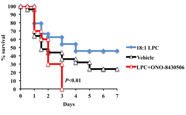
CLP was performed on C57BL6/J adult male mice, as described in Methods. The effects of LPC injection on CLP-induced sepsis were studied as reported previously6). Intraperitoneally injected 18:1 LPC at 6 h, 12 h, 24 h, 36 h and 48 h after CLP provided slight protection against sepsis-induced lethality. Interestingly, this LPC-provided slight protection was abrogated completely by ATX inhibitor (ONO-8430506) daily administration (P<0.01) and ATX inhibitor induced severe sepsis as judged by the survival rate (Fig.1), suggesting the possibility of ATX-dependently produced endogenous LPA in protection against CLP-induced sepsis.
Figure 1. Abrogated protective effects of LPC in CLP-induced sepsis by ATX inhibitor. CLP mice were intraperitoneally injected with 18:1 LPC at 6 h, 12 h, 24 h, 36 h and 48 h after surgery. ATX inhibitor (ONO-8430506) was orally administrated daily at the dose of 100 mg/kg mice body. Survival was monitored for 7 d. n=10-20 mice per group.
0 10 20 30 40 50 60 70 80 90 100 0 1 2 3 4 5 6 7 % s u rvi val Days 18:1 LPC Vehicle LPC+ONO-8430506 P<0.01
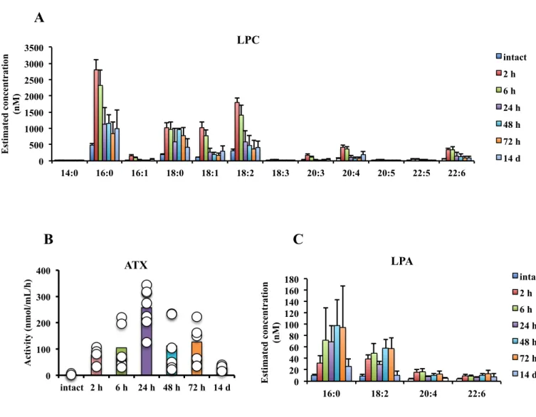
3.2.2. Analysis of lysophospholipids amount and ATX activity in peritoneal cavity of CLP-subjected mice
In order to determine the effects of endogenous LPA in sepsis, firstly the levels of LPA and other lysophospholipids in CLP model were measured. Peritoneal fluids were sampled by 1 mL sterile PBS injection after 2 h, 6 h, 24 h, 48 h, 72 h and 14 d after CLP induction. Levels of LPA, LPC and the other lysophospholipids were extracted by one-step methanol precipitation and quantified by LC-MS/MS. ATX activities of peritoneal fluids were also examined by LysoPLD activity assay. The levels of all species of lysophospholipids measured (lysophosphatidic acid, lysophosphatidylcholine, lysophosphatidylethanolamine, lysophosphatidylinositol, lysophosphatidylglycerol and lysophosphatidylserine) that have different polar heads were significantly increased at the early stage of 6 h after CLP induction (Fig.2). The time-course analysis for LPA and LPC were performed. The increase of LPA and LPC started from only 2 h after CLP and returned to a lower level after 2 weeks for recovery (Fig.3A, C). Meanwhile, there are much higher activities of ATX in the
ATX activity peaked at 24 h after CLP and returned to the basal level after 14 d (Fig.3B). These results suggested that the endogenous LPA in CLP-induced sepsis could be produced from the other lysophospholipids, especially LPC, in the ATX dependent manner.
Figure 2. Increased levels of various lysophospholipids in peritoneal cavity of CLP-subjected mice. Peritoneal lavage fluids were collected 6 h after CLP and were subjected to LC-MS/MS. Fold changes are shown relative to intact mice.
0 5 10 15 20 25 30 S1P LPC LPA LysoP S LPG LPE LPI F ol d c h an ge Intact 6 h after CLP
Figure 3. Increased levels LPC, LPA and ATX activity in peritoneal cavity of CLP-subjected mice. Time course analysis of LPC, LPA and ATX activity in peritoneal lavage fluids in CLP-treated mice. Levels of LPC and LPA were measured by LC-MS/MS. ATX activity was measured by LysoPLD activity assay. Data are mean±S.D.. 0 500 1000 1500 2000 2500 3000 3500 14:0 16:0 16:1 18:0 18:1 18:2 18:3 20:3 20:4 20:5 22:5 22:6 Es ti mate d c on ce n tr ati on (n M ) LPC intact 2 h 6 h 24 h 48 h 72 h 14 d 0 20 40 60 80 100 120 140 160 180 16:0 18:2 20:4 22:6 Es ti mate d c on ce n tr ati on (n M ) LPA intact 2 h 6 h 24 h 48 h 72 h 14 d 0 100 200 300 400 intact 2 h 6 h 24 h 48 h 72 h 14 d A cti vi ty (n mol /mL/ h ) ATX
A
B
C
3.2.3. Effects of ATX inhibitor administration in CLP mice
To determine whether the increased endogenous LPA in peritoneal cavity of CLP-subjected mice is dependent on ATX or not, ATX inhibitor (ONO-8430506) was administrated orally at the dose of 100 mg/kg body weight, once a day since mice suffered from CLP. ATX activities and LPA levels in peritoneal fluids were examined from 6 h to 72 h after CLP (Fig.4A-D). Survival was monitored every 12 h for 7 d (Fig.5). Partially reduced ATX activities of peritoneal fluids of CLP mice after ATX inhibitor administration were confirmed (Fig. 4A). ATX inhibitor also reduced the level of LPA molecules with acyl chains of 18:2 and 20:4 partially (Fig.4C,D). However, ATX inhibitor had no effects on the amount of 16:0 LPA (Fig.4B). ATX inhibitor administrated mice suffered significantly higher mortality than the control group (Fig.5), suggesting that ATX-dependently produced endogenous LPA is protective in experimental sepsis.
Figure 4. ATX inhibitor partially reduces ATX activity and levels of LPA in peritoneal cavity of CLP-subjected mice. CLP mice were orally administrated with or without ATX inhibitor. Time course analysis of ATX activity (A) and levels of LPA (B-D) in peritoneal lavage fluids were performed.
0 100 200 300 400 500 - 6h + - 24h + - 48h + - 72h + A cti vi ty (n mol /mL/ h ) ATX 0.000 0.010 0.020 0.030 - + - + - + - + 6h 24h 48h 72h A re a r ati o 16:0 LPA 0.000 0.001 0.002 - + - + - + - + 6h 24h 48h 72h A re a r ati o 20:4 LPA 0.000 0.006 0.012 0.018 - + - + - + - + 6h 24h 48h 72h A re a r ati o 18:2 LPA
A
B
C
D
ONO-8430506 ONO-8430506Figure 5. ATX inhibitor leads to higher mortality in CLP-induced sepsis. ATX inhibitor (ONO-8430506) was orally administrated once a day since mice suffered from CLP. Survival was monitored every 12 h. n=7 mice per group.
0 20 40 60 80 100 0 1 2 3 4 5 6 7 % s u rvi val Days Vehicle ONO-8430506 P<0.05
3.2.4. Effects of LPA treatment in experimental sepsis
The effects of 18:1 LPA in experimental sepsis were examined. Intraperitoneally injected LPA five times at 6 h, 12 h, 24 h, 36 h and 48 h after CLP provided significant protection against sepsis-induced lethality (Fig.6A). In contrast, subcutaneously injected LPA had no significant effect on the 7-d survival rate in CLP mice (Fig.6B), suggesting that it is necessary for LPA to be delivered to the peritoneal cavity to exist its function in sepsis. Changes of sepsis-induced cytokine levels affected by LPA treatment were also examined. First of all, since LPA is unstable and easily be graded in vivo, a stable LPA analogue (T13, 19.08 mg/kg body weight, i.p.) was utilized (Fig.7). T13 is one of the ‘T-series’ compounds, which structures are similar to 2-acyl-LPA. A ring structure derived from carbohydrates is introduced as the scaffold in T-series compounds instead of the glycerol backbone of LPA28). Plasma was sampled after LPS and T13 co-injection, and 12 kinds of cytokines were measured by MAGPIX system (Fig.7). T13 induced the changes in the level of certain cytokines. The anti-inflammatory cytokines IFN-γ, TNF-α and IL-1β were decreased (Fig.7). Interestingly, the level of the prototypical anti-inflammatory cytokine IL-10
was dramatically increased by T13 (Fig.7). Next, the effects of LPA treatment on CLP-induced cytokine levels were also examined. Peritoneal fluids sampled from mice that treated with 18:1 LPA (4 mg/kg body weight, i.p.) three times at 6-h intervals since mice suffered from CLP. Only very small changes of IL-1β, TNFα, IL-17A and IFNγ levels were observed (Fig.8). However, LPA induced the increasing of IL-10 level significantly at 6 h after CLP (Fig.8). These findings highlighted the possibility of LPA in regulating IL-10 level in sepsis.
Figure 6. LPA treatment protects against CLP-induced lethality. (A) CLP mice were intraperitoneally injected with 18:1 LPA five times at 6 h, 12 h, 24 h, 36 h and 48 h after CLP. Survival was monitored for 7 d. n=18 mice per group. (B) CLP mice were subcutaneously injected with 18:1 LPA at the same experimental conditions with (A). n=7 mice per group.
0 20 40 60 80 100 0 1 2 3 4 5 6 7 % s u rvi val Days 18:1 LPA Vehicle P<0.05
A
B
0 20 40 60 80 100 0 1 2 3 4 5 6 7 % s u rvi val Days Vehicle 18:1 LPA s.c.Figure 7. Changes of levels of cytokines induced by a stable LPA analogue in the model of LPS-induced sepsis. Time course analysis of levels of IL-10, IFNγ, TNFα, IL-1β, RANTES, IL-12p40, IL-17, IL-9, IL-6, MCP-1, IP-10 and IL-12p70 in plasma of mice by MAGPIX system after LPS plus LPA analogue (T13) co-injection.
Figure 8. Changes of levels of cytokines induced by 18:1 LPA in the model CLP-induced sepsis. Time course analysis of levels of IL-10, IL-1β, IL-6, TNFα, IL-2, IL-17A and IFNγ by ELISA in plasma of mice treated with 18:1 LPA 6 h, 12 h, 24 h after CLP. Data are mean±S.D.. *P<0.05
0 2000 4000 6000 8000 10000 0 6 12 18 24 IL-10 (p g/ mL) Time after CLP (h) Vehicle LPA 0 200 400 600 800 1000 1200 0 6 12 18 24 IL-1 β ( pg /mL) Time after CLP (h) Vehicle LPA 0 50 100 150 200 250 0 6 12 18 24 TN Fα ( pg /mL) Time after CLP (h) Vehicle LPA 0 10000 20000 30000 40000 50000 60000 0 6 12 18 24 IL-6 (p g/ mL) Time after CLP (h) Vehicle LPA 0 5 10 15 20 25 30 35 0 6 12 18 24 IL-2 (p g/ mL) Time after CLP (h) Vehicle LPA 0 100 200 300 400 500 600 700 0 6 12 18 24 IL-17A (p g/ mL) Time after CLP (h) Vehicle LPA 0 5 10 15 20 25 30 35 40 45 0 6 12 18 24 IFN γ ( pg /mL) Time after CLP (h) Vehicle LPA
*
3.2.5. Dependency of ATX-LPA on IL-10 to protect against CLP-induced sepsis To examine the regulation of IL-10 by ATX, IL-10 level in the peritoneal fluid of CLP mice with ATX inhibitor administration was measured. ATX inhibitor significantly reduced the high 24-h IL-10 level in peritoneal fluids (Fig.9). Rescue experiment with mouse IL-10 recombinant protein was performed next. Mouse IL-10 recombinant protein was prepared and purified as described in Methods. The activity was confirmed by using it to rescue CLP-induced lethality with the same effect at the same dose as reported24) (Fig.10A). 3µg/body of mouse IL-10 recombinant protein was injected subcutaneously once at 6 h after surgery to ATX inhibitor daily-administrated CLP mice. ATX inhibitor-induced sever sepsis, which has higher mortality, was rescued by IL-10 recombinant protein (Fig.10B). Proved that the protective effect of ATX-LPA axis is working through IL-10.
Figure 9. ATX inhibitor reduces the level of IL-10 in peritoneal cavity of CLP-subjected mice. CLP mice were administrated with ATX inhibitor. Levels of IL-10 in peritoneal lavage fluids 24 h after CLP were measured by ELISA. ***P<0.001 0 500 1000 1500 2000 2500 3000 3500 4000 4500 Vehicle ONO-8430506 IL-10 (p g/ mL)
IL-10 in peritoneal fluid
Figure 10. Rescue of the higher mortality in ATX inhibitor treated CLP mice by IL-10 recombinant protein. (A) CLP mice were subcutaneously injected once with 1 or 3 µg/body IL-10 recombinant protein at the timing of 6 h after CLP. n=10-14 mice per group. (B) ATX inhibitor daily-administrated CLP mice were subcutaneously
0 20 40 60 80 100 0 1 2 3 4 5 6 7 % s u rvi val Days ONO-8430506 + rIL-10 Vehicle ONO-8430506 P<0.05 0 20 40 60 80 100 0 1 2 3 4 5 6 7 % s u rvi val Days 3 ug/body rIL-10 1 ug/body rIL-10 Vehicle
A
B
3.3. Discussion
All the results of Chapter 3 proved the protective effect of ATX-LPA in experimental sepsis, which functions through the prototypical anti-inflammatory cytokine IL-10. Firstly, the significant increase of LPA and also other lysophospholipids including LPC, which could be the substrates for LPA production29), was observed in the peritoneal cavity of CLP mice. It is consistent with previous reports that metabolic dysfunctions including alternations in fatty acid metabolism are critical on survival in patients with sepsis26). Higher concentrations of PC are observed in sepsis patients27).
Furthermore, the increase of the lysophospholipase D activity of ATX, which has been demonstrated to catalyze the production of LPA from LPC9), was also observed. ATX inhibitor (ONO-8430506) inhibited lysophospholipase D activity and LPA production in peritoneal fluids of CLP mice. However, the time course of production of LPA was not completely consistent with its main substrate LPC and the inhibition of lysophospholipase D activity and LPA production by ATX inhibitor are only partially. We cannot exclude the possibility that there are other pathways, other than
the ATX-dependent manner, for the production of LPA in the peritoneal cavity in sepsis.
Secondly, it is important that ATX inhibitor significantly increased CLP-induced mortality, indicates that the endogenous LPA, which produced by ATX is essential for survival in sepsis. Both LPA and one stable LPA analogue treatment in CLP model and LPS model respectively induced the changes of levels of certain cytokines. IFNγ, TNFα and IL-1β were decreased, and IL-10 was increased in LPS model. In CLP model, although only slight changes of levels of cytokines were observed, the pattern of IL-10 regulation, which increased at the very early stages, was similar to LPS model. Targeting of several cytokines has been reported be beneficial to sepsis, including the monoclonal antibodies to TNF prevented sepsis4), treated sepsis with combining TNFα inhibitor plus antagonist of IL-1β receptor30), and IFNγ KO mice are resisted to CLP-induced mortality31), but the clinical trials were not overtly successful5). The protective effects of IL-10 in experimental sepsis also have been
level of IL-10 by LPA injection highlighted the relationship between LPA and IL-10 in sepsis. IL-10 recombinant protein also rescued mice from the severe sepsis caused by ATX inhibitor. Thus, ATX-LPA functioned dependent on the anti-inflammatory cytokine IL-10 to protect against CLP-induced lethality. Although the dependency was confirmed, since IL-10 recombinant protein administration alone is effective enough for rescue25), we cannot simply conclude that IL-10 is the direct downstream component of ATX-LPA. It will be interesting to look for other possible targets that mediating the protective effects ATX- LPA in sepsis.
3.4. Conclusion
In this chapter, the protective effects of ATX dependently produced endogenous LPA and the therapeutic effects of LPA administration in experimental sepsis were demonstrated. Importantly, the increase of the level of endogenous LPA and the activity of its producing enzyme ATX were observed. As far as I am concerned, it is first time that someone has proposed and proved the idea that endogenous LPA could protect against sepsis in in vivo mice model. IL-10, the prototypical anti-inflammatory cytokine that has already been reported to have beneficial effects in sepsis, is regulated by LPA endogenously and could rescue the severe sepsis when LPA production is inhibited. Thus, the protective effects of ATX-LPA are working through IL-10.
Chapter 4. Mechanism of LPA enhanced IL-10 production: mediated
by LPA
5in peritoneal macrophage
4.1. Introduction
To explore the underlying mechanism in more detail, the responsible cell type and the involved LPA receptor were examined. Macrophage is a major effector of innate immunity and the main population in peritoneal cavity. Previous studies proved the protection from experimental sepsis, suppression on TNFα production19) in peritoneal macrophage and reduced organ injury20) by exogenous LPA administration. The
regulations of LPA on LPS-induced maturing dendritic cell capacity including cytokines expression32) and on pro-inflammatory phenotypic change of microglia through LPA533) are reported. ATX over-expressed microglia also reduced
LPS-induced pro-inflammatory cytokines production34). However, the mechanism of regulation of LPA on IL-10 production is totally unknown.
4.2. Results
4.2.1.Isolated peritoneal macrophage responded to LPA for IL-10 enhancement The response of whole peritoneal cavity cells to LPA and LPS co-stimulation was examined firstly. Peritoneal cavity cells that harvested from adult male C57BL6/J mice were seeded in 96-well plate and added with 10 ng/mL LPS and doses of LPA as indicated for 4 h. IL-10 levels in the cell supernatant were increased in a LPA dose dependent manner (Fig.11). IL-10 enhancement could not be observed without LPS addition, indicated that LPA synergistically induces IL-10 with LPS (Fig.11). Mice peritoneal cavity cells were harvested and further sorted by FACS. After staining with the distinct markers that specific to macrophage, B cell and T cell, which are the three major cell populations in the mouse peritoneal cavity, the isolated cells were co-stimulated with LPS with or without LPA for 4 hours. LPA only enhanced IL-10 production from F4/80 positive cells (Fig. 12). Time-course analysis (1 h, 2 h, 4 h, 8 h and 24 h) of both IL-10 mRNA and protein levels of FACS-sorted peritoneal cavity cells with LPS and LPA co-stimulation was performed next. LPA-enhanced IL-10
IL-10 message RNA level increased at an earlier timing (Fig.13A,B). For the F4/80 negative cells, there is much less IL-10 induction compared with positive ones (Fig.13C,D).
Figure 11. Peritoneal cavity cells respond to LPS and LPA co-stimulation to induce IL-10 production. Whole peritoneal cavity cells were harvested and co-stimulated with LPS plus LPA for 4 h. Levels of IL-10 in supernatant were measured by ELISA. Data are mean±S.D..
0 200 400 600 800 1000 1200 1400 - + - + + + 0 0 1 0.01 0.1 1 IL-10 p g/ mL LPS (10 ng/mL) LPA (µM)
Figure 12. Isolated peritoneal macrophage responds to LPS and LPA co-stimulation for IL-10 induction. Peritoneal cavity cells were harvested, stained with anti-F4/80 antibody, anti-CD19 antibody and anti-CD3 antibody, sorted by FACS and seeded in 96-well plate. After co-stimulated with LPS and LPA, levels of IL-10 in supernatant were measured by ELISA. Data are means±S.D..
0 50 100 150 200 250 300 350 400 - + + - + + - + + - - + - - + - - + F4/80+ CD19+ CD3+ IL-10 p g/ mL ND ND LPS (10 ng/mL) LPA (1 µM)
Figure 13. Increased IL-10 protein and mRNA levels by LPS and LPA co-stimulation of isolated peritoneal macrophage. FACS-sorted F4/80 positive or negative cells were co-stimulated with LPS and LPA. Time course measurement of
0 200 400 600 800 1000 1200 - + - + - + - + - + 1h 2h 4h 8h 24h IL-10 (p g/ mL) IL-10 protein F4/80+ 0 200 400 600 800 1000 1200 - + - + - + - + - + 1h 2h 4h 8h 24h IL-10 (p g/ mL) F4/80 -0 0.1 0.2 0.3 0.4 0.5 0.6 0.7 - + - + - + - + - + 1h 2h 4h 8h 24h Il -10/ G apdh (r ati o) 0 0.1 0.2 0.3 0.4 0.5 0.6 0.7 - + - + - + - + - + 1h 2h 4h 8h 24h Il -10/ G apdh (r ati o) Il-10 mRNA
A
B
C
D
4.2.2. Effects of macrophage in vivo depletion in LPA-enhanced IL-10 production To confirm that macrophage is the responsible cell, macrophage in vivo depletion with clodronate liposome injection was performed. The success of macrophage depletion was confirmed by flow cytometry that no F4/80 positive cell could be detected one day after clodronate injection (Fig.14A). The effects of LPA treatment on LPS-induced IL-10 levels one day after macrophage depletion were examined. LPA enhanced IL-10 levels in both plasma and peritoneal fluids were significantly reduced in macrophage depleted LPS-injected mice (Fig.14B,C). The effects of macrophage depletion in CLP mice were also examined. LPA-enhanced IL-10 in the peritoneal cavity of CLP-subjected mice was also reduced by macrophage depletion (Fig.15).
Figure 14. In vivo macrophage depletion abrogated LPA induced IL-10 enhancement in LPS model. Mice were i.p. injected with 150 µL/body Clophosome®-A-Clodronate Liposomes (Anionic) 24 h before LPS injection. LPA was co-injected with LPS intraperitoneally. Plasma and peritoneal fluids were sampled after 2 h. (A) Peritoneal cavity cells were stained with anti-F4/80 antibody
0 200 400 600 800 1000 1200 1400 1600 1800 2000 - - - - + - - + + + - - + + - + + + IL-10 (p g/ mL) 0 500 1000 1500 2000 2500 3000 - - - - + - - + + + - - + + - + + + IL-10 (p g/ mL)
***
Clodronate LPS LPAPB
Anti-F4/80 APC Clophosome®-A C ou n ts PBS***
Clodronate LPS LPAA
B
C
Figure 15. In vivo macrophage depletion abrogated LPA induced IL-10 enhancement in CLP model. Mice were i.p. injected with 150 µL/body Clophosome®-A-Clodronate Liposomes (Anionic) 24 h before CLP. LPA was injected at the same time with CLP surgery. Peritoneal fluids were sampled after 6 h and levels of IL-10 were measure by ELISA. *P<0.05
0 1000 2000 3000 4000 5000 6000 7000 8000 9000 10000 - - + + - + - + 6 h after CLP IL-10 (p g/ mL)
*
Clodronate LPA4.2.3. LPA5 receptor regulated LPA-enhanced IL-10 production
To investigate which LPA receptor is involved in the LPA-enhanced IL-10 from peritoneal macrophage, the expression of six LPA receptors on isolated F4/80+ peritoneal macrophage were judged by RT-PCR. Except LPA3 and LPA4, the
expression other LPA receptors were detectable (Fig.16). Therefore, response to LPS and LPA co-stimulation of peritoneal cavity cells from LPA1, LPA2, LPA5 and LPA6
single knockout mice were examined. Peritoneal cavity cells from LPA5 knockout
mice significantly reduced IL-10 level when stimulated with LPS and LPA in vitro (Fig.17C). IL-10 was not affected by lacking of LPA1 and LPA6 receptors
(Fig.17A,D). Decreasing tendency of IL-10 production of peritoneal cavity cells from LPA2 knockout mice was observed (Fig.17B), but no effect of one LPA2 specific
agonist (RP-10-83) in enhancing IL-10 production (Fig.18A). The ability and specificity of RP-10-83 to LPA2 receptor were already confirmed by the
TGFα-shedding assay (data not shown), an assay that could sensitively detect GPCR activity35). Although there is no LPA5 specific agonist, one of the LPA analogues
and with extremely low affinity to LPA2 (data not shown), enhanced IL-10 production
comparably to LPA (Fig.18C). A commercial LPA5 antagonist (TC LPA5 4) was also
examined. The LPA dose-dependently induced IL-10 enhancement was partially reduced by TC LPA5 4 (Fig.18D).
Figure 16. mRNA expression of LPA receptors in mice peritoneal macrophage. qRT-PCR quantification of Lpar1, Lpar 2, Lpar3, Lpar4, Lpar5, and Lpar6 in FACS-sorted F4/80+ cells. Data are means±S.D.. ND: not detected.
0.000 0.001 0.002 0.003 0.004 0.005
Lpar1 Lpar2 Lpar3 Lpar4 Lpar5 Lpar6
L par /G apdh (r ati o) F4/80+ ND ND
Figure 17. Response of peritoneal cavity cells from each LPA receptor single knockout mice to LPA for IL-10 induction. Peritoneal cavity cells harvested from LPA1, LPA2, LPA5 and LPA6 single knockout mice were stimulated with LPS and
LPA for 4 h. Levels of IL-10 in the supernatant were measured by ELISA. Data are means±S.D.. 0 300 600 900 1200 1500 1800 - + + - + + + + 0 0 1 0 0 0.01 0.1 1 HT KO IL-10 (p g/ mL) LPA1 0 300 600 900 1200 1500 1800 - + + - + + + + 0 0 1 0 0 0.01 0.1 1 HT KO IL-10 (p g/ mL) LPA2 0 300 600 900 1200 1500 1800 - + + - + + + + 0 0 1 0 0 0.01 0.1 1 WT KO IL-10 (p g/ mL) LPA5 0 300 600 900 1200 1500 1800 - + + - + + + + 0 0 1 0 0 0.01 0.1 1 WT KO IL-10 (p g/ mL) LPA6 LPS (10 ng/mL) LPA (µM) LPS (10 ng/mL) LPA (µM) LPS (10 ng/mL) LPA (µM) LPS (10 ng/mL) LPA (µM)
A
B
C
D
0 300 600 900 1200 1500 1800 2100 0.001 0.01 0.1 1 IL-10 (p g/ mL) LPA doses (µM) LPS+LPA LPS+LPA+TC LPA5 4 0 200 400 600 800 1000 1200 1400 0.01 0.1 1 IL-10 (p g/ mL) Doses (µM) LPA RP-10-83 O O O P O HO HO Oleoyl T7 O O O P O OH OH Oleoyl T8 O O O P O OH OH Oleoyl T10 O O O P O HO HO Oleoyl T16 O O O P O OH OH Oleoyl T17 T19 O O O P O OH OH Oleoyl O O O P O SH OH Oleoyl T13 0 300 600 900 1200 1500 1800 2100 0.001 0.01 0.1 1 IL-10 (p g/ mL) Doses (µM) LPA T19
***
A
B
C
D
Figure 18. Effects of LPA receptor agonists and antagonist on IL-10 induction. Peritoneal cavity cells were co-stimulated with LPS and LPA2 specific agonist (A),
LPA analogue T19 (B), LPA plus LPA5 antagonist TC LPA5 4 (C). Levels of IL-10
in the supernatant after 4 h stimulation were measured by ELISA. Data are means± S.D.. ***P<0.001
4.3. Discussion
All the experiment data presented in Chapter 4 indicated that LPA enhanced IL-10 production through LPA5 receptor from mice peritoneal macrophage. Firstly,
according to the results obtained using the CLP model, all the events observed occurred at the very early stages in the peritoneal cavity. Thus, we proposed the targets of LPA, at least in part, exist in mice peritoneal cavity. The results showed that LPA addition dose dependently increased IL-10 level from the whole peritoneal cavity cells of mice. The enhancement could not be observed without LPS co-stimulation suggested that LPA synergized with TLR to induce IL-10 production. Several co-stimulators have already been reported to reinforce IL-10 production after TLR-dependent production36). For instance, the prostaglandin-2 (PGE-2) promoted
macrophage IL-10 in combination with LPS37,38). Type I IFN is required for LPS-induced IL-10 up-regulation39). Vitamin D3 acts synergistically with TLR4 and TLR2 to induce IL-10 production in dendritic cells40). These co-stimulatorsmay act on different target cells in different patho-physiological conditions. In this study, we
and functioned as one co-stimulator for inducing IL-10 in peritoneal macrophage.
Secondly, LPA5 receptor was identified as the responsible receptor for LPA-enhanced
IL-10 production. LPA5 mRNA, together with LPA1, LPA2 and LPA6, is expressed in
peritoneal macrophage. LPA could not induce IL-10 in the peritoneal macrophage of LPA5 knockout mice. In contrast, there is no influence on IL-10 production of
peritoneal macrophage from LPA1 and LPA6 single knockout mice suggesting that
LPA1 and LPA6 are not related to the induction of IL-10 by LPA. Moreover, although
IL-10 level showed decreasing tendency in peritoneal macrophage derived from LPA2
knockout mice, no response observed using one LPA2 specific agonist. This finding
suggests that it may be due to the changes of the characteristics of peritoneal cavity cells, not through LPA2 receptor. The results of inducing the anti-inflammatory
cytokine IL-10 in this study is contradictory to the previous report that LPA promotes M1-like phenotypic change of microglia, characterized by the production of pro-inflammatory cytokines, through LPA5 receptor33). However, the differences may
in this study. The protective effects of LPA have been reported in several LPS-induced inflammatory models15-20). Thus, in pathological conditions, the endogenous LPA5 receptor may function protectively. In addition, it was reported that
LPA5 negatively regulates B cell activation and inhibits Ab response in vivo41). LPA5
suppresses CD8 T cells activation and LPA5-deficient tumor-specific CD8 T cells are
better at controlling tumor growth in vivo42). Thus, it is reasonable to assume that LPA5 mediates the inhibition of immune responses in various immune cells including
4.4. Conclusion
In this chapter, the precise mechanism for the phenomenon observed in vivo was investigated in more detail. Peritoneal macrophage, rather than other cell populations in mice peritoneal cavity, was identified as the target cell by both using the isolated pure macrophage in vitro and macrophage depletion in vivo. LPA5 receptor, which
expressed in peritoneal macrophage, was the involved receptor for LPA-enhanced IL-10 production as confirmed with LPA5 knockout mice, LPA5 receptor agonist and
Chapter 5. Summary
In this study, we observed the high levels of various lysophospholipids including LPA by LC-MS/MS in the peritoneal cavity of CLP-subjected mice. The activity of LPA-producing enzyme, ATX was also increased significantly. The increased endogenous LPA in sepsis could be reduced after ATX inhibitor administration. ATX administration also induced severe sepsis with lower survival rate. On the other hand, the intraperitoneal injection of LPA protected against sepsis-induced lethality. After measuring the changes of cytokines level with direct LPA injection in CLP-subjected mice, the dramatic increasing of IL-10 was observed. ATX inhibitor administration reduced IL-10 in vivo, and the ATX inhibitor-induced sever sepsis could be rescued by recombinant IL-10 protein. Proved the protective effect of ATX-LPA-IL-10 in sepsis. In addition, peritoneal macrophage was identified as the responsible cell for LPA-enhanced IL-10 production since LPS and LPA co-stimulated FACS-sorted F4/80+ cell showed high IL-10 level. The macrophage depletion in vivo by clodronate also abrogated IL-10. Furthermore, peritoneal cavity cells that harvested from LPA5
possibility of LPA5 as the involver receptor was further confirmed with high IL-10
induced by LPA5 agonist and canceled LPA response by LPA5 antagonist.
In conclusion, we have demonstrated the protective effects of ATX dependently produced endogenous LPA in experimental sepsis. It functioned through enhancing IL-10 induction from peritoneal macrophage mediated by LPA5 receptor. (Fig.19)
Figure 19. Proposed model for LPA-provided protection against sepsis through ATX-LPA5-IL-10 axis. LPA is produced in the ATX-dependent manner in the
peritoneal cavity when sepsis occurred. LPA5 activation in peritoneal macrophage
synergistically with PAMPs reinforces IL-10 production and protects against sepsis-induced lethality. Peritoneal sepsis
LPA
IL-10
LPLs
ATX
TLR etc.LPA
5 PAMPsMφ
Sepsis
Acknowledgements
I would like to thank to my supervisor, Professor Junken Aoki for his advices and suggestions, and for giving me the chance to carry out my research in Japan. I am very grateful to Assistant Professor Kuniyuki Kano for supervising my work and always putting forward his valuable comments and suggestions. From the preparation of this thesis, he has spent so much time. I do appreciate his patience and professional instructions. Without his help and generous encouragement, it would never be possible to finish this work. I would also like to thank to Associate Professor Asuka Inoue and all the lab members for their guidance and support.
In addition, I would like to give my thanks to Professor Satoshi Ishi and Associate Professor Takashi Maruyama in Akita University for generously providing LPA5
knockout mice, and to Assistant Professor Daisuke Saigusa in Medical Megabank Organization, Tohoku University for the guidance of LC-MS/MS analysis. My sincere thanks are also given to Professor Noriyasu Hirasawa and Professor Atsushi Matsuzawa in Department of Pharmaceutical Science, Tohoku University for
reviewing this paper. My thanks also go to Kobayashi International Scholarship Foundation for offering financial support for my living expenses.
Last but not least, I am deeply indebted to my father, my mother, all my family members, my best friend and all my friends for their continuous support and endless love, help me overcome all the difficulties to live and study in a foreign country.
Reference
1) M. Singer, C. S. Deutschman, C. W. Seymour, M. Shankar-Hari, D. Annane,
M. Bauer, R. Bellomo, G. R. Bernard, J. D. Chiche, C. M. Coopersmith, R. S. Hotchkiss, M. M. Levy, J. C. Marshall, G. S. Martin, S. M. Opal, G. D. Rubenfeld, T. van der Poll, J. L. Vincent, D. C. Angus. The Third International Consensus Definitions for Sepsis and Septic Shock (Sepsis-3). Jama 315: 801-810, 2016.
2) T. van der Poll, F. L. van de Veerdonk, B. P. Scicluna, M. G. Netea. The immunopathology of sepsis and potential therapeutic targets. Nature reviews. Immunology 17: 407-420, 2017.
3) C. S. Deutschman, K. J. Tracey. Sepsis: current dogma and new perspectives. Immunity 40: 463-475, 2014.
4) K. J. Tracey, Y. Fong, D. G. Hesse, K. R. Manogue, A. T. Lee, G. C. Kuo, S. F. Lowry, A. Cerami. Anti-cachectin/TNF monoclonal antibodies prevent septic shock during lethal bacteraemia. Nature 330: 662-664, 1987.
sepsis. Nature reviews. Drug discovery 2: 391-405, 2003.
6) J. J. Yan, J. S. Jung, J. E. Lee, J. Lee, S. O. Huh, H. S. Kim, K. C. Jung, J. Y. Cho, J. S. Nam, H. W. Suh, Y. H. Kim, D. K. Song. Therapeutic effects of lysophosphatidylcholine in experimental sepsis. Nature medicine 10: 161-167, 2004.
7) J. W. Choi, D. R. Herr, K. Noguchi, Y. C. Yung, C. W. Lee, T. Mutoh, M. E.
Lin, S. T. Teo, K. E. Park, A. N. Mosley, J. Chun. LPA receptors: subtypes and biological actions. Annual review of pharmacology and toxicology 50: 157-186, 2010.
8) S. Aikawa, T. Hashimoto, K. Kano, J. Aoki. Lysophosphatidic acid as a lipid mediator with multiple biological actions. Journal of biochemistry 157: 81-89, 2015.
9) M. Umezu-Goto, Y. Kishi, A. Taira, K. Hama, N. Dohmae, K. Takio, T. Yamori, G. B. Mills, K. Inoue, J. Aoki, H. Arai. Autotaxin has lysophospholipase D activity leading to tumor cell growth and motility by
2002.
10) S. Aikawa, K. Kano, A. Inoue, J. Wang, D. Saigusa, T. Nagamatsu, Y. Hirota, T. Fujii, S. Tsuchiya, Y. Taketomi, Y. Sugimoto, M. Murakami, M. Arita, M. Kurano, H. Ikeda, Y. Yatomi, J. Chun, J. Aoki. Autotaxin-lysophosphatidic acid-LPA3 signaling at the embryo-epithelial boundary controls decidualization pathways. The EMBO journal 36: 2146-2160, 2017.
11) A. Inoue, N. Arima, J. Ishiguro, G. D. Prestwich, H. Arai, J. Aoki. LPA-producing enzyme PA-PLA(1)alpha regulates hair follicle development by modulating EGFR signalling. The EMBO journal 30: 4248-4260, 2011.
12) H. Yukiura, K. Hama, K. Nakanaga, M. Tanaka, Y. Asaoka, S. Okudaira, N.
Arima, A. Inoue, T. Hashimoto, H. Arai, A. Kawahara, H. Nishina, J. Aoki. Autotaxin regulates vascular development via multiple lysophosphatidic acid (LPA) receptors in zebrafish. The Journal of biological chemistry 286: 43972-43983, 2011.
13) T. Nishioka, N. Arima, K. Kano, K. Hama, E. Itai, H. Yukiura, R. Kise, A. Inoue, S. H. Kim, L. Solnica-Krezel, W. H. Moolenaar, J. Chun, J. Aoki.
ATX-LPA1 axis contributes to proliferation of chondrocytes by regulating fibronectin assembly leading to proper cartilage formation. Scientific reports 6: 23433, 2016.
14) Y. C. Yung, N. C. Stoddard, H. Mirendil, J. Chun. Lysophosphatidic Acid signaling in the nervous system. Neuron 85: 669-682, 2015.
15) K. Mirzoyan, C. Denis, A. Casemayou, M. Gilet, D. Marsal, D. Goudouneche,
S. Faguer, J. L. Bascands, J. P. Schanstra, J. S. Saulnier-Blache. Lysophosphatidic Acid Protects Against Endotoxin-Induced Acute Kidney Injury. Inflammation 40: 1707-1716, 2017.
16) D. He, Y. Su, P. V. Usatyuk, E. W. Spannhake, P. Kogut, J. Solway, V. Natarajan, Y. Zhao. Lysophosphatidic acid enhances pulmonary epithelial barrier integrity and protects endotoxin-induced epithelial barrier disruption and lung injury. The Journal of biological chemistry 284: 24123-24132, 2009. 17) A. Sturm, T. Sudermann, K. M. Schulte, H. Goebell, A. U. Dignass.
18) A. Sturm, J. Zeeh, T. Sudermann, H. Rath, G. Gerken, A. U. Dignass. Lisofylline and lysophospholipids ameliorate experimental colitis in rats. Digestion 66: 23-29, 2002.
19) H. Fan, B. Zingarelli, V. Harris, G. E. Tempel, P. V. Halushka, J. A. Cook. Lysophosphatidic acid inhibits bacterial endotoxin-induced pro-inflammatory response: potential anti-inflammatory signaling pathways. Molecular medicine (Cambridge, Mass.) 14: 422-428, 2008.
20) O. Murch, M. Collin, C. Thiemermann. Lysophosphatidic acid reduces the organ injury caused by endotoxemia-a role for G-protein-coupled receptors and peroxisome proliferator-activated receptor-gamma. Shock (Augusta, Ga.) 27: 48-54, 2007.
21) M. Okudaira, A. Inoue, A. Shuto, K. Nakanaga, K. Kano, K. Makide, D. Saigusa, Y. Tomioka, J. Aoki. Separation and quantification of 2-acyl-1-lysophospholipids and 1-acyl-2-lysophospholipids in biological samples by LC-MS/MS. Journal of lipid research 55: 2178-2192, 2014. 22) S. Q. Latifi, M. A. O'Riordan, A. D. Levine. Interleukin-10 controls the onset
of irreversible septic shock. Infection and immunity 70: 4441-4446, 2002. 23) T. van der Poll, A. Marchant, W. A. Buurman, L. Berman, C. V. Keogh, D. D.
Lazarus, L. Nguyen, M. Goldman, L. L. Moldawer, S. F. Lowry. Endogenous IL-10 protects mice from death during septic peritonitis. Journal of immunology (Baltimore, Md. : 1950) 155: 5397-5401, 1995.
24) T. Kato, A. Murata, H. Ishida, H. Toda, N. Tanaka, H. Hayashida, M. Monden, N. Matsuura. Interleukin 10 reduces mortality from severe peritonitis in mice. Antimicrobial agents and chemotherapy 39: 1336-1340, 1995.
25) M. Howard, T. Muchamuel, S. Andrade, S. Menon. Interleukin 10 protects mice from lethal endotoxemia. The Journal of experimental medicine 177: 1205-1208, 1993.
26) R. J. Langley, E. L. Tsalik, J. C. van Velkinburgh, S. W. Glickman, B. J. Rice, C. Wang, B. Chen, L. Carin, A. Suarez, R. P. Mohney, D. H. Freeman, M. Wang, J. You, J. Wulff, J. W. Thompson, M. A. Moseley, S. Reisinger, B. T. Edmonds, B. Grinnell, D. R. Nelson, D. L. Dinwiddie, N. A. Miller, C. J.
Massaro, R. M. Baron, A. M. Choi, G. R. Corey, G. S. Ginsburg, C. B. Cairns, R. M. Otero, V. G. Fowler, Jr., E. P. Rivers, C. W. Woods, S. F. Kingsmore. An integrated clinico-metabolomic model improves prediction of death in sepsis. Science translational medicine 5: 195ra195, 2013.
27) D. Schmerler, S. Neugebauer, K. Ludewig, S. Bremer-Streck, F. M.
Brunkhorst, M. Kiehntopf. Targeted metabolomics for discrimination of systemic inflammatory disorders in critically ill patients. Journal of lipid research 53: 1369-1375, 2012.
28) Y. Tamaruya, M. Suzuki, G. Kamura, M. Kanai, K. Hama, K. Shimizu, J. Aoki, H. Arai, M. Shibasaki. Identifying specific conformations by using a carbohydrate scaffold: discovery of subtype-selective LPA-receptor agonists and an antagonist. Angewandte Chemie (International ed. in English) 43: 2834-2837, 2004.
29) J. Aoki, A. Taira, Y. Takanezawa, Y. Kishi, K. Hama, T. Kishimoto, K. Mizuno, K. Saku, R. Taguchi, H. Arai. Serum lysophosphatidic acid is produced through diverse phospholipase pathways. The Journal of biological
chemistry 277: 48737-48744, 2002.
30) D. G. Remick, D. R. Call, S. J. Ebong, D. E. Newcomb, P. Nybom, J. A. Nemzek, G. E. Bolgos. Combination immunotherapy with soluble tumor necrosis factor receptors plus interleukin 1 receptor antagonist decreases sepsis mortality. Critical care medicine 29: 473-481, 2001.
31) C. R. Romero, D. S. Herzig, A. Etogo, J. Nunez, R. Mahmoudizad, G. Fang, E. D. Murphey, T. Toliver-Kinsky, E. R. Sherwood. The role of interferon-gamma in the pathogenesis of acute intra-abdominal sepsis. Journal of leukocyte biology 88: 725-735, 2010.
32) E. Panther, M. Idzko, S. Corinti, D. Ferrari, Y. Herouy, M. Mockenhaupt, S. Dichmann, P. Gebicke-Haerter, F. Di Virgilio, G. Girolomoni, J. Norgauer. The influence of lysophosphatidic acid on the functions of human dendritic cells. Journal of immunology (Baltimore, Md. : 1950) 169: 4129-4135, 2002. 33) I. Plastira, E. Bernhart, M. Goeritzer, H. Reicher, V. B. Kumble, N. Kogelnik,