MAP4K3 mediates amino acid-dependent
regulation of autophagy via phosphorylation of
TFEB
Cynthia L. Hsu
1
, Elian X. Lee
1
, Kara L. Gordon
1
, Edwin A. Paz
2
, Wen-Chuan Shen
2
, Kohta Ohnishi
1
,
Jill Meisenhelder
3
, Tony Hunter
3
& Albert R. La Spada
1,2
Autophagy is the major cellular pathway by which macromolecules are degraded, and amino
acid depletion powerfully activates autophagy. MAP4K3, or germinal-center kinase-like
kinase, is required for robust cell growth in response to amino acids, but the basis for
MAP4K3 regulation of cellular metabolic disposition remains unknown. Here we identify
MAP4K3 as an amino acid-dependent regulator of autophagy through its phosphorylation of
transcription factor EB (TFEB), a transcriptional activator of autophagy, and through amino
acid starvation-dependent lysosomal localization of MAP4K3. We document that MAP4K3
physically interacts with TFEB and MAP4K3 inhibition is suf
ficient for TFEB nuclear
locali-zation, target gene transactivation, and autophagy, even when mTORC1 is activated.
More-over, MAP4K3 serine 3 phosphorylation of TFEB is required for TFEB interaction with
mTORC1-Rag GTPase-Ragulator complex and TFEB cytosolic sequestration. Our results
uncover a role for MAP4K3 in the control of autophagy and reveal MAP4K3 as a central node
in nutrient-sensing regulation.
DOI: 10.1038/s41467-018-03340-7
OPEN
1Department of Pediatrics, University of California, San Diego, La Jolla, CA 92093, USA.2Departments of Neurology, Neurobiology, and Cell Biology, Duke Center for Neurodegeneration and Neurotherapeutics, Duke University School of Medicine, Durham, NC 27710, USA.3Molecular and Cellular Biology Laboratory, Salk Institute for Biological Studies, La Jolla, CA 92037, USA. Correspondence and requests for materials should be addressed to A.R.L.S. (email:[email protected])
123456789
A
utophagy refers to a set of three cellular processes, i.e.,
macroautophagy, chaperone-mediated autophagy, and
microautophagy, each of which achieve the sequestration
and delivery of cytosolic cargoes to the lysosome for degradation.
Macroautophagy (hereafter referred to as autophagy) is a tightly
regulated cellular process by which long-lived proteins,
macro-molecules, and organelles are degraded
1. Autophagy can be
selective or non-selective in terms of which cargoes are directed to
the lysosome for degradation and the basis for substrate selection
remains an area of active research with many underlying
prin-ciples yet to be elucidated. The regulation of autophagy activation
and autophagosome formation, on the other hand, is better
worked out, with specific protein complexes implicated in the
process of initiation, nucleation, and expansion of the
phago-phore isolation membrane (reviewed in ref.
2). One critical
fea-ture of autophagy regulation is its incredibly dynamic nafea-ture,
with autophagy activation status constantly responding to cellular
nutrient levels and stress conditions. As autophagy-mediated
protein degradation yields free amino acids for protein synthesis
and energy production, amino acid depletion is a very powerful
activator of autophagy. The importance of the autophagy
path-way for promoting physiological processes supported by amino
acids has been demonstrated in knockout (k.o.) mice lacking
critical autophagy genes, as Atg5- and Atg7-null mice exhibit
embryonic and neonatal lethality linked to depletion of amino
acids, due to impaired protein synthesis and diminished
tri-carboxylic acid (TCA) cycle function
3–5.
Mitogen-activated protein kinases (MAPKs) comprise a large
family of highly conserved proteins that control a wide range of
cellular processes in all eukaryotes
6. MAP4K3, also known as
germinal-center kinase-like kinase, is a member of the Ste20
sub-family of MAPKs
7and has been implicated in autoimmune
dis-ease via activation of protein kinase C-θ
8, activation of c-Jun
N-terminal kinase (JNK) to promote apoptosis
7, and the amino
acid-stimulated activation of the mechanistic target of rapamycin
complex 1 (mTORC1), a multi-protein subunit complex
con-sisting of the catalytic mTOR subunit, mLST8, DEPTOR, the
Tti1–Tel2 complex, Raptor, and PRAS40
9. Studies in mammalian
cell lines and in Drosophila have shown that MAP4K3 is
abso-lutely required for activation of mTORC1 in response to amino
acids
9–11and amino acid levels principally determine the
acti-vation status of mTORC1
12, 13. Furthermore, MAP4K3 is
ubi-quitously expressed, as MAP4K3 RNA and protein are detected in
all human tissues
7,14. Thus, MAP4K3 probably has a central role
in regulating the metabolic disposition of the cell, but nothing is
known as to how MAP4K3 achieves this regulation.
We recently discovered that knock-down of MAP4K3 is
suf-ficient to induce autophagy
15and so considered the current
model of amino-acid-dependent autophagy regulation. According
to this model, in response to amino acid stimulation, mTORC1 is
recruited to the cytosolic surface of lysosomes via a physical
interaction between Raptor, a set of membrane-bound lysosomal
proteins known as the Ragulator complex, and the Rag GTPases,
which function as heterodimers wherein the active complex
consists of GTP-bound RagA or B complexed with GDP-bound
RagC or D
16,17. When amino acids are plentiful, GATOR1, the
GTPase-activating protein for Rag A/B, is inactive
18, whereas
Folliculin, the GTPase-activating protein for Rag C/D, is turned
on
19. Of the various amino acid inputs to mTORC1, leucine and
arginine appear to be the most potent
20. Leucine is sensed in the
cytosol by Sestrin 1 and 2, which physically interact with and
inhibit GATOR2 when leucine levels drop
21; however, when
leucine is abundant, Sestrin binding to GATOR2 is abrogated,
permitting GATOR2 to promote mTORC1 activation through
the Rag GTPases, possibly via its inhibition of GATOR1. Arginine
is sensed in the cytosol by CASTOR1, which binds to and inhibits
GATOR2 when arginine levels diminish
22. Similar to the model
for Sestrin regulation, arginine abundance promotes release of
CASTOR1 from GATOR2, favoring GATOR2 activation of
mTORC1
23. The lysosomal amino acid transporter SLC38A9, a
transmembrane protein, also serves as a sensor of arginine, but in
the lysosomal membrane, where it binds to the Rag GTPases and
Ragulator
to
favor
mTORC1
activation
upon
arginine
satiety
24, 25. Bringing mTORC1 to lysosomes is critical for the
activation of its kinase activity by Rheb, a lysosome-enriched
GTPase that is regulated by TSC2
26.
At the lysosome, mTORC1 directly represses autophagy by
phosphorylating
and
inhibiting
transcription
factor
EB
(TFEB)
27–29. TFEB is a helix-loop-helix transcription factor that
recognizes a 10-base pair motif (5′-GTCACGTGAC-3′) enriched
in the promoter regions of numerous lysosomal genes
30. Activation
of TFEB not only induces the expression of genes associated with
lysosomal function but also transactivates genes necessary for
autophagosome formation, autophagosome–lysosome fusion, and
cargo degradation
31, 32. Under conditions of amino acid satiety,
TFEB interacts with active Rag GTPases, which recruit TFEB to the
lysosomal surface, where mTORC1 phosphorylation of TFEB at
serine 211 creates a binding site for 14-3-3, a chaperone that
sequesters TFEB in the cytosol
27–29. However, the sufficiency of
mTORC1 phosphorylation of TFEB at serine 211 has been
chal-lenged by the observation that the amino-terminal region of TFEB
must be present for proper regulation of TFEB subcellular
locali-zation and function
33, although the nature of this crucial TFEB
amino-terminal regulation is currently lacking.
To determine the role of MAP4K3 in the control of amino
acid-mediated autophagy regulation, we generated MAP4K3 k.o.
cells in this study and found that MAP4K3 k.o. is sufficient to
promote TFEB nuclear localization, resulting in a remarkable
upregulation of TFEB-regulated genes and productive induction
of autophagy. We then examined the amino-terminal region of
TFEB and noted that the phosphorylation status of the serine 3
residue of TFEB supersedes mTORC1 phosphoregulation of
TFEB and autophagy. We found evidence for a direct physical
interaction between MAP4K3 and TFEB, and for MAP4K3
phosphorylation of TFEB at serine 3. We also documented that
the TFEB serine 3 phosphorylation is required for its inhibitory
phosphorylation by mTORC1 and observed amino
acid-dependent subcellular localization of MAP4K3. These results
thus establish MAP4K3 as a key node in the amino acid-mediated
control of autophagy and reveal MAP4K3 as a putative
nutrient-sensing regulator in the cell.
Results
Knockout of MAP4K3 promotes autophagy induction and
flux. Knock-down of MAP4K3 is sufficient to induce
autop-hagy
15. However, to fully determine the role of MAP4K3 in
autophagy regulation, we chose to derive MAP4K3 k.o. cell lines
via CRISPR-Cas9 gene editing with guide RNAs targeting two
different MAP4K3 exon sequences in HEK293 cells. This
approach yielded two distinct sets of clones (M1 and M4) with
frameshift mutations at either of the two targeted sites, resulting
in a complete loss of MAP4K3 protein expression (Fig.
1
a and
Supplementary Fig.
1
). To evaluate the status of the autophagy
pathway in clonal MAP4K3 k.o. cell lines, we cultured MAP4K3
k.o. cells and control HEK293 cells in either nutrient replete
complete media (CM) or under conditions of amino acid
depri-vation, and noted increased levels of LC3-II in MAP4K3 k.o. cells
upon LC3 immunoblot analysis (Fig.
1
b). To assess
flux through
the autophagy pathway, we also measured LC3-II levels after
treatment with the lysosomal inhibitor ammonium chloride and
documented a further increase in LC3-II levels in both MAP4K3
k.o. cells and control HEK293 cells (Fig.
1
b), indicative of
autophagy
induction
and
progression
to
autophagosome–lysosome fusion. Calculation of LC3-II flux,
based upon LC3 immunoblotting, revealed significantly increased
autophagy activation in MAP4K3 k.o. cell lines in normal media
in comparison with control HEK293 cells cultured under the
same conditions (Fig.
1
c). To corroborate these
findings, we also
performed LC3 immunostaining analysis of various MAP4K3 k.o.
cell lines and control HEK293 cells, and observed a remarkably
increased LC3 puncta formation in cells lacking MAP4K3
(Fig.
1
d, e). To directly evaluate autophagy
flux via
immunos-taining, we transfected MAP4K3 k.o. cells and control HEK293
cells with a GFP-mCherry-LC3 tandem-tagged reporter
con-struct, quantified autophagosome and autolysosome formation in
normal media and upon amino acid starvation, and observed
significant increases in the numbers of autophagosomes and
autolysosomes per cell in MAP4K3 k.o. cell lines (Fig.
1
f-h),
indicative of increased autophagy induction and
flux in the
absence of MAP4K3.
MAP4K3 regulation of TFEB is upstream of mTORC1. TFEB is
a transcriptional activator of autophagy and is regulated by its
subcellular localization
30. As TFEB entry into the nucleus is
required for transactivation of its target genes and inhibition of
TFEB by mTORC1 phosphorylation restricts TFEB to the cytosol,
we examined the effect of MAP4K3 loss-of-function on TFEB
subcellular localization. Under conditions of amino acid
depri-vation, TFEB localizes to the nucleus in control HEK293 cells as
well as in MAP4K3 k.o. cells (Fig.
2
a, b and Supplementary
Fig.
1
b). When amino acids are supplied to control HEK293 cells
previously subjected to amino acid starvation, TFEB no longer
localizes to the nucleus but instead mostly remains in the cytosol.
However, in MAP4K3 k.o. cells, the amino acid-induced
sequestration of TFEB in the cytosol is dramatically blunted, as
the vast majority of MAP4K3 k.o. cells display TFEB nuclear
WT
d
M1-1 M4-6 M4-21f
WT MAP4K3 k.o. –AA CM ** *** ***e
MAP4K3 k.o.LC3-II puncta area
per cell 14% 12% 10% 8% 6% 4% 2% 0% WT M1-1 M4-6 M4-21 0 2 4 6 8 WT 0 5 *** *** ***
g
h
*** ** *** ***No. of APs per cell
16 10 12 14 CM –AA M4-6 WT M4-6
No. of ALs per cell
40 35 30 25 20 15 10 CM –AA M4-6 WT WT M4-6 WT M1-1 M4-6 MAP4K3
a
b
0 1 2 3 – + – + – + CM LC3-II / actin NH Cl4 – + –AA CM –AAWild-type MAP4K3 k.o. LC3-I LC3-II 0 0.5 1.0 1.5 2.0 LC3-II flux * * WT CM WT –AA M1-1 CM M4-6 CM *
c
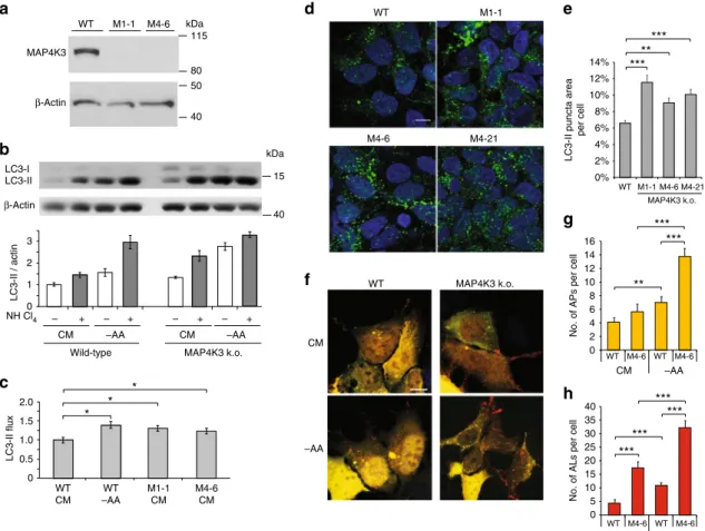
80 115 40 50 kDa 15 40 kDa β-Actin β-ActinFig. 1 Knockout of MAP4K3 promotes autophagy induction andflux. a Validation of MAP4K3 knockout (k.o.) cell lines. Wild-type (WT) and HEK293A cells gene-edited with either of two different sgRNAs (M1 and M4) were lysed, and protein lysates were immunoblotted for MAP4K3. Immunoblotting of β-actin served as a loading control. b, c Knockout of MAP4K3 promotes autophagy flux. WT HEK293A cells, M1-1 MAP4K3 k.o. cells, and M4-6 MAP4K3 k.o. cells (not shown) were cultured in complete media (CM) or subjected to amino acid starvation (– AA), and remained untreated or were treated with ammonium chloride. Protein lysates were immunoblotted for LC3 andβ-actin, which served as a loading control b. The ratio of LC3-II:actin was determined by densitometry using ImageJ and normalized to WT CM, which was arbitrarily set to 1c. One-way ANOVA with post-hoc Tukey’s test; *P < 0.05. d, e Knockout of MAP4K3 promotes autophagy induction. LC-3 immunostaining of WT HEK293A cells and three different MAP4K3 k.o. cell lines, all cultured in CMd. Quantification of LC3 puncta area per cell area was determined using ImageJ. n > 100 cells per genotype. One-way ANOVA with post-hoc Tukey’s test; **P < 0.01, ***P < 0.001. f–h Knockout of MAP4K3 promotes autophagy flux. WT HEK293A cells and MAP4K3 k.o. cells were cultured in CM or amino acid starved, and were transfected with a GFP-mCherry-LC3 expression construct (f). Note the predominance of red puncta indicative of autolysosomes in MAP4K3 k.o. cells. Quantification of autophagosome number per cell was determined by counting yellow puncta GFP-mCherry-LC3-expressing cell (g). Quantification of autolysosome number per cell was determined by counting red puncta/GFP-mCherry-LC3-expressing cell (h). n > 50 cells per condition. One-way ANOVA with post-hoc Tukey’ test; **P < 0.01, ***P < 0.001. All experiments were performed in triplicate. Error bars = SEM. Scale bars = 10 μm
localization (Fig.
2
a, b). To confirm that this difference in TFEB
subcellular localization is solely attributable to MAP4K3
loss-of-function, we transfected MAP4K3 k.o. cells with a MAP4K3
expression construct, and observed rescue of amino-acid induced
TFEB cytosolic sequestration in MAP4K3 k.o. cells expressing
MAP4K3 (Fig.
2
c, d). Failure of complete rescue upon MAP4K3
overexpression can likely be attributed to the fact that MAP4K3
induces the JNK signaling pathway and caspase activation when
overexpressed, resulting in cellular stress
34.
To further evaluate TFEB function in MAP4K3 k.o. cells, we
measured the expression of TFEB target genes in MAP4K3 k.o.
cells and in control HEK293 cells, and documented a marked
induction of TFEB target genes in nutrient replete cells lacking
MAP4K3—at levels comparable to those obtained upon
treat-ment of control HEK293 cells with the mTORC1 inhibitor Torin1
(Fig.
3
a). We evaluated MAP4K3 regulation of TFEB target gene
expression in TFEB k.o. cells, which were derived by CRISPR/
Cas9 gene editing, and confirmed that the presence of TFEB is
required to yield this effect (Supplementary Fig.
2
a). MAP4K3 is
known to activate mTORC1 under conditions of amino acid
satiety
9, raising the question of whether MAP4K3 regulation of
TFEB is upstream or downstream of mTORC1 regulation. To
address this question, we repeated the amino-acid induced TFEB
translocation experiment, but transfected HEK293 cells and
MAP4K3 k.o. cells with the constitutively active Rheb-Q64L
mutant (CA-Rheb)
35, which potently activates mTORC1 even in
the absence of amino acids (Supplementary Fig.
2
b). Although
many control HEK293 cells expressing CA-Rheb exhibited TFEB
cytosolic localization despite amino acid starvation, most
MAP4K3 k.o. cells expressing CA-Rheb retained TFEB in the
nucleus upon amino acid starvation (Fig.
3
b). CA-Rheb
expression similarly failed to elicit a marked change in TFEB
subcellular localization in MAP4K3 k.o. cells cultured in normal
CM (Supplementary Fig.
3
), demonstrating that MAP4K3
regulation of TFEB is a key input. To confirm the role of
MAP4K3 regulation of TFEB vis-à-vis mTORC1 activation status,
a
WT MAP4K3 k.o. –AA for 120 min –AA for 120 min +AA for 10 min TFEB TFEB CM WT MAP4K3 k.o. CM –AAc
+MAP4K3DAPI-TFEB DAPI-TFEB-MAP4K3
d
0
Untransfected MAP4K3 k.o. cells
*
**
0
–AA –AA +AA
***
***
b
% TFEB nuclear localization 100 80 60 40 20 WT M1-1 M4-6 WT M1-1 M4-6% TFEB nuclear localization
100 80 60 40 20 CM –AA WT HEK293A cells
MAP4K3 k.o. cells expressing MAP4K3
Fig. 2 MAP4K3 regulates TFEB subcellular localization. a Knockout of MAP4K3 yields TFEB nuclear localization. WT HEK293A cells and MAP4K3 k.o. cells were transfected with a TFEB-FLAG expression construct and cultured in complete media (CM) or starved of amino acids for 120 min, and then restimulated with amino acids for 10 min. Here we see representative images of cells immunostained with anti-FLAG antibody.b Quantification of cells with predominantly TFEB nuclear localization from experiment shown ina.n > 100 cells per condition. One-way ANOVA with post-hoc Tukey’s test; ***P < 0.001.c TFEB nuclear localization in MAP4K3 k.o. cells is rescued by MAP4K3 expression. WT HEK293A cells and MAP4K3 k.o. cells were transfected with a TFEB-FLAG expression construct and cultured in CM or amino-acid starved, and MAP4K3 k.o. cells were co-transfected with a MAP4K3-mCherry expression construct. Cells were stained with DAPI and immunostained with anti-FLAG antibody to permit visualization of nuclei, TFEB, and MAP4K3, as indicated. TFEB remains in the cytosol in CM in WT HEK293A cells and in MAP4K3 k.o. cells transfected with the MAP4K3 vector (white arrows); however, TFEB exhibits nuclear localization in untransfected MAP4K3 k.o. cells (orange arrows). Under conditions of amino acid starvation, TFEB translocates to the nucleus in WT cells, MAP4K3-transfected MAP4K3 k.o. cells (white arrow), and untransfected MAP4K3 k.o. cells (orange arrow).d Quantification of cells with predominantly TFEB nuclear localization from experiment shown in c. n > 50 cells per condition. One-way ANOVA with post-hoc Tukey’s test; *P < 0.05, **P < 0.01. All experiments were performed in triplicate. Error bars = SEM. Scale bars = 10 μm
we also measured the expression of TFEB target genes in
MAP4K3 k.o. cell lines expressing CA-Rheb and documented
marked induction of TFEB target genes at levels that were
significantly higher than the expression levels of TFEB target
genes obtained in HEK293 cells expressing CA-Rheb (Fig.
3
c).
Importantly, levels of TFEB target gene expression in MAP4K3 k.
o. cells were unchanged upon transfection with CA-Rheb
(Supplementary Fig.
4
).
MAP4K3 regulation of TFEB interaction with
mTORC1-Ragulator. Retention of TFEB in the cytosol is determined
by its phosphorylation status, as phospho-TFEB complexes with
14-3-3, thereby precluding TFEB nuclear entry, whereas
0 1 2 3 4 5*
*
*
*
*
*
*
*
**
**
**
**
**
**
*
*
*
*
Relative expression level
WT WT + Torin1 M1-1 (MAP4K3 k.o.) M4-6 (MAP4K3 k.o.)
a
b
WT MAP4K3 k.o. TFEB TFEB Rheb Rheb 0**
WT + CA-Rheb MAP4K3 k.o. + CA-Rheb% TFEB nuclear localization
100 80 60 40 20
c
0 1 2Relative expression level
WT WT + CA-Rheb M1-1 + CA-Rheb M4-6 + CA-Rheb
*
**
***
*
**
*
**
**
*
*
*
**
*
**
*
**
*
*
2.5 1.5 0.5TFEB ATP6V1H CLCN7 CTSD CTSF GLA MCOLN1 MCOLN1 TFEB ATP6V1H CLCN7 CTSD GLA
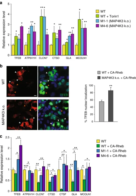
Fig. 3 MAP4K3 regulation of TFEB is upstream of mTORC1. a Knockout of MAP4K3 promotes TFEB-mediated transactivation of its target genes. WT HEK293A cells, untreated, or treated with Torin1, and two different MAP4K3 k.o. cell lines were cultured in CM. Quantitative RT-PCR of isolated RNAs for these cell lines was performed for six TFEB target genes. One-way ANOVA with post-hoc Tukey’s test; *P < 0.05, **P < 0.01. b Activation of mTORC1 does not alter TFEB localization in MAP4K3 k.o. cells. WT HEK293A cells and MAP4K3 k.o. cells were transfected with an expression construct for constitutively active Rheb, epitope-tagged with myc, and starved of amino acids for 120 min. Although untransfected WT HEK293A cells exhibit TFEB nuclear localization (orange arrows), many WT HEK293A cells expressing constitutively active Rheb display TFEB cytosolic localization (white arrows). Although
untransfected MAP4K3 k.o. cells also exhibit TFEB nuclear localization (orange arrows) as expected, most MAP4K3 k.o. cells expressing constitutively active Rheb show that TFEB still localizes to the nucleus (white arrows). Scale bar= 10 μm. Quantification of cells with predominantly TFEB nuclear localization for this experiment is shown in the adjacent graph.n > 50 cells per condition. **P < 0.01; two-tailed t-test. c Activation of mTORC1 does not prevent TFEB-mediated target gene activation in MAP4K3 k.o. cells. WT HEK293A cells, mock transfected, or transfected with constitutively active Rheb, and two different MAP4K3 k.o. cell lines, each transfected with constitutively active Rheb, were cultured in CM. Quantitative RT-PCR of isolated RNAs for these cell lines was performed for seven TFEB target genes. One-way ANOVA with post-hoc Tukey’s test; *P < 0.05, **P < 0.01. All experiments were performed in triplicate. Error bars= SEM
dephosphorylated TFEB readily translocates to the nucleus
28.
TFEB interacts with activated Rag GTPases, which promote
recruitment of TFEB to the lysosomal surface, where mTORC1
phosphorylates TFEB on serine 211 to enforce its cytosolic
retention and inactivation
33. One previous study found that the
first 30 amino acids of TFEB are required for TFEB localization to
lysosomes and documented that mutagenesis of serine 3 and
arginine 4 to alanines (S3A/R4A) completely prevented TFEB
lysosomal localization
33. To confirm the importance of the TFEB
amino-terminal region for regulation of its subcellular
localiza-tion and in particular the role of serine 3, we transfected control
HEK293 cells with WT TFEB and confirmed robust interaction of
TFEB with Raptor, Rag A, Rag C, and Lamtor1 (Fig.
4
a).
How-ever, when we transfected HEK293 cells with a version of TFEB
with serine 3 mutated to alanine (TFEB-S3A), or with the
first 30
amino acids of TFEB deleted (TFEB-Δ30), we found that both the
S3A mutation and the deletion of the
first 30 amino acids of
TFEB abrogated its interaction with mTORC1, the Rag GTPases,
and the Ragulator complex (Fig.
4
a).
The mechanistic basis for serine 3 regulation of TFEB
interaction with the mTORC1 complex is yet to be determined.
As serine residues are subject to phosphorylation and MAP4K3 is
a kinase, we considered the possibility that MAP4K3 regulation of
TFEB is occurring through phosphorylation of TFEB. To initially
test this hypothesis, we evaluated the physical interaction of TFEB
with mTORC1 and its associated regulatory proteins in complex
with it at the lysosome. Although immunoprecipitation of TFEB
provided evidence for robust interactions with Raptor, Rag A, and
Lamtor1 in control HEK293 cells, interaction of TFEB with these
mTORC1 complex components was dramatically reduced in two
different MAP4K3 k.o. cell lines (Fig.
4
b). Concern has been
raised that CRISPR-Cas9 gene editing may produce off-target
alterations in unrelated genes throughout the genome. Although
our results were obtained in distinct MAP4K3 k.o. cell lines
derived with different guide RNAs, to exclude off-target effects as
a potential explanation for our
findings, we repeated the TFEB
interaction studies in MAP4K3 k.o. cell lines transfected with
MAP4K3, and noted that exogenous MAP4K3 expression
restored TFEB interaction with Raptor, Rag GTPases, and
Lamtor1 in two different MAP4K3 k.o. cell lines (Fig.
4
b).
Noticeably, reduced interaction of TFEB-S3A with mTORC1
complex components was similarly observed in control HEK293
cells (Fig.
4
c). These results indicate that MAP4K3 is required for
TFEB interaction with the mTORC1 complex, and that mutation
of serine 3 to an alanine, which cannot be phosphorylated,
renders TFEB incapable of fully interacting with the mTORC1
complex, highlighting serine 3 as a potential site for TFEB
phosphoregulation. To further evaluate this hypothesis, we
expressed either wild-type TFEB or TFEB S3E, which features a
phosphomimetic amino acid substitution of glutamate for serine
3, in control or MAP4K3 k.o. cells, and we found that expression
of TFEB-S3E partially restored TFEB interaction with mTORC1
complex components (Fig.
4
d). These
findings thus establish a
role for MAP4K3 and TFEB serine 3 phosphorylation in the
regulation of TFEB interactions with the mTORC1 complex.
MAP4K3 interacts with and phosphorylates TFEB at serine 3.
To determine whether MAP4K3 and TFEB interact, we
trans-fected HEK293 cells with a kinase dead version of MAP4K3
(KD-MAP4K3) and with normal MAP4K3 (WT-(KD-MAP4K3). When we
a
Input IP: FLAG
WT S3A Δ30 WT S3A Δ30 TFEB-FLAG: RagC Raptor RagA Lamtor1 TFEB Input WT MAP4K3: – + – + – + Cell line: M1-1 M4-6 RagA RagC Lamtor1 TFEB Raptor TFEB-FLAG: WT – + – + – + M1-1 M4-6 + + + + + + + + + + + + IP: FLAG
c
140 40 30 50 25 15 50 kDa 40 30 50 25 15 50 140 kDab
d
Input WT Cell line: M1-1 M4-6 TFEB: WT M1-1 M4-6 IP: FLAGWT S3E WT S3E WT S3E WT S3E WT S3E WT S3E
RagA Lamtor1 Raptor
TFEB
WTS3AWT WT WT WT S3A WT WT WT
Input IP: FLAG
WT MAP4K3 k.o. TFEB-FLAG: RagA Lamtor1 Raptor TFEB WT MAP4K3 k.o. 25 15 50 140 40 30 kDa 25 15 50 140 40 30 kDa
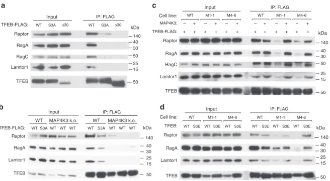
Fig. 4 MAP4K3 and TFEB serine 3 phosphorylation are required for interaction of TFEB with the mTORC1-Rag GTPase complex. a TFEB serine 3 is required for its interaction with the mTORC1-Rag GTPase complex. HEK293A cells were transfected with either WT TFEB, TFEB-S3A, or TFEB-Δ30, each FLAG-tagged, and cell lysates and FLAG immunoprecipitates were subjected to immunoblotting.b MAP4K3 is required for TFEB interaction with the mTORC1-Rag GTPase complex. WT HEK293A cells and MAP4K3 k.o. cells were transfected with either WT TFEB or TFEB-S3A, each FLAG-tagged, and cell lysates and FLAG immunoprecipitates were subjected to immunoblotting.c TFEB interaction with the mTORC1-Rag GTPase complex is rescued by MAP4K3 in MAP4K3 k.o. cells. WT HEK293A cells, M1-1 MAP4K3 k.o. cells, and M4-6 MAP4K3 k.o. cells were transfected with TFEB-FLAG alone, or co-transfected with TFEB-FLAG and MAP4K3, and cell lysates and FLAG immunoprecipitates were subjected to immunoblotting.d TFEB phosphomimetic S3E enhances TFEB interaction with the mTORC1-Rag GTPase complex in MAP4K3 k.o. cells. WT HEK293A cells, M1-1 MAP4K3 k.o. cells, and M4-6 MAP4K3 k.o. cells were transfected with either WT TFEB or TFEB-S3E, each FLAG-tagged, and cell lysates and FLAG immunoprecipitates were subjected to immunoblotting. All experiments were performed in triplicate
immunoprecipitated endogenous TFEB and immunoblotted for
MAP4K3, we detected a physical interaction between TFEB and
MAP4K3, but not between TFEB and an unrelated control
pro-tein, HDAC6, with the identical epitope tag (Fig.
5
a).
Interest-ingly, the interaction between TFEB and KD-MAP4K3 was
significantly stronger than the interaction between TFEB and
WT-MAP4K3, suggesting that MAP4K3 kinase activity dictates
the nature of its interaction with TFEB, such that failure of
KD-MAP4K3 to phosphorylate substrate may favor an extended
physical interaction. To determine whether the MAP4K3–TFEB
interaction is direct, we generated in vitro transcribed and
translated TFEB protein, and performed pull-down assays with
purified FLAG-tagged WT-MAP4K3, KD-MAP4K3, or HDAC6.
WT-MAP4K3 and KD-MAP4K3 were both pulled down with an
anti-TFEB antibody, but the pull-down of KD-MAP4K3 was
clearly stronger (Fig.
5
b). Finally, to determine whether the
physical interaction of TFEB with MAP4K3 involves the
terminal region of TFEB, we generated a recombinant
amino-terminal fragment of TFEB (TFEB:aa1-37) linked to glutathione
S-transferase (GST) and documented that TFEB:aa1–37 is capable
of pulling down WT-MAP4K3 or KD-MAP4K3 (Fig.
5
c). These
results provide evidence for a direct interaction between MAP4K3
and TFEB, and indicate that the
first 37 amino acids of TFEB are
critical for this physical interaction.
As MAP4K3 is a kinase and our results implicate the serine 3
residue of TFEB as a key site for its phosphoregulation, we sought
to test the hypothesis that MAP4K3 is phosphorylating TFEB at
serine 3. Although mass spectrometry is a powerful method for
mapping phosphorylation sites, we did not employ this approach,
as trypsin digestion of TFEB is predicted to yield a tiny
four-amino-acid amino-terminal fragment, which would not be
detectable. Rather, to determine whether MAP4K3 can
phos-phorylate TFEB, we performed in vitro phosphopeptide mapping
in reaction mixtures containing the mTOR inhibitor Torin1 and a
TFEB HDAC6-FLAGa
MAP4K3-FLAG KD MAP4K3-FLAG: HDAC6-FLAG: WT MAP4K3-FLAG: IB: FLAG IB: TFEB 50 140 115 80 + – – – + – – – + + – – – + – – – + IP: anti-TFEB Input kDa TFEB HDAC6-FLAG MAP4K3-FLAGb
IB: FLAG IB: TFEBd
50 WT MAP4K3 + WT TFEB WT MAP4K3 + S3A TFEB KD MAP4K3 + WT TFEB KD MAP4K3 + S211A TFEB 140 115 80 HDAC6-FLAG: WT MAP4K3-FLAG: KD MAP4K3-FLAG: IVTT TFEB: – – – + + – – – – + – – – – + – + – – + – + – + – – + + IP: anti-TFEB Input kDa HDAC6-FLAG: KD MAP4K3-FLAG: WT MAP4K3-FLAG: anti-GST-TFEB-aa1-37 anti-GST-empty Input: anti-GST GST-TFEB aa 1-37 GST-empty HDAC6-FLAG MAP4K3-FLAG GST-TFEB GSTc
e
IB: FLAG 30 140 115 80 25 + – – – + – – – + + – – – + – – – + + – – – + – – – + InputWT MAP4K3 + TFEB KD MAP4K3 + TFEB kDa
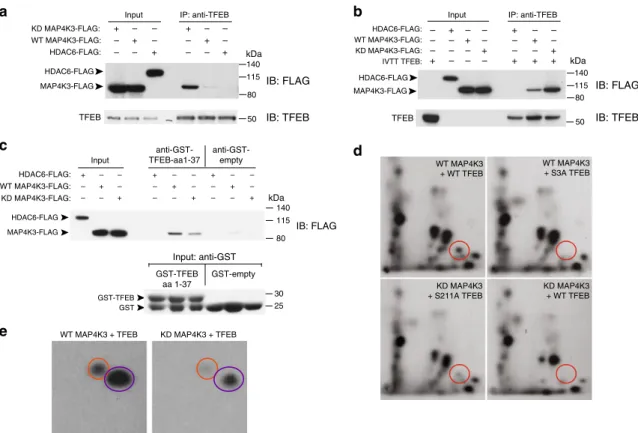
Fig. 5 MAP4K3 phosphorylates TFEB on serine 3. a MAP4K3 physically interacts with TFEB. WT HEK293A cells were transfected with an expression vector for either kinase dead (KD)-MAP4K3-FLAG, WT-MAP4K3-FLAG, or HDAC6-FLAG, and cell lysates and FLAG immunoprecipitates were subjected to immunoblotting.b MAP4K3 directly interacts with TFEB. WT HEK293A cells were transfected with an expression vector for either HDAC6-FLAG, WT-MAP4K3-FLAG, or kinase dead (KD)-WT-MAP4K3-FLAG, and FLAG immunoprecipitates were incubated with recombinant TFEB generated by in vitro transcription and translation (IVTT TFEB). Cell lysates and TFEB immunoprecipitates were then subjected to immunoblotting.c MAP4K3 directly interacts with the N-terminal 37 amino acids of TFEB. WT HEK293A cells were transfected with an expression vector for either kinase dead (KD)-MAP4K3-FLAG, WT-MAP4K3-FLAG, or HDAC6-FLAG, and FLAG immunoprecipitates were incubated with recombinant GST-TFEB amino acids 1-37 or recombinant GST alone, before mixing with GST-containing beads. Cell lysates and the eluate obtained from GST-bound fractions were subjected to anti-FLAG and anti-GST immunoblotting, as indicated.d MAP4K3 phosphorylates TFEB at serine 3. WT HEK293A cells were transfected with WT-MAP4K3-FLAG or KD-MAP4K3-FLAG, and either TFEB-FLAG, TFEB-S3A-FLAG, or TFEB-S211A-FLAG, as indicated. FLAG immunoprecipitates were subjected to in vitro kinase reactions withγ-P32-ATP, with Torin1 and the general kinase inhibitor FSBA included in the reaction mixture. Phosphopeptide mapping was performed after enzymatic digestion with thermolysin by spotting the resulting peptide mix onto cellulose thin layer chromatography plates, followed by 2D gel electrophoresis and chromatography, andfinally autoradiography to visualize phospho-labeled peptides. Circles indicate location of phospho-S3-TFEB. Note the absence of phospho-S3-TFEB for TFEB-S3A and for kinase-dead (KD) MAP4K3.e MAP4K3 heavily phosphorylates TFEB on serines and threonines. WT HEK293A cells were transfected with WT-MAP4K3-FLAG or KD-MAP4K3-FLAG, and TFEB-FLAG, as indicated. FLAG immunoprecipitates were subjected to in vitro kinase reactions withγ-P32-ATP, and phospho-amino acid mapping performed by matching the resultant spots on the autoradiograph with ninhydrin-stained standards. Orange circles indicate phospho-serine, and purple circles indicate phospho-threonine. All experiments were performed in triplicate
general kinase inhibitor. After two-dimensional gel fractionation,
we observed a phosphopeptide fragment that was present upon
co-incubation of WT-MAP4K3 with either WT TFEB or
TFEB-S211A, but absent upon co-incubation of WT-MAP4K3 with
TFEB-S3A, or KD-MAP4K3 with WT TFEB under these
phosphorylation conditions (Fig.
5
d and Supplementary Fig.
5
).
Phospho-amino acid analysis of these peptides confirmed that the
level of phosphoserine was lower for TFEB in the KD-MAP4K3
+ TFEB reaction, in comparison with the MAP4K3 +
WT-TFEB reaction (Fig.
5
e). These
findings thus illustrate that
MAP4K3 may phosphorylate TFEB on serine 3.
Serine 3 phosphorylation precedes TFEB serine 211
phos-phorylation. mTORC1 phosphorylation of TFEB at serine 211 is
viewed as a crucial regulatory event for TFEB repression
27,28. To
determine the regulatory relationship between MAP4K3
phos-phorylation of serine 3 and mTORC1 phosphos-phorylation of serine
211, we examined the effect of TFEB serine 3 phosphorylation
status upon TFEB serine 211 phosphorylation. Immunoblotting
analysis of control HEK293 cells transfected with either normal
TFEB or S3A revealed a remarkable reduction in
TFEB-S3A serine 211 phosphorylation that was comparable to the
reduction in TFEB serine 211 phosphorylation with Torin1
treatment (Fig.
6
a). When we performed TFEB phosphoserine
211 immunoblotting on MAP4K3 k.o. cells transfected with
either normal TFEB, TFEB-S3A, or TFEB S3E, we only detected
TFEB serine 211 phosphorylation in MAP4K3 k.o. cells
trans-fected with the phosphomimetic TFEB S3E mutant (Fig.
6
a),
suggesting that serine 3 phosphorylation is required for
sub-sequent phosphorylation of TFEB serine 211 by mTORC1. To
confirm that MAP4K3 phosphorylation of serine 3 dictates the
ability of mTORC1 to phosphorylate TFEB at serine 211, we
performed an additional TFEB phosphoserine 211
immunoblot-ting experiment in control HEK293 cells and MAP4K3 k.o. cells
transfected with normal TFEB in combination with either
MAP4K3 or KD-MAP4K3. We found that the expression of
WT-MAP4K3 in WT-MAP4K3 k.o. cells rescued TFEB serine 211
phos-phorylation, but the expression of KD-MAP4K3 in MAP4K3 k.o.
cells did not yield appreciable TFEB serine 211 phosphorylation
(Fig.
6
b). We also noted that KD-MAP4K3 expression in control
HEK293 cells that possess endogenous MAP4K3 resulted in a
decrease in TFEB serine 211 phosphorylation, probably reflecting
a dominant-negative effect, as MAP4K3 requires
transautopho-sphorylation at serine 170 within its kinase enzymatic domain for
full activation
36. Taken together, these results indicate that
MAP4K3 kinase activity occurs before and is necessary for TFEB
serine 211 phosphorylation by mTORC1.
TFEB serine 3 phosphorylation regulates autophagy activation.
To assess the physiological relevance of TFEB serine 3
phos-phorylation
for
regulation
of
autophagy,
we
generated
tetracycline-inducible TFEB-WT and TFEB-S3A expression
constructs. We then created TFEB k.o. cell lines stably transfected
with either inducible TFEB-WT or inducible TFEB-S3A, and
confirmed inducible expression of TFEB at close to endogenous
levels to validate the utility of these cell lines (Supplementary
Fig.
6
a). To establish the regulatory significance of the serine 3
phosphorylation for TFEB repression, we examined the role of
TFEB serine 3 phosphorylation in dictating the interaction of
TFEB with 14-3-3, as mTORC1 phosphorylation of TFEB at
serine 211 has been shown to promote TFEB binding to 14-3-3
and its sequestration in the cytosol
27,28. We found that induction
of TFEB expression in TFEB k.o. HeLa cells cultured in normal
media resulted in a productive interaction between TFEB and
14-3-3, based upon 14-3-3 immunoblot analysis of TFEB
immunoprecipitates (Fig.
6
c). However, 14-3-3 immmunoblot
analysis of TFEB immunoprecipitates prepared from TFEB k.o.
HeLa cells expressing TFEB-S3A protein yielded barely detectable
signal, akin to results obtained in TFEB k.o. cells induced to
express normal TFEB but in the presence of the mTORC1
inhi-bitor Torin1 (Fig.
6
c). To further assess the role of serine 3
phosphorylation in dictating TFEB subcellular localization and
function, we cultured TFEB k.o. cells induced to express either
normal TFEB or TFEB-S3A in CM or in media lacking amino
acids, and then performed TFEB immunostaining analysis to
examine subcellular localization. We observed nearly complete
nuclear localization of TFEB when cells were starved of amino
acids, as expected, but documented nearly complete nuclear
localization of TFEB-S3A in amino acid-replete media, in striking
contrast to complete cytosolic localization of TFEB-WT in amino
acid-replete media (Fig.
6
d). These results confirm that the
phosphorylation status of serine 3 of TFEB is the primary
determinant of its subcellular localization.
As overexpression of TFEB is sufficient to induce productive
autophagy
32, we next tested the effect of TFEB serine 3
phosphorylation on autophagy activation by expressing
GFP-mCherry-LC3 in TFEB k.o. cells stably transfected with inducible
TFEB-WT or inducible TFEB-S3A. When we treated these TFEB
k.o. cells with doxycycline and monitored autophagic
flux, we
detected a much more robust activation of autophagy in TFEB k.
o. cells expressing TFEB-S3A in comparison with TFEB k.o. cells
expressing normal TFEB, based upon our observation of
significantly greater numbers of autophagosomes and
autolyo-somes in the HeLa cells expressing TFEB-S3A (Fig.
6
e). These
results demonstrate that TFEB serine 3 phosphorylation
deter-mines TFEB function and autophagy activation status. To further
corroborate the physiological relevance of increased autophagy
engagement in TFEB-S3A expressing TFEB k.o. cells, we
compared cell growth between induced and uninduced TFEB k.
o. cells expressing either normal TFEB or TFEB-S3A, and noted
that TFEB k.o. cells induced to express TFEB-S3A exhibited
significantly reduced cellular proliferation over time
(Supple-mentary Fig.
6
b), culminating in markedly reduced cell numbers
at 72 h after doxycycline induction (Supplementary Fig.
6
c).
Impaired cell proliferation of TFEB-S3A-expressing cells is
consistent with the greater catabolic disposition of these cells,
which display elevated autophagy pathway activation.
MAP4K3 localizes to lysosomes upon amino acid starvation.
As our
findings indicate that MAP4K3 is a key node in autophagy
regulation, an important question is: how does amino acid
star-vation prevent MAP4K3 repression of TFEB in the cytosol? To
address this question, we examined the subcellular localization of
MAP4K3 by performing a subcellular fractionation of HEK293A
cells expressing FLAG-tagged MAP4K3, employing a protocol
that permitted isolation of a gradient fraction highly enriched for
lysosomes (Supplementary Fig.
7
). Immunoblotting confirmed
enrichment for lysosomes in the P1 fraction, based upon the
presence of abundant Lamp1 and the absence of cytosolic and
mitochondrial proteins (Fig.
7
a). Immunoblotting for MAP4K3
revealed that MAP4K3 is abundant in the lysosome-enriched P1
fraction, and that MAP4K3 abundance in the lysosomal fraction
is increased when HEK293A cells are subjected to amino acid
starvation (Fig.
7
a). To further examine the subcellular
localiza-tion of MAP4K3, we transfected nutrient-replete HEK293A cells
with MAP4K3-mNeonGreen and then switched the HEK293A
cells expressing MAP4K3-mNeonGreen to media lacking amino
acids, which resulted in pronounced colocalization of MAP4K3
with Lamp2 in discrete cytosolic puncta (Fig.
7
b). We also
per-formed live-cell imaging of MAP4K3 subcellular localization in
HEK293A cells subjected to amino acid starvation and found that
upon amino acid starvation, MAP4K3 transitions from a diffuse
cytosolic appearance to discrete localization into cytosolic puncta
(Fig.
7
c and Supplementary Movie
1
). Furthermore, upon
resupply of amino acids to amino acid-starved HEK293A cells,
MAP4K3 no longer remains in cytosolic puncta but instead
returns to a diffuse cytosolic localization (Fig.
7
d). Formation of
cytosolic puncta containing MAP4K3 upon amino acid depletion
was not restricted to HEK293A cells, but was also documented in
primary
retinal
pigmented
epithelial
(RPE)
cells
and
HEK293T cells (Supplementary Fig.
8
). To verify the nature of the
cytosolic puncta to which MAP4K3 localizes upon amino acid
starvation, we performed live-cell imaging and observed
coloca-lization of MAP4K3 cytosolic puncta with Lysotracker Red
(Fig.
7
e and Supplementary Movie
2
). When we supplied amino
acids to starved HEK293A cells, we noted that MAP4K3
lysoso-mal localization decreased within minutes (Fig.
7
e and
Supple-mentary Movie
2
).
WT + WT – S3A – S3E – WT – S3A – S3E – 0.04 ±0.00 1.00 ±0.36 0.36 ±0.06 0.86 ±0.15 0.17 ±0.04 0.14 ±0.05 0.84 ±0.06 WT MAP4K3 k.o. TFEB-FLAG: Torin1: P-S211 / total TFEB:a
65 65 kDa α-P-S211-TFEB α-FLAG 0 WT S3A WT S3A CM –AA TFEB –AA CM TFEB-WT TFEB-S3A *** *** ***d
% TFEB nuclear localization
100 20 40 60 80
b
– WT KD – WT KD 1.00 ±0.02 0.96 ±0.03 0.62 ±0.08 0.17 ±0.04 0.52 ±0.03 0.02 ±0.01 WT P-S211 / total TFEB: MAP4K3: MAP4K3 k.o. TFEB-FLAG: WT WT WT WT WT WT 65 65 115 kDa α-P-S211-TFEB α-TFEB α-MAP4K3 – – + – – 0 1 IP: anti-TFEB Input TFEB: Torin1:c
14-3-3 IP : Input ** * *TFEB k.o. (untransfected) TFEB-WT + Torin1 TFEB-WT TFEB-S3A 65 25 kDa WT WT S3A – – + – – WT WT S3A α-14–3–3 α-TFEB 1.4 1.2 0.8 0.6 0.4 0.2 0 2 4 6 8 14 WT –dox
e
TFEB-WT TFEB-S3A –Dox +Dox * ** * ** **No. of vesicles per cell
12 10 S3A –dox WT +dox S3A +dox WT –dox S3A –dox WT +dox S3A +dox APs ALs
Fig. 6 Phosphorylation of TFEB at serine 3 is a key determinant of TFEB cellular regulation and autophagy function. a TFEB serine 3 phosphorylation is required for mTORC1 phosphorylation at serine 211. WT HEK293A and MAP4K3 k.o. cells were transfected with FLAG, S3A-FLAG, or TFEB-S3E-FLAG, and Torin1 treated. FLAG immunoprecipitates were immunoblotted and TFEB serine 211 phosphorylation as a fraction of total TFEB was quantified by densitometry. b MAP4K3 phosphorylation of TFEB is required for mTORC1 phosphorylation at serine 211. WT HEK293A and MAP4K3 k.o. cells were transfected with TFEB-FLAG and either no MAP4K3 (--), WT-MAP4K3, or KD-MAP4K3. FLAG immunoprecipitates were immunoblotted, and TFEB serine 211 phosphorylation as a fraction of total TFEB was quantified by densitometry. c TFEB serine 3 phosphorylation is required for interaction with 14-3-3. TFEB k.o. cells were transfected with no TFEB (--), inducible TFEB-WT-FLAG, or inducible TFEB-S3A-FLAG, and Torin1 treated, whereas all cells received doxycycline to induce TFEB-WT or TFEB-S3A expression. Cell lysates and TFEB immunoprecipitates were immunoblotted and
immunoprecipitated 14-3-3 was quantified by densitometry. One-way ANOVA with post-hoc Tukey’s test; *P < 0.05, **P < 0.01. d TFEB serine 3 phosphorylation regulates TFEB nuclear localization. TFEB k.o. cells were transfected with inducible TFEB-WT-FLAG or TFEB-S3A-FLAG, and cultured in CM or amino-acid starved (– AA). Under amino acid deprivation, TFEB localizes to nucleus, regardless of serine 3 status; however, upon amino acid satiety, mutation of TFEB serine 3 to phospho-resistant alanine prevents retention of TFEB in the cytosol. Quantification of TFEB nuclear localization to right. n > 100 cells per condition. One-way ANOVA with post-hoc Tukey’s test; ***P < 0.001. e TFEB serine 3 phosphorylation regulates autophagy activation. TFEB k.o. cells were transfected with GFP-mCherry-LC3 and either inducible TFEB-WT-FLAG or TFEB-S3A-FLAG, and cultured in CM and doxycycline, as indicated. Note the red puncta indicative of autolysosomes in cells expressing TFEB-S3A. Autophagosome number per cell was determined by counting yellow puncta per expressing cell, and autolysosome number per cell was determined by counting red puncta per GFP-mCherry-LC3-expressing cell.n > 50 cells per condition. One-way ANOVA with post-hoc Tukey’s test; *P 0< 0.05, **P < 0.01. All experiments performed in triplicate. Error bars= SEM. Scale bars = 20 μm
To confirm the physiological relevance of MAP4K3 localization
to
lysosomes
and
the
effect
of
nutrient
status
upon
MAP4K3 subcellular localization, we again subjected HEK293A
cells to amino acid starvation, but this time immunostained for
endogenous MAP4K3 and Lamp2, noting extensive colocalization
of MAP4K3 with Lamp2 in punctate structures (Fig.
8
a). When
we resupplied amino acids, we observed a significant reduction in
this MAP4K3–Lamp2 colocalization (Fig.
8
a-b). The recruitment
of TFEB and mTORC1 to the lysosome involves the Rag
GTPases, whose activation is dictated by the amino acid status
of the cell
23. To determine whether MAP4K3 association with the
lysosome might involve the Rag GTPases, we performed a series
of immunoblots and documented a physical interaction between
MAP4K3 and RagC (Fig.
8
c). To evaluate the effect of amino acid
status on the interaction of MAP4K3 with the Rag GTPases, we
performed co-immunoprecipitation of MAP4K3 with RagA in
HEK293A cells grown under nutrient replete conditions or under
conditions of amino acid deprivation. We found that the
interaction of MAP4K3 with RagA was comparable in amino
acid-starved HEK293A cells and in nutrient-replete HEK293A
cells (Fig.
8
d), indicating that other components of the amino
acid sensing circuitry likely regulate MAP4K3 recruitment to
lysosomes.
Discussion
Given the tight linkage between amino acid supply and the
capacity of the cell to survive and support anabolic growth, amino
acid sensing has emerged as a key determinant of autophagy
status. Over the last decade, our understanding of how amino
acid satiety regulates autophagy has advanced dramatically and
has led to a model wherein certain amino acids are sensed in the
cytosol and lysosome (reviewed in ref.
37). These sensors, when
–AA for 60 min +AA for 15 min MAP4K3 Lysotracker
c
e
MAP4K3 MAP4K3 +AA for 60 min –AA for 20 min –AA for 60 min +AA for 10 mind
Merge Homogenate P1 P2CM –AA CM –AA CM –AA Shorter exposure Longer exposure FLAG/Lamp1
a
b
MAP4K3 Lamp2 Hoechst
115 kDa 115 40 15 30 115 Merge α-GAPDH α-Cox IV α-14–3–3 α-Lamp1 α-FLAG 1.2 1.0
Fig. 7 MAP4K3 exhibits lysosomal localization. a HEK293A cells were transfected with FLAG-tagged MAP4K3 for 16 h, then cultured in complete media (CM) or subjected to amino acid starvation (– AA) for 1 h. Cells were subjected to subcellular organelle fractionation via sucrose gradient density ultracentrifugation. The whole homogenate and isolated lysosomal fractions (P1 or P2) were collected for immunoblotting analysis, as indicated. Densitometry of MAP4K3 and Lamp1 was performed on the immunoblot of the P1 lysosomal fraction to quantify MAP4K3 in the lysosomal fraction, normalized to Lamp1. The relative ratio of MAP4K3 in the P1 fraction for CM-cultured HEK293A cells and amino-acid starved HEK293A cells is given below their respective lanes, with MAP4K3 in CM-cultured HEK293A cells arbitrarily set to 1.b HEK293A cells were transfected with MAP4K3-mNeonGreen and maintained under conditions of amino acid satiety for at least 60 min, before being switched to media lacking amino acids for 60 min, after which cells were fixed and immunostained for Lamp2. Note numerous puncta (arrows indicate representative examples) revealing colocalization of MAP4K3 with Lamp2. Scale bar= 10 μm. c HEK293A cells were transfected with MAP4K3-mNeonGreen and maintained under conditions of amino acid satiety for at least 60 min, before being switched to media lacking amino acids. Note the marked increase in MAP4K3 localization to cytosolic puncta upon amino acid starvation. Scale bar= 10 μm (see Supplementary Movie1for live cell imaging).d HEK293A cells were transfected with MAP4K3-mNeonGreen and starved of amino acids for 60 min, before being switched to amino acid-replete media. Note the MAP4K3 disassociation from cytosolic puncta to diffuse cytosolic localization upon supplying amino acids. Scale bar= 10 μm. e HEK293A cells were transfected with MAP4K3-mNeonGreen, treated with Lysotracker Red, and starved of amino acids for 60 min, before being switched to amino acid-replete media. Note the prominent MAP4K3 colocalization with Lysotracker Red in cytosolic puncta during amino acid starvation, then upon amino acid supplementation, and MAP4K3 movement from cytosolic puncta and Lysotracker Red colocalization to a more diffuse cytosolic localization as well. Scale bar= 10 μm (see Supplementary Movie2for live-cell imaging). All experiments were performed in triplicate
amino acid stimulated, promote the activation of the Rag
GTPases that recruit the mTORC1 complex to the lysosome, and
there interact with and are regulated by a set of membrane-bound
proteins known as the Ragulator complex. The mTORC1-Rag
GTPase-Ragulator complex, once at the lysosome, places
mTORC1 in close proximity to Rheb, which thereby activates it.
To fully achieve effective autophagy repression when amino acids
are abundant, the Rag GTPases recruit TFEB to the lysosomal
surface, where activated mTORC1 resides, to promote mTORC1
inhibition of TFEB
32,33. According to this model, once mTORC1
phosphorylation of TFEB at serine 211 occurs, TFEB leaves the
lysosomal surface and is bound by 14-3-3 in the cytosol, which
renders TFEB sequestered and inactive—until the metabolic
disposition or stress level of the cell changes.
We recently determined that MAP4K3 knock-down is
suffi-cient to induce productive autophagy
15. As MAP4K3 is an
upstream regulator in the amino acid response pathway
9, we
considered the existing model for amino acid-dependent
autop-hagy regulation and the basis for TFEB repression, as the amino
acid-induced translocation of TFEB to the lysosome depends
upon the amino-terminal region of TFEB
33, which suggests that
TFEB inhibition is not entirely mTORC1-dependent. We derived
two different MAP4K3 k.o. cell lines and found that MAP4K3
absence yielded TFEB nuclear localization despite amino acid
abundance. We generated phosphoresistant and phosphomimetic
mutations of the serine 3 residue in TFEB, and documented that
MAP4K3 presence and TFEB serine 3 phosphorylation are
required for the interaction of TFEB with the mTORC1-Rag
GTPase-Ragulator complex at the lysosome. We discovered that
when amino acids are plentiful, MAP4K3 and TFEB physically
interact, and MAP4K3 may phosphorylate TFEB at serine 3; this
TFEB serine 3 phosphorylation appears necessary for mTORC1’s
inhibitory phosphorylation at serine 211 of TFEB. We also
determined that amino acid levels affect MAP4K3 subcellular
localization, as amino acid depletion favors MAP4K3 lysosomal
localization. Our
findings support a model of amino
acid-mediated autophagy regulation where MAP4K3 is acting
upstream of mTORC1 in the control of TFEB localization and
activation, and MAP4K3 subcellular localization itself is
influ-enced by amino acid levels (Fig.
9
). However, although our results
indicate that MAP4K3 initiates TFEB repression, MAP4K3 also
promotes robust mTORC1 activation upon amino acid
stimula-tion
9–11; hence, MAP4K3 and mTORC1 must ultimately work
together to achieve robust suppression of autophagy (Fig.
9
).
Indeed, when we compare TFEB target gene induction achieved
in MAP4K3 k.o. cells expressing CA-Rheb with TFEB target gene
induction in MAP4K3 k.o. cells alone, we note that TFEB target
gene expression is lower in the former situation (Fig.
3
c),
sug-gesting that mTORC1 inhibition contributes to the robust
autophagy activation observed in MAP4K3 k.o. cells alone
(Fig.
3
a). Undoubtedly, the regulatory interactions between
MAP4K3, mTORC1, and TFEB are likely to be complex, as a
recent study found that TFEB paradoxically promotes mTORC1
activation through the induction of RagD, which facilitates
mTORC1 localization to the lysosome
38. Although we
docu-mented a physical interaction between MAP4K3 and the Rag
b
–AA –AA +AA*
0 20 40 60 80 IB: FLAG IB: FLAG IB: RagC IB: FLAG IB: RagC IB: RagAc
HDAC6-FLAG MAP4K3-FLAG MAP4K3-FLAG Hey1-FLAG RagC RagC RagAd
115 50 40 40 140 115 80 Hey1-FLAG: WT MAP4K3-FLAG: KD MAP4K3-FLAG: + – – – + – – – + + – – – + – – – + IP: anti-RagC Input HDAC6-FLAG: WT MAP4K3-FLAG: + – – + – + Input CM CM –AA IP: anti-RagA + – – + – + CM CM –AA kDa kDa 50 40 40 30a
–AA for 60 min +AA for 15 minMAP4K3 Lamp2 Hoechst Merge
MAP4K3-Lamp2 colocalization
(mean puncta count)
Fig. 8 MAP4K3 preferentially localizes to lysosomes upon amino acid depletion and interacts with Rag GTPases. a HEK293A cells were starved of amino acids for 60 min and then switched to amino acid-replete media for 15 min, after which cells werefixed and immunostained for endogenous MAP4K3 and Lamp2. It is noteworthy that extensive MAP4K3–Lamp2 colocalization upon amino acid starvation diminishes with resupply of amino acids. Scale bar = 10 μm. b Quantification of MAP4K3–Lamp2 colocalization in a. We counted the number of colocalized puncta in 10 cells per field for 3 fields per condition, performed in triplicate, and determined the mean puncta count per condition. *P < 0.05; two-tailed t-test. c MAP4K3 physically interacts with RagC. WT HEK293A cells were transfected with an expression vector for Hey1-FLAG, WT-MAP4K3-FLAG, or kinase dead (KD)-MAP4K3-FLAG as indicated, and cell lysates and RagC immunoprecipitates were subjected to immunoblotting.d MAP4K3 interaction with RagA is dependent on amino acid status. WT HEK293A cells cultured in complete media (CM) or subjected to amino acid starvation (– AA) were transfected with an expression vector for either HDAC6-FLAG or WT-MAP4K3-FLAG as indicated, and cell lysates and RagA immunoprecipitates were subjected to immunoblotting. All experiments were performed in triplicate
GTPases, interaction of MAP4K3 with the Rag GTPases did not
appear to be amino-acid dependent; hence, the regulation of
MAP4K3 recruitment to lysosomes likely involves other
com-ponents of the amino acid-sensing machinery. Our recognition of
MAP4K3 as a key node in the regulation of autophagy in
response to amino acids, however, underscores its role as a central
player in nutrient sensing in the cell.
In 2007, MAP4K3 was identified as a regulator of mTORC1
activation in response to amino acid satiety in an RNA
inter-ference screen in Drosophila, and was shown to be required for
amino acid activation of mTORC1 in mammalian cell lines
9.
Initial studies focused on the MAP4K3
fly ortholog happyhour
(hppy) and revealed that hppy
flies exhibit defects in wing cell
growth, suggestive of impaired mTORC1 activation
10. Although
MAP4K3 loss-of-function phenotypes in Drosophila were
MAP4K3 MAP4K3 TFEB Lysosome Rheb Ragulator mTORC1 + Amino acids MAP4K3 P TFEB S3 Lysosome mTORC1 Rheb Ragulator GDP GTP Lysosome mTORC1 Rheb Ragulator GDP GTP P S211 P TFEB S3 Lysosome mTORC1 Rheb Ragulator Rag C/D GDP GTP MAP4K3 P S211 14–3–3 P TFEB S3 Autophagy OFF – Amino acids
Nucleus
Lysosome Rheb Ragulator mTORC1 MAP4K3 TFEB TFEB AutophagyON GTP GDP Rag C/D Rag A/B Rag C/D Rag A/B Rag A/B Rag A/B Rag C/D Rag A/B Rag C/DFig. 9 Model for MAP4K3 regulation of TFEB activation-dependent autophagy. Right: when amino acids are abundant, MAP4K3 phosphorylates TFEB on serine 3 in the cytosol. TFEB serine 3 phosphorylation enables the Rag GTPases to recruit TFEB, which may still be in complex with MAP4K3, to the surface of the lysosome via the interaction of Rag GTPases with the Ragulator complex. Recruitment of TFEB to the lysosomal surface facilitates mTORC1 interaction with TFEB and mTORC1 phosphorylation of TFEB on serine 211. Upon serine 211 phosphorylation, TFEB is released from the lysosome to the cytosol, where 14-3-3 binds to TFEB and retains inactive TFEB sequestered in the cytosol. Left: when amino acids are scarce, MAP4K3 localizes to the lysosome and TFEB is thus no longer phosphorylated, permitting TFEB to translocate into the nucleus and activate the expression of genes that promote autophagy-lysosome pathway function
attributed to impaired mTORC1 activation, our results implicate
overactive autophagy in the retarded growth, reduced size, and
markedly decreased fat mass observed in
flies with low levels of
MAP4K3 expression, although reduced cell growth and size in the
context of MAP4K3 loss-of-function likely also results from
decreased anabolic function due to mTORC1 inhibition. Our
observation of increased autophagy
flux in combination with
reduced cell growth in cell lines expressing only the TFEB S3A
isoform are consistent with a role for over-exuberant autophagy
activation in the reduced cell growth phenotype. Additional
evi-dence for the physiological importance of MAP4K3 regulation of
autophagy comes from studies of certain cancers, where
decreased
or
absent
expression
of
MAP4K3
has
been
documented
39,40, and an extensive literature has indicated a role
for increased autophagy activation in supporting the altered
metabolism of thriving cancer cells
41. MAP4K3 repression of
autophagy may also contribute to the development of
auto-immune diseases, such as rheumatoid arthritis or systemic lupus
erythematosus, as autoimmune disease patients exhibit elevated
expression of MAP4K3
42, and k.o. of MAP4K3 in mice protects
against experimental autoimmune encephalomyelitis
8, a series of
findings that are reminiscent of the association between a Atg16L
variant yielding impaired autophagy function and increased risk
for Crohn’s disease in humans
43.
Our discovery of MAP4K3 as a central regulator of autophagy
places it upstream of mTORC1 regulation at the lysosome. How
MAP4K3 is activated by amino acids remains to be determined,
as MAP4K3 has not yet been linked to any of the identified amino
acid sensors or signal transducers. Defining the basis for
MAP4K3 sensing of amino acid satiety and determining how
MAP4K3 is trafficked will be the focus of further study. MAP4K3
is present in the cytosol and mediates the regulation of TFEB
there through a direct physical interaction with TFEB, and it is
the serine 3 phosphorylation that is required for TFEB’s
sub-sequent interaction with the Rag GTPases that recruit it to the
lysosome. Hence, MAP4K3-controlled recruitment of TFEB to
lysosomes likely precedes the negative regulation of TFEB by the
mTORC1 complex. The regulation of TFEB reactivation after
MAP4K3 and mTORC1 phosphoinhibition is not entirely clear,
but may involve calcium signaling from lysosomes, as release of
calcium from lysosomes can activate the phosphatase calcineurin,
which
dephosphorylates
TFEB
to
promote
its
nuclear
localization
44.
As mTORC1 activity is determined by a vast array of inputs,
and autophagy activation status is dictated by various anabolic
and catabolic inputs, MAP4K3 may represent an appealing target
for pharmacological modulation. Indeed, although inhibition of
mTORC1 can upregulate TFEB, mTORC1 is not an ideal drug
target, due to its central role in regulating cell growth and
mac-romolecule synthesis, as long-term mTORC1 inhibition results in
immunosuppression and impaired wound-healing
45. MAP4K3
inhibition could thus prove to be a highly effective therapy for
diseases where enhanced autophagy activation would be
bene-ficial, including neurodegenerative disorders, lysosomal storage
diseases, and possibly autoimmune disorders. Hence,
identifica-tion of MAP4K3 as an upstream regulator of autophagy offers an
attractive target for therapeutically enhancing the clearance of
protein aggregates and dysfunctional organelles.
Methods
Materials and reagents. HEK293A cells (Thermo Fisher R70507) and HeLa cells (ATCC CCL-2) were grown in Dulbecco’s modified Eagle’s medium (DMEM) media with 10% fetal bovine serum (FBS). For amino acid deprivation (– AA), cells were treated with Earle’s balanced salt solution for 2 h. Restimulation was formed in by adding DMEM with 10% FBS for 10 min. Transfections were per-formed using Lipofectamine 2000, according to the manufacturer’s instructions (Invitrogen). For quantitative reverse transcriptase-PCR experiments, transfection
was performed with 2.4μg of DNA per 10 cm2of cells. For immunofluorescence experiments, transfection was performed with 0.08μg of DNA per 0.7 cm2of cells. After 6 h, the media was replaced. When indicated, cells were treated with 250 nM Torin1 for 2 h.
Generation of MAP4K3 and TFEB k.o. cells. The 20-nucleotide guide sequences targeting human TFEB and MAP4K3 were designed using the CRISPR design tool athttp://crispr.mit.edu/46and cloned into a bicistronic expression vector (pX330) containing human codon-optimized Cas9 and RNA components (Addgene, 42230).
The guide sequences targeting Exon 1 of human MAP4K3 and Exon 3 of TFEB are as follows:
MAP4K3: 5′–TACCTTGTAGACGTCGCCGT–3′ TFEB: 5′–GAGTACCTGTCCGAGACCTA–3′
The single guide RNAs in the pX330 vector (1 µg) were mixed with enhanced greenfluorescent protein (EGFP) (0.1 µg; Clontech) and co-transfected into HEK293A (for MAP4K3) or HeLa (for TFEB) cells using Lipofectamine 2000 (Life Technologies) according to the manufacturer’s instructions. Twenty-four hours post transfection, the cells were trypsinized, washed with phosphate-buffered saline (PBS), and re-suspended influorescence-activated cell sorting (FACs) buffer (PBS, 5 mM EDTA, 2% FBS, and Pen/Strep). GFP-positive cells were single-cell sorted by FACs (UCSD; Human Embryonic Stem Cell Core, BDInflux) into 96-well plate format into DMEM containing 20% FBS and 50 µg ml/L penicillin/streptomycin. Single clones were expanded, and screened for MAP4K3 and TFEB by protein immunoblotting. Genomic DNA was purified from clones using the DNeasy Blood & Tissue Kit (QIAGEN, 69504), and the region surrounding the protospacer adjacent motif was amplified with Phusion High-Fidelity DNA Polymerase (New England Biolabs, M0530) using the following primers:
MAP4K3: Forward: 5′–GGAGCCGGGTGATTGTGA–3′ Reverse: 5′–AGAAGGGAGGTGGCAAAAAT–3′ TFEB: Forward: 5′–CGTCACGCATAGGGTTGC–3′ Reverse: 5′–CGTCCAGACGCATAATGTTG–3′
PCR products were purified using the QIAquick PCR Purification Kit (QIAGEN, 28104) and cloned using the TOPO TA Cloning (ThermoFisher, K457502). To determine the specific mutations for the individual alleles, at least 10 bacterial colonies were expanded and the plasmid DNA purified and sequenced. Creation of doxycycline inducible WT and S3A-TFEB HeLa cells. Generation of doxycycline-inducible cell lines was accomplished by generating multi-cistronic pCAM vectors utilizing the TetOn3G (Clontech) expression cassette. Vectors are expressing puroR-2A-TetOn3G under the control of the chickenβ-actin promoter or EGFP-2a-TFEB(WT)-FLAG or TFEB(S3A)-FLAG under the control of the Tetracycline Responsive Element promoter. The plasmids were linearized and transfected into the TFEB k.o. HeLa cells using Lipofectamine 2000 to generate stable cell lines. Cells were selected using puromycin and FACs sorted (UCSD; Human Embryonic Stem Cell Core, BD Influx) for GFP-expressing cells post-doxycycline addition with very high expressing cells excluded from the sort, to ensure near-endogenous expressing cell lines. Dilutions of doxycycline treatment followed by immunoblotting for TFEB was performed to determine the doxycy-cline concentration that would induce the WT or S3A TFEB expression in cells lines at near-endogenous levels. In all experiments, WT TFEB cells were treated with 20 ng/μL doxycycline and S3A TFEB cell lines were treated with 200 ng/μL doxycline.
Cell lysis and immunoprecipitation. Cells were rinsed twice with ice-cold PBS and lysed in ice-cold lysis buffer (25 mM HEPES-KOH pH 7.4, 150 mM NaCl, 5 mM EDTA, 1% Triton X-100 40 mM, 1 tablet of EDTA-free protease inhibitors (Roche, 11873580001) per 10 mL of lysis buffer, and 1 tablet of PhosStop phos-phatase inhibitor (Roche, 4906845001), as necessary. The soluble fractions from cell lysates were isolated by centrifugation at 8,000 r.p.m. for 10 min in a microfuge. Protein lysates were quantified using Pierce BCA Protein Assay Kit (ThermoFisher, 23225) following the manufacturer’s protocol. For immunoprecipitations, primary antibodies were incubated with Dynabeads (Invitrogen) overnight, then washed with sterile PBS. Antibodies bound to Dynabeads were then incubated with lysates with rotation for 2 h at 4 °C. Immunoprecipitates were washed three times with lysis buffer. Immunoprecipitated proteins were denatured by the addition of 20 µL of sample buffer and boiling for 10 min at 70 °C, resolved by SDS-polyacrylamide gel electrophoresis (PAGE), and analyzed via western blot analysis.
Western blot analysis. After SDS-PAGE, proteins were transferred to a 0.45 mm polyvinylidene difluoride Immobilon-P membrane (ThermoFisher, IPVH00010) and blocked for 1 h at room temperature (RT) with 5% phosphate-buffered saline-Tween-20 (PBS-T) milk. Membranes were incubated overnight with primary antibodies against the following: LC3 (Novus Biologicals, NB100-2220) 1/3000; β-actin (Abcam, #ab8226) 1/10,000; Map4k3 (Cell Signaling, 9613) 1/1000; RagA (Cell Signaling, 4357) 1/1000; RagC (Cell Signaling, 5466) 1/1000; Lamtor1 (Cell Signaling, 8975) 1/1000; TFEB (Cell Signaling, 4240) 1/1000; pan 14-3-3 (Santa