Influence of solvent evaporation on ultimate
tensile strength of contemporary dental
adhesives
Atsushi Kameyama
1,2*, Akiko Haruyama
2, Hirokazu Abo
3, Masashi Kojima
2, Yuichi Nakazawa
2and Takashi Muramatsu
2Introduction
Dental practice has been significantly revolutionized by the development of composite filling materials [1]. These materials not only satisfy the esthetic demands of the patients, but also allow for easy manipulation and contouring of the material to replace or fill the lost or damaged tooth. Additionally, the use of a dental adhesive is much less invasive and conserves the tooth structure, with minimal visibility. A resin-based composite fill-ing material can be used routinely in the clinic to restore caries and worn structures, close diastemas, and remodel malformed or fractured teeth [2]. It is essential that a durable bond is formed between the adhesive and the dental tissue during direct com-posite restoration. Therefore, adhesive resin monomers should properly integrate and polymerize with the demineralized dental tissue.
Abstract
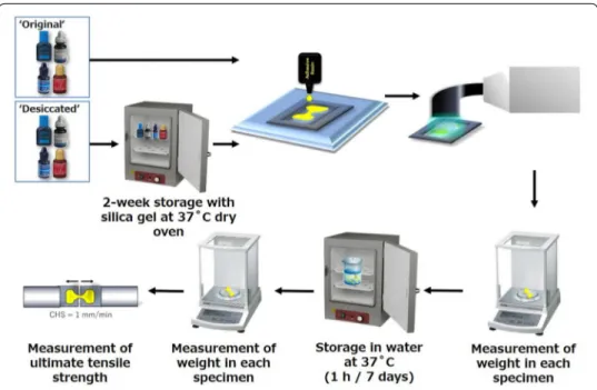
The aim of this study was to evaluate the influence of solvent evaporation on the ultimate tensile strength (UTS) of commercial adhesives. Two 1-step (OptiBond All-In-One and G-Premio Bond) and two 2-step (Clearfil SE Protect, OptiBond XTR) adhesives were selected. Two bottles of each adhesive were opened and stored at 37 °C in a dry oven with silica gel shielded from light for 2 weeks (“Desiccated”). Two unopened bottles were stored at room temperature (“Original”). After 2 weeks, the adhesives were used to fill an hour-glass shaped, metallic matrix mold and light-cured. Samples were weighed, and then immersed in a 37 °C water bath for 1 h or 7 days. The UTS of each sample was then measured at a cross-head speed of 1 mm/min (n = 10). The UTS for the Clearfil SE Protect was higher in the “Original” than “Desiccated” samples (p < 0.05). For the OptiBond XTR, no significant difference was found between the ‘Original’ and ‘Desiccated’ samples (p > 0.05). Neither of the two “Original” 1-step samples could be hardened, even after light-curing, yet the ‘Desiccated’ OptiBond All-In-One samples obtained high UTS values. Both OptiBond All-In-One and Clearfil SE Protect had an increase in weight after the 7-day immersion in water. In conclusion, residual solvent reduces the mechanical strength of the adhesive. The hydrophilicity of the adhesive resin might also affect its mechanical strength.
Keywords: Dental adhesive, Solvent evaporation, Ultimate tensile strength, Water
sorption, HEMA
Open Access
© The Author(s) 2019. This article is distributed under the terms of the Creative Commons Attribution 4.0 International License (http://creat iveco mmons .org/licen ses/by/4.0/), which permits unrestricted use, distribution, and reproduction in any medium, provided you give appropriate credit to the original author(s) and the source, provide a link to the Creative Commons license, and indicate if changes were made.
RESEARCH
*Correspondence: atsushi.kameyama@mdu. ac.jp 1 Department of Operative Dentistry, Endodontology, and Periodontology, School of Dentistry, Matsumoto Dental University, 1780 Gobara Hirooka, Shiojiri, Nagano 399-0781, Japan Full list of author information is available at the end of the articleContemporary adhesives have been categorized as “etch-and-rinse” and “self-etch” [3]. Among them, the two-step self-etch adhesives (2-SEAs) are popular because of their ease of use and rapid application [4]. Self-etching primers generally contain acidic func-tional monomers that are dissolved in water. Therefore, they do not require a separate rinse step after etching, as the acidic and hydrophilic functional monomer simultane-ously demineralizes and infiltrates the enamel/dentin substrate [5]. These hydrophilic monomers promote wettability and infiltration of the adhesive resin, which is mainly composed of hydrophobic resin monomers; for example, 2,2-bis[4-(2-hydroxy-3-meth-acryloyloxypropoxy)phenyl]propane (Bis-GMA), urethane dimethacrylate (UDMA), and triethylene glycol dimethacrylate (TEGDMA).
Recent formulations have shifted to one-step self-etch adhesive (1-SEA) systems, in which all components are combined into a single solution. Despite their user-friendli-ness, the lack of a hydrophobic bonding resin in these 1-SEA formulations reduces the stability of the bond over time, because the bonded interfaces behave as semipermeable membranes that allow the movement of water across them, expediting hydrolytic degra-dation [6, 7].
Since almost all 1-SEA reagents contains solvents such as water, ethanol or acetone, evaporation of the solvent affects monomer conversion and the mechanical proper-ties of the agent after setting [8, 9]. However, some 2-SEA agents have a solvent as part of the ingredients of the bonding agent, and some 1-SEA and 2-SEA products contain 2-hydroxyethyl methacrylate (HEMA), a hydrophilic compound that can affect water absorption by the cured adhesive [10].
Therefore, the purpose of this study was to evaluate the influence of solvent evapora-tion on the ultimate tensile strength (UTS) of commercial adhesives. The null hypotheses of this study were that (1) residual solvent does not influence the mechanical properties of the cured adhesive regardless of the period of water storage, and (2) mechanical prop-erties of the cured adhesive are influenced by the presence of HEMA regardless of the period of water storage.
Methods
Study design and adhesives tested
This study followed a factorial 4×2×2 design for “adhesive”, desiccation” and “storage period”.
Two 2-SEAs (Clearfil SE Protect, Kuraray Noritake Dental, Tokyo, Japan; OptiBond XTR, Kerr, Orange, CA, USA) and two 1-SEAs (OptiBond All-In-One, Kerr; G-Pre-mio Bond, GC, Tokyo, Japan) were used in this study (Table 1). For each adhesive, four bottles with the same batch number were used and randomly divided into two groups (“Original” and “Desiccated”).
Active solvent evaporation for “Desiccated” group
Two bottles of each adhesive were opened and the nozzle removed using pliers in a dark room. The adhesives were stored in a dark dry oven at 37 °C with silica gel (Wako Pure Chemical, Osaka, Japan) shielded from light for 2 weeks to aggressively evaporate any residual solvent. After 2-weeks, the nozzle and the bottle cap were replaced and the adhesive was stored at a controlled temperature (25 °C) until specimen preparation. The
other two bottles were stored at a controlled temperature (25 °C) until specimen prepa-ration with the bottle cap sealed.
Measurements of specimen weight and ultimate tensile strength (UTS)
Figure 1 shows a schematic representation of specimen preparation and the method of UTS testing. A stainless steel, hour glass-shaped split mold of 1 mm thickness (Fig. 2) was constructed and fixed onto a glass slide. The mold was then filled with adhesive, pressed with a second 1-mm-thick glass slide, and then light cured for 60 s through the top glass slide using an LED light-curing unit (Demi Plus, Kerr) [11, 12]. Light intensity was controlled using a hand-held dental radiometer (Model L.E.D. Table 1 Materials used in this study
Bis-GMA, 2,2-bis[4-(2-hydroxy-3-methacryloyloxypropoxy)phenyl]propane; CQ, dl-camphorquinone; GPDM, glycerol-phosphate-dimethacrylate; HEMA; hydroxyethylmethacrylate; 4-MET, 4-methacryloyloxyethyl trimellitate; 10-MDP, methacryloyloxydecyl dihydrogen phosphate; MDTP, methacryloyloxydecyl dihydrogen thiophosphate
a Also known as “Clearfil Megabond FA” in Japan
Adhesive (manufacturer) Ingredients Batch no.
2-step self-etch adhesive Clearfil SE Protect (Kuraray
Noritake Dental)a Bis-GMA, HEMA, 10-MDP, Hydrophobic aliphatic methacrylate, colloidal silica, CQ, initiators and accelerators, sodium fluoride, others CK0014
OptiBond XTR (Kerr) Hydrophobic monomers, ethanol, camphorquinone, 0.4-μm barium
glass, nano-silica filler, sodium hexafluorosilicate 5143490 1-step self-etch adhesive
OptiBond All-In-One (Kerr) GPDM, glycerol dimethacrylate, HEMA, water, acetone, ethanol, CQ-based photo-initiator, nano-sized fillers, sodium hexafluorosilicate, ytterbium fluoride
5164557 G-Premio Bond (GC) 4-MET, MDP, MDTP, methacrylate monomer, acetone, water, photo
initiators, silica 1605241
Fig. 1 Schematic illustration of the experimental set-up for ultimate tensile strength (UTS) testing. CHS,
Radiometer, SDS Kerr, Middleton, WI, USA) to ensure a light output of at least 1000 mW/cm2 [13]. After light curing, the samples were carefully removed from the mold.
The flash was removed with a surgical blade #15C. The weight of each specimen was immediately measured using a digital analytical balance (HR-202i, A&D Co., Tokyo, Japan). The specimens were then randomly divided and assigned to one of two stor-age conditions: 1 h or 7 days in 37 °C water bath shielded from light. After incuba-tion, each specimen was weighed again, and the cross-sectional area at the narrowest part of the specimen was measured using a digital caliper (Mitutoyo, Tokyo, Japan) to the nearest 0.01 mm. The remaining specimens were attached to the two free-sliding parts of a purpose-built holding device (Micro Tensile Test Jaw, Bisco, Schaumburg, IL, USA) using cyanoacrylate glue (Model Repair II Blue, Dentsply-Sankin, Ohtawara, Tochigi, Japan), and a tensile load was applied with a Micro Tensile Tester (Bisco, Schaumburg, IL, USA) at a cross-head speed of 1 mm/min until the specimen frac-tured (n = 10). The UTS of the cured adhesive was calculated as:
where F is the tensile load force at failure (N), and A is the cross-sectional area of the narrowest part of the specimen (mm2). The ultimate tensile strength (N/mm2) was
expressed in MPa [11, 12]. Statistical analysis
Data were analyzed using a three-way analysis of variance (ANOVA). If differences were found, pair-wise testing was performed using Tukey’s HSD test. The significance level was set to α = 0.05. Statistical analysis was performed using IBM SPSS 18 statistical soft-ware (SPSS, Chicago, IL, USA).
Results
Ultimate tensile strength
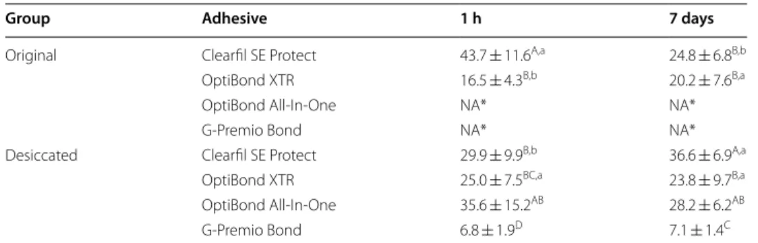
The result for the UTS tests is shown in Table 2. No effect was found for “storage period” (p = 0.072), whereas a significant effect was found for “adhesive” (p = 0.000) and “des-iccation” (p = 0.000). A significant interaction was also found for the three factors (p = 0.000).
Neither of the 1-SEAs (OptiBond All-In-One and G-Premio Bond) hardened in their “Original” states, even after sufficient light exposure. Therefore, these results were elimi-nated for statistical analysis.
The UTS of the “Desiccated” OptiBond All-In-One was 35.6 ± 15.2 MPa after 1 h and 28.2 ± 6.2 MPa after 7 days, respectively. In contrast, G-Premio Bond showed the signifi-cantly lower UTS values than OptiBond All-In-One at both time points (p < 0.05).
Differ to 1-SEAs, both 2-SEAs hardened in their “Original” and “Desiccated” forms. The UTS of the “Original” Clearfil SE Protect was significantly higher than the “Desic-cated” adhesive samples at 1 h (43.7 ± 11.6 vs. 29.9 ± 9.9, respectively; p = 0.008); in con-trast, by 7 days, “Desiccated” group was significantly higher than the “Original” group (24.8 ± 6.8 vs. 36.6 ± 6.9, respectively; p = 0.002). No significant differences were found between the “Original” and “Desiccated” OptiBond XTR samples (p = 0.298).
(1) UTS = F/A
Changes in specimen weight after water storage
The results for the percentage change in weight after water storage are shown in Table 3. According to the 3-way ANOVA analysis and Tukey’s post hoc test, a sig-nificant effect was found for all three factors (p = 0.000). A sigsig-nificant interaction was also found among the three factors (p = 0.000).
There was a significant increase in weight for both the “Original” and “Desiccated” Clearfil SE Protect specimens after storage for 7 days than storage for 1 h (p < 0.05). The “Original” OptiBond XTR showed slightly increase in weight after 1-h storage; however, this rather decreased after 7 days in storage. On the other hand, for the “Desiccated” OptiBond XTR samples, the percentage weight increase was higher after 7 days than 1 h. No weight measurements were taken for either of the 1-SEAs (Opti-Bond All-In-One and G-Premio (Opti-Bond) in their “Original” forms. The weight of the “Desiccated” G-Premio Bond specimen decreased after storage in water, whereas the weight of the “Desiccated” OptiBond All-In-One specimen increased after storage, both in a time-dependent manner.
Fig. 2 Hour glass-shaped stainless split mold for specimen preparation for ultimate tensile strength (UTS)
testing
Table 2 Ultimate tensile strengths
Data are the mean ± S.D., MPa; n = 10
Same capital superscript letters indicate that ultimate tensile strength values were not significantly different among each group in the same water storage period (p > 0.05)
Same small superscript letters indicate that ultimate tensile strength values were not significantly different among each adhesive in the same water storage period (p > 0.05)
ANOVA results could not be detected the significant difference for “storage period” (horizontal raws) * Not applicable because adhesive could not be hardened even after sufficient light exposure
Group Adhesive 1 h 7 days
Original Clearfil SE Protect 43.7 ± 11.6A,a 24.8 ± 6.8B,b
OptiBond XTR 16.5 ± 4.3B,b 20.2 ± 7.6B,a
OptiBond All-In-One NA* NA* G-Premio Bond NA* NA* Desiccated Clearfil SE Protect 29.9 ± 9.9B,b 36.6 ± 6.9A,a
OptiBond XTR 25.0 ± 7.5BC,a 23.8 ± 9.7B,a
OptiBond All-In-One 35.6 ± 15.2AB 28.2 ± 6.2AB
Discussion
The purpose of this study was to evaluate the influence of solvent evaporation on the mechanical properties of commercially available 2-SEAs and 1-SEAs. The mechanical property was measured in terms of ultimate micro-tensile strength (UTS), a variable that is frequently used to evaluate the performance of an adhesive in terms of its ten-sile bond strength with the local tissue [14, 15], which differs to the Young’s modulus or the nano-indentation value [16].
Two representative 2-SEAs were compared in this study: Clearfil SE Protect and OptiBond XTR. The Clearfil SE Protect contains the same proprietary self-etching, light-curing technology as the “gold-standard” Clearfil SE Bond, which has been well described in the literature [17], but the Clearfil SE Protect has some additional ingre-dients: an MDPB antibacterial monomer in the self-etching primer, and sodium fluo-ride and HEMA in the adhesive agent [18, 19]. HEMA has been reported to affect water absorption even after the adhesive is polymerized. In contrast, OptiBond XTR contains ethanol but does not contain HEMA. Our results found differences in the behaviors of these two adhesives.
The “Original” Clearfil SE Protect adhesive showed high UTS after 1-h storage in water, which decreased after 7 days of storage in water. Both the “Original” and “Des-iccated” specimens were also significantly heavier after 7 days in water, presumably because the Clearfil SE Protect contains HEMA monomers, which are hydrophilic and would have encouraged water sorption. For the “Original” specimens, this increased water sorption may be correlated with its reduced mechanical strength at 7 days; how-ever, this is not the same for the “Desiccated” specimens, which showed no difference in UTS at 1 h or 7 days. Desiccation at 37 °C might accelerate the hydrolysis of hydrophilic monomers, such as HEMA and MDP [20]. However, we surmise that the 2-week desic-cation period at 37 °C might have led to some deterioration of the adhesive’s composi-tion, and this may explain the difference in the UTS at the 1-h time point. Yet, without desiccation, this reduction in UTS in response to water sorption would, by extension, Table 3 Percentage increase in weight after storage in water
Data are the mean ± S.D., %; n = 10
Same capital superscript letters indicate that ultimate tensile strength values were not significantly different among each group in the same water storage period (p > 0.05)
Same small superscript letters indicate that ultimate tensile strength values were not significantly different among each adhesive in the same water storage period (p > 0.05)
S, detection of the significant difference between 1 h and 7 days (p > 0.05); NS, not detection of the significant difference between 1 h and 7 days (p > 0.05); NA, not applicable because adhesive could not be hardened even after sufficient light exposure
Group Adhesive 1 h 7 days S/NS
Original Clearfil SE Protect 0.8 ± 0.4B,a 6.7 ± 0.3A,a S
OptiBond XTR 0.7 ± 0.7B,a −1.4 ± 1.5B,b S
OptiBond All-In-One NA* NA* – G-Premio Bond NA* NA* – Desiccated Clearfil SE Protect 0.9 ± 0.3B,a 5.9 ± 0.3B,a S
OptiBond XTR 1.0 ± 0.3AB,a 3.0 ± 1.0C,a S
OptiBond All-In-One 2.2 ± 0.6A 8.1 ± 0.7A S
suggest a similar reduction in bond strength of the adhesive to the tooth substrate as it was exposed to water over time [8]. Collectively, these results point to the need for the optimization of solvent evaporation for specific adhesive compounds.
Previous studies have shown that, although insufficient air-drying of Clearfil SE Bond primer can result in lower UTS of the Clearfil SE Bond adhesive, the UTS of the pure adhesive is very high [14, 21], presumably because of its high filler loading and polym-erization efficacy [22]. It has also been reported that the adhesive layer thickness of the Clearfil SE Bond (which is of similar composition to Clearfil SE Protect) does not affect the bond strength [23]. Our results therefore confirm that strong air-blowing is unneces-sary after applying the Clearfil SE Protect adhesive.
According to the manufacturer’s report, filler loading of OptiBond XTR is higher than that of Clearfil SE Bond. However, the UTS of the “Original” OptiBond XTR was lower than that of the Clearfil SE Protect after 1 h of water storage. Also, in contrast to the Clearfil SE Protect, the “Original” OptiBond XTR had a lower specimen weight after water storage for 1 week. This phenomenon can be explained in two ways: Unlike the Clearfil SE Protect, the OptiBond XTR agent contains ethanol in the adhesive agent. Therefore, the percentage of the residual unpolymerized monomer within the light-cured OptiBond XTR specimens might be larger than that of Clearfil SE Protect. In addition, because OptiBond XTR does not contain HEMA, the water absorption of the OptiBond XTR is likely to be lower than that of Clearfil SE Protect. For the OptiBond XTR, the difference in the UTS between the “Original” and “Desiccated” specimens was smaller than that for Clearfil SE Protect. These results suggest that there is less likely to be a time-dependent degradation in bond strength, because the polymerized adhesives are less susceptible to water absorption [24]. The slight increase in the specimen weight may be due to the addition of glycerol-phosphate-dimethacrylate (GPDM) monomer [25].
For OptiBond XTR, desiccation did not improve the UTS. Others have shown that the addition of 10% to 20% ethanol to the experimental resin blends improves the degree of conversion [26]. Although the evaporation of the remaining ethanol in the OptiBond XTR might increase conversion, some ethanol remains trapped in the polymer and this would promote water sorption, which may lower the mechanical properties of the poly-mer [27, 28]. Therefore, in the clinic, excess evaporation of the OptiBond XTR adhesive might negatively affect the mechanical properties of polymer.
The “Original” OptiBond All-In-One adhesive did not hard even when light curing was used. This is probably due to the presence of water and other solvents, which inhibits the polymerization of the monomers in the adhesive resin [29]. The “Desiccated” OptiBond All-In-One adhesive, however, showed high UTS values. Almost all commercially avail-able 1-SEAs contain water as a solvent to promote wetting of the substrate and improve the homogeneity of the liquid. Hydrophilic monomers enhance the wettability and infil-tration of the hydrophobic resin monomers into the demineralized dentin matrix. How-ever, insufficient air-blowing might result in the presence of residual solvents from the adhesive layer, and thereby lead to insufficient polymerization. According to the report of Fu and colleagues, the microtensile bond strength of OptiBond All-In-One applied to dentin was high (more than 75 MPa) if air-blowing was performed for more than 15 s. If air-blowing was omitted, all specimens failed before reaching the microtensile testing
phase [30]. Others show that solvent evaporation of OptiBond All-In-One using warm air-blowing (60 °C ± 2 °C) led to significantly higher bond strength than after using cold air-blowing (20 °C ± 1 °C) [31]. In the clinic, it is difficult to evaporate the solvent com-pletely [32], because the relative humidity of the oral environment is higher than that of an experimental setting (23 °C, 50%), and this is independent of whether the resin is applied to an incisal site or a molar site [33–35]. Resin bonding in a high humidity envi-ronment has reduced bond strength [36]. Therefore, it is recommended to control the intra-oral humidity using rubber dam isolation or an intra-oral vacuum device.
“Desiccated” G-Premio Bond, unlike the “Desiccated” OptiBond All-In-One, did not obtain adequate hardness even if with sufficient light curing, leaving a “gummy” speci-men. In addition, whereas the weight of the OptiBond All-In-One specimens increased after 7 days of water storage, the weight of G-Premio Bond specimens decreased. Papa-dogiannis and colleagues [37] recently reported that Vickers hardness could not be measured in G-Premio Bond, since the material demonstrated a soft, gel consistency, contrary to all other 1-SEA (Adhese Universal, Ivoclar-Vivadent; All-Bond Universal, Bisco; Clearfil Universal Bond Quick, Kuraray Noritake Dental; Prelude One, Danville Materials; Scotchbond Universal, 3M ESPE). They also reported that the degree of C=C conversion of G-Premio Bond (67.2 DC%) was significantly lower than the all other 1-SEAs (77.7–82.0 DC%) [37]. G-Premio Bond has been reported the strong acidity (pH 1.5) than the other contemporary 1-SEAs, possibly associated with the presence of many acidic monomers (MDP, 4-MET, and MDTP) [30, 37]. Therefore, it can be suggested that the “gummy” G-Premio Bond specimen was resulted as the interference of polym-erization. Ikeda et al. [14] reported that the UTS of G-Bond HEMA-free 1-SEA (GC) was significantly lower than that of HEMA-containing 1-SEA (Clearfil S3 Bond, Kuraray
Noritake Dental), despite the significantly higher amount of evaporation that occurs by air-drying for the G-Bond than the Clearfil S3 Bond. They also observed numerous
lets in the G-Bond, regardless of the length of air-drying (0 s, 5 s, and 10 s). These drop-lets represent a phase-separation between water and the other adhesive ingredients [38,
39]. The authors explained that, because longer air-drying did not eliminate the droplets and thus the volatile parts of the adhesive, the droplets were likely encapsulated within the adhesive upon curing. The lower UTS and reduced specimen weight for the G-Pre-mio Bond in our study supports their explanation; we surmise that similar droplets must have inhibited polymerization of adhesive and reduced its UTS. Tsujimoto et al. [40] recently reported numerous cracks in the fractured adhesive interfaces of G-Premio Bond adhesive after shear fatigue strength tests through SEM analysis. These defects at the adhesive interface might accelerate degradation at the resin-tooth adhesive interface. Therefore, further studies should be conducted to establish the relationship between residual solvents and bond durability.
Conclusion
Based on the limitations imposed within this work, it can be concluded that the removal of residual solvent is important for polymerization of adhesives, particularly 1-SEAs. However, the presence of HEMA can negatively affect the mechanical properties of the cured adhesive, and desiccation for some types of adhesives can reduce the mechanical
strength after contact with water for an extended length of time. It is recommended that clinicians remove residual solvent to achieve optimal mechanical properties of the cured adhesive but perhaps consider the type of material and solvent before performing aggressive desiccation.
Abbreviations
1-SEA: one-step self-etch adhesive; 2-SEA: two-step self-etch adhesive; ANOVA: analysis of variance; Bis-GMA: 2,2-bis[4-(3-methacryloyloxypropoxy)phenyl]propane; GPDM: glycerol-phosphate-dimethacrylate; HEMA: 2-hydroxy-ethyl methacrylate; MDP: methacryloyloxydecyl dihydrogen phosphate; TEGDMA: tri2-hydroxy-ethylene glycol dimethacrylate; Tukey’s HSD test: Tukey’s honestly significant difference test; UDMA: urethane dimethacrylate; UTS: ultimate tensile strength.
Acknowledgements
The authors thank Rebecca Jackson, PhD, from Edanz Group (https ://www.edanz editi ng.com/ac) for editing a draft of this manuscript.
Authors’ contributions
AK designed the study, prepared the specimens, and carried out the experimental measurements. AH carried out the experimental measurements and performed data analysis. HA, MK and YN carried out the experimental measurements. TM mainly reviewed the paper critically for content and approved it for submission. All authors read and approved the final manuscript.
Funding
This work was supported by JSPS KAKENHI Grant Numbers JP 17K11715 and JP 16K20464.
Availability of data and materials
The datasets generated during and/or analyzed during the current study are not publicly available, but are available from the corresponding author on reasonable request.
Ethics approval and consent to participate
This article does not contain any studies with human participants or animals performed by any of the authors.
Consent for publication
Not applicable.
Competing interests
The authors declare that they have no competing interests.
Author details
1 Department of Operative Dentistry, Endodontology, and Periodontology, School of Dentistry, Matsumoto Dental
Uni-versity, 1780 Gobara Hirooka, Shiojiri, Nagano 399-0781, Japan. 2 Department of Operative Dentistry, Cariology and Pulp
Biology, Tokyo Dental College, 2-9-18 Kanda-Misakicho, Chiyoda-ku, Tokyo 101-0061, Japan. 3 ABO Dental Clinic, 3-76-6
Keyaki-dai, Sanda, Hyogo 669-1321, Japan. Received: 21 March 2019 Accepted: 10 June 2019
References
1. Zimmerli B, Strub M, Jeger F, Stadler O, Lussi A. Composite materials: composition, properties and clinical applica-tions. A literature review. Schweiz Monatsschr Zahnmed. 2010;120:972–86.
2. Kim M, Suh B-I, Shin D, Kim K-M. Comparison of the physical and mechanical properties of resin matrix with two photoinitiator systems in dental adhesives. Polymers. 2016;8:250. https ://doi.org/10.3390/polym 80702 50. 3. De Munck J, Van Landuyt K, Peumans M, Poitevin A, Lambrechts P, Braem M, Van Meerbeek B. A critical review
of the durability of adhesion to tooth tissue: methods and results. J Dent Res. 2005;84:118–32. https ://doi. org/10.1177/15440 59105 08400 204.
4. Van Meerbeek B. Dentin/enamel bonding. J Esthet Restor Dent. 2010;22:157. https ://doi.org/10.111 1/j.1708-8240.2010.00329 .x.
5. Van Meerbeek B, Yoshihara K, Yoshida Y, Mine A, De Munck J, Van Landuyt KL. State of the art of self-etch adhesives. Dent Mater. 2011;27:17–28. https ://doi.org/10.1016/j.denta l.2010.10.023.
6. Van Landuyt KL, Mine A, De Munck J, Jaecques S, Peumans M, Lambrechts P, Van Meerbeek B. Are one-step adhesives easier to use and better performing? Multifactorial assessment of contemporary one-step self-etching adhesives. J Adhes Dent. 2009;11:175–90. https ://doi.org/10.3290/j.jad.a1562 3.
7. Hashimoto M. A review—micromorphological evidence of degradation in resin-dentin bonds and potential preven-tional solutions. J Biomed Mater Res B Appl Biomater. 2010;92:268–80. https ://doi.org/10.1002/jbm.b.31535 . 8. Hosaka K, Nakajima M, Takahashi M, Itoh S, Ikeda M, Tagami J, Pashley DH. Relationship between mechanical
proper-ties of one-step self-etch adhesives and water sorption. Dent Mater. 2010;26:360–7. https ://doi.org/10.1016/j.denta l.2009.12.007.
9. Emamieh S, Sadr A, Ghasemi A, Torabzadeh H, Akhavanzanjani V, Tagami J. Effects of solvent drying time and water storage on ultimate tensile strength of adhesives. J Investig Clin Dent. 2014;5:51–7. https ://doi.org/10.111 1/j.2041-1626.2012.00146 .x.
10. Itoh S, Nakajima M, Hosaka K, Okuma M, Takahashi M, Shinoda Y, Seki N, Ikeda M, Kishikawa R, Foxton RM, Tagami J. Dentin bond durability and water sorption/solubility of one-step self-etch adhesives. Dent Mater J. 2010;29:623–30. https ://doi.org/10.4012/dmj.2010-028.
11. Manso AP, Bedran-Russo AK, Suh B, Pashley DH, Carvalho RM. Mechanical stability of adhesives under water storage. Dent Mater. 2009;35:744–9. https ://doi.org/10.1016/j.denta l.2008.12.006.
12. Kameyama A, Kato J, De Munck J, Hatayama H, Haruyama A, Yoshinari M, Takase Y, Van Meerbeek B, Tsunoda M. Light-curing efficiency of dental adhesives by gallium nitride violet-laser diode determined in terms of ultimate micro-tensile strength. Bio-Med Mater Eng. 2011;21:347–56. https ://doi.org/10.3233/BME-2012-0682.
13. Kameyama A, Haruyama A, Asami M, Takahashi T. Effect of emitted wavelength and light guide type on irra-diance discrepancies in hand-held dental curing radiometer. Sci World J. 2013;2013:647941. https ://doi. org/10.1155/2013/64794 1.
14. Ikeda T, De Munck J, Shirai K, Hikita K, Inoue S, Sano H, Lambrechts P, Van Meerbeek B. Effect of fracture strength of primer-adhesive mixture on bonding effectiveness. Dent Mater. 2005;21:413–20. https ://doi.org/10.1016/j.denta l.2004.07.006.
15. Takahashi A, Sato Y, Uno S, Pereira PNR, Sano H. Effects of mechanical properties of adhesive resins on bond strength to dentin. Dent Mater. 2002;18:263–8. https ://doi.org/10.1016/S0109 -5641(01)00046 -X.
16. Freitas PH, Giannini M, França R, Correr AB, Correr-Sobrinho L, Consani S. Correlation between bond strength and nanomechanical properties of adhesive interface. Clin Oral Investig. 2017;21:1055–62. https ://doi.org/10.1007/s0078 4-016-1847-7.
17. Van Meerbeek B, Van Landuyt K, De Munck J, Hashimoto M, Peumans M, Lambrechts P, Yoshida Y, Inoue S, Suzuki K. Technique-sensitivity of contemporary adhesives. Dent Mater J. 2005;24:1–13. https ://doi.org/10.4012/dmj.24.1. 18. Imazato S, Kuramoto A, Takahashi Y, Ebisu S, Peters MC. In vitro antibacterial effects of the dentin primer of Clearfil
Protect Bond. Dent Mater. 2006;22:527–32. https ://doi.org/10.1016/j.denta l.2005.05.009.
19. Kameyama A, Tsumori M, Ushiki T, Muto Y, Koga H, Matsukubo T, Hirai Y. Fluoride release from newly developed dental adhesives. Bull Tokyo Dent Coll. 2002;43:193–7. https ://doi.org/10.2209/tdcpu blica tion.43.193. 20. Ma S, Fujita K, Nishiyama N. Effects of storage temperature on the shelf life of one-step and two-step self-etch
adhesives. Oper Dent. 2009;34:472–80. https ://doi.org/10.2341/08-010-L.
21. Kameyama A, Kato J, Yoshinari M, Kotoku Y, Akashi G, Hirai Y. Ultimate micro-tensile strength of dental adhesives cured at different light sources. J Photopolym Sci Technol. 2008;21:31–5. https ://doi.org/10.2494/photo polym er.21.31.
22. Cadenaro M, Antonialli F, Sauro S, Tay FE, Di Lenarda R, Prati C, Biasotto M, Contardo L, Breschi L. Degree of conversion and permeability of dental adhesives. Eur J Oral Sci. 2005;113:525–30. https ://doi.org/10.111 1/j.1600-0722.2005.00251 .x.
23. Coelho PG, Calamia C, Harsono M, Thompson VP, Silva NR. Laboratory and FEA evaluation of dentin-to-composite bonding as a function adhesive layer thickness. Dent Mater. 2008;24:1297–303. https ://doi.org/10.1016/j.denta l.2008.02.007.
24. Shinoda Y, Nakajima M, Hosaka K, Otsuki M, Foxton RM, Tagami J. Effect of smear layer characteristics on dentin bonding durability of HEMA-free and HEMA-containing one-step self-etch adhesives. Dent Mater J. 2011;30:501–10. https ://doi.org/10.4012/dmj.2011-001.
25. Hoshika S, Kameyama A, Suyama Y, De Munck J, Sano H, Van Meerbeek B. GPDM- and 10-MDP-based self-etch adhe-sives bonded to bur-cut and uncut enamel—”Immediate” and “aged” µTBS. J Adhes Dent. 2018;20:113–20. https :// doi.org/10.3290/j.jad.a4030 7.
26. Cadenaro M, Breschi L, Antoniolli F, Navarra CO, Mazzoni A, Tay FR, Di Lenarda R, Pashley DH. Degree of conver-sion of resin blends in relation to ethanol content and hydrophilicity. Dent Mater. 2008;24:1194–200. https ://doi. org/10.1016/j.denta l.2008.01.012.
27. Ye Q, Spencer P, Wang Y, Misra A. Relationship of solvent to the photopolymerization process, properties, and struc-ture in model dentin adhesives. J Biomed Mater Res. 2007;80A:342–50. https ://doi.org/10.1002/jbm.a.30890 . 28. Cadenaro M, Breschi L, Rueggeberg FA, Suchko M, Grodin E, Agee K, Di Lenarda R, Tay FR, Pashley DH. Effects of
residual ethanol on the rate and degree of conversion of five experimental resins. Dent Mater. 2009;25:621–8. https ://doi.org/10.1016/j.denta l.2008.11.005.
29. Wang Y, Spencer P. Continuing etching of an all-in-one adhesive in wet dentin tubules. J Dent Res. 2005;84:350–4. https ://doi.org/10.1177/15440 59105 08400 411.
30. Fu J, Saikaew P, Kawano S, Carvalho RM, Hannig M, Sano H, Selimovic D. Effect of air-blowing duration on the bond strength of current one-step adhesives to dentin. Dent Mater. 2017;33:895–903. https ://doi.org/10.1016/j.denta l.2017.03.015.
31. Moura SK, Murad CG, Reis A, Klein-Júnior CA, Grande RM, Loguercio AD. The influence of air temperature for solvent evaporation on bonding of self-etch adhesives to dentin. Eur J Dent. 2014;8:205–10. https ://doi.org/10.4103/1305-7456.13060 2.
32. Abo H, Kameyama A, Haruyama A. Clinical observation of the tooth surface during air-drying of self-etching primer under 3D video microscope. Appl Adhes Sci. 2016;4:7. https ://doi.org/10.1186/s4056 3-016-0064-6.
33. Kameyama A, Asami M, Noro A, Abo H, Hirai Y, Tsunoda M. The effects of three dry-field techniques on intraoral tem-perature and relative humidity. J Am Dent Assoc. 2011;142:274–80. https ://doi.org/10.14219 /jada.archi ve.2011.0166. 34. Haruyama A, Kameyama A, Tatsuta C, Ishii K, Sugiyama T, Sugiyama S, Takahashi T. Influence of different rubber
dam application on intraoral temperature and relative humidity. Bull Tokyo Dent Coll. 2014;55:11–7. https ://doi. org/10.2209/tdcpu blica tion.55.11.
35. Saraiva LO, Aguiar TR, Costa L, Cavalcanti AN, Giannini M, Mathias P. Influence of intraoral temperature and relative humidity on the dentin bond strength: an in situ study. J Esthet Restor Dent. 2015;27:92–9. https ://doi.org/10.1111/ jerd.12098 .
36. Chiba Y, Yamaguchi K, Miyazaki M, Tsubota K, Takamizawa T, Moore BK. Effect of air-drying time of single-application self-etch adhesives on dentin bond strength. Oper Dent. 2006;31:233–9. https ://doi.org/10.2341/05-19.
37. Papadogiannis D, Dimitriadi M, Zafiropoulou M, Gaintantzopoulou M-D, Eliades G. Universal adhesives: setting characteristics and reactivity with dentin. Materials. 2019;12:1720. https ://doi.org/10.3390/ma121 01720 . 38. Van Landuyt KL, De Munck J, Snauwaert J, Coutinho E, Poitevin A, Yoshida Y, Inoue S, Peumans M, Suzuki K,
Lambrechts P, Van Meerbeek B. Monomer-solvent phase separation in one-step self-etch adhesives. J Dent Res. 2005;84:183–8. https ://doi.org/10.1177/15440 59105 08400 214.
39. Van Landuyt KL, Snauwaert J, De Munck J, Coutinho E, Poitevin A, Yoshida Y, Suzuki K, Lambrechts P, Van Meerbeek B. Origin of interfacial droplets with one-step adhesives. J Dent Res. 2007;86:739–44. https ://doi.org/10.1177/15440 59107 08600 810.
40. Tsujimoto A, Barkmeier WW, Takamizawa T, Watanabe H, Johnson WW, Latta MA, Miyazaki M. Comparison between universal adhesives and two-step self-etch adhesives in terms of dentin bond fatigue durability in self-etch mode. Eur J Oral Sci. 2017;125:215–22. https ://doi.org/10.1111/eos.12346 .
Publisher’s Note
![For copies of the original instructions see [7]](data:image/gif;base64,R0lGODlhAQABAIAAAP///wAAACH5BAEAAAAALAAAAAABAAEAAAICRAEAOw==)