Japan Advanced Institute of Science and Technology
JAIST Repository
https://dspace.jaist.ac.jp/Title
Comparative analysis of the cellular entry of polystyrene and gold nanoparticles using the freeze concentration method
Author(s) Ahmed, Sana; Okuma, Koyo; Matsumura, Kazuaki Citation Biomaterials Science, 6: 1791-1799
Issue Date 2018-04-30
Type Journal Article
Text version author
URL http://hdl.handle.net/10119/15878
Rights
Copyright (C) 2018 Royal Society of Chemistry. Sana Ahmed, Koyo Okuma and Kazuaki Matsumura, Biomaterials Science, 6, 2018, 1791-1799.
http://dx.doi.org/10.1039/C8BM00206A - Reproduced by permission of The Royal Society of Chemistry Description
1
Comparative analysis of the cellular entry of polystyrene and gold nanoparticles 1
using the freeze concentration method 2
Sana Ahmed, Koyo Okuma, and Kazuaki Matsumura*
3 4
School of Materials Science, Japan Advanced Institute of Science and Technology, Nomi,
5
Ishikawa 923-1292, Japan
6 7
KEYWORDS: freeze concentration, gold, polystyrene, nanoparticles, endocytic 8 mechanism 9 E mail: mkazuaki@jaist.ac.jp 10 11 12 13 14 15 16 17 18 19 20 21 22 23
2 Abstract
1
Despite advances in nanoparticle delivery, established physical approaches, such as
2
electroporation and sonication, result in cell damage, limiting their practical applications.
3
In this study, we proposed a unique freeze concentration-based technique and evaluated
4
the efficacy of the method using two types of nanoparticles, citrate-capped gold
5
nanoparticles and carboxylated polystyrene nanoparticles. We further compared the
6
internalisation behaviour of particles of various sizes with and without freezing. Confocal
7
microscopic images showed that the uptake efficacy for nanomaterials of 50 nm was
8
greater than that of l00-nm particles. Polystyrene nanoparticles of 50 nm had more
9
favourable adsorption and internalisation behaviours compared to those of gold
10
nanoparticles after freeze concentration. We also examined the possible endocytic
11
pathways involved in the uptake of gold and polystyrene nanoparticles, and found that
12
the route differed between unfrozen and frozen conditions. Overall, we determined the
13
influence of the freeze concentration strategy on both nanomaterial internalisation and
14
the endocytic uptake pathway. Our findings provide a mechanistic understanding of the
15
internalisation of nanoparticles using a freezing approach and thereby contribute to
16
further developments in nanotherapeutic applications.
17 18 19 20 21 22 23 24 25 26 27
3 Introduction
1
Recently, functional nanomaterials have gained substantial interest as diagnostic tools
2
and for the development of delivery systems.1 Among nanoparticles, gold2 (Au) and 3
polystyrene3 (PS) nanoparticles are common nanocarriers for the delivery of a variety of 4
biomacromolecules inside cells. Au nanoparticles are beneficial for various applications,
5
such as biosensors,4 antimicrobial agents,5 tumour imaging,6 and drug delivery.7 PS with 6
fluorescence plays an important role in dynamic interactions between nano-bio
7
interfaces, has good biocompatibility and high stability, and is useful for cell imaging.8 8
However, nanomaterials have to cross the cell membrane to enter cells. To achieve this,
9
physical approaches, such as electroporation9 and sonication,10 have been used for the 10
delivery of nanomaterials of different sizes. A few studies have indicated that the use of
11
electric fields might damage cells.11,12 Therefore, we have developed a new freeze 12
concentration-based strategy for the safe and effective delivery of proteins.13-16 Freeze 13
concentration is a physical process in which extremely low temperatures result in the
14
transformation of water into ice crystals and the ejection of the protein-nanocarrier
15
complex, thereby increasing the concentration of the protein-nanocarrier complex at the
16
peripheral cell membrane. We previously demonstrated the benefits of the enhanced
17
concentration of the protein-nanocarrier in immunotherapy15 as well as in gene delivery 18
systems.16 However, the influence of the size and concentration of particles induced by 19
freezing on the endocytic pathway has not been evaluated, and comparisons between
20
non-frozen and frozen conditions are needed.
21
Endocytosis is a well-characterised pathway by which particles efficiently enter living
22
cells. Plasma membranes are selectively permeable. Materials are usually enclosed by a
23
small portion of the plasma membrane, initially invaginated, and then pinched off to form
24
an endocytic vesicle that includes the ingested substance. 17,18 Nearly all types of cells 25
utilise the endocytic pathway to communicate with viruses and nanoparticles.19 Particles 26
can be internalised into cells via two major pathways, i.e. phagocytosis and pinocytosis.20 27
Large particles with a diameter of greater than 200 nm are usually taken up by
28
phagocytosis. Some internalised vesicles never reach lysosomes because they are
29
recovered by phagosomes vesicles and returned to the plasma membrane.21 The majority 30
of small particles enter via pinocytosis pathways, including macropinocytosis,
4
mediated endocytosis, and caveolae-mediated endocytosis.22 Typically, for clathrin 1
mediated endocytosis, the endocytic cycle starts at clathrin-coated pits in small areas of
2
the plasma membrane. Caveolae are present in the plasma membrane of all cell types.
3
They are formed from lipid rafts (patches of plasma membrane) containing cholesterol,
4
glycosphingolipids, and GPI-anchored membrane proteins. Macropinocytosis begins with
5
the deformation of the plasma membrane; linear or curved ruffles form on cell membrane,
6
and these constrict and become separated from the plasma membrane to form
7
macropinosomes inside of cells.23,24 8
In general, physical approaches, e.g. electroporation and sonication, promote
9
nanomaterial uptake via non-endocytic pathways, resulting in high oxidative stress and
10
changes in cell stimuli.25,26 The enhanced concentration of materials in the peripheral 11
membrane using the freezing strategy enables uptake by endocytic pathways, as
12
demonstrated in our previous research.14 However, the precise effects of particle size and 13
frozen or non-frozen conditions are unclear. Therefore, to elucidate the effect of particle
14
concentration using freezing and non-freezing systems and the influence of size, we used
15
citrate-capped Au nanoparticles and carboxylated PS nanoparticles as model systems.
16
In this study, we evaluated two types of particles, i.e. inorganic nanoparticles and
17
polymeric nanoparticles. A number of inorganic particles, such as silver nanoparticles
18
(Ag), are used in biomedical applications owing to their beneficial properties, including
19
their antibacterial effects.27 For example, the nanohybrid Ag@PS complex with a 20
nitroxide linker provides efficient activity against pathogenic bacteria.28 Many previous 21
studies have assessed the influence of various factors associated with the cellular uptake
22
of fluorescent latex particles (PS beads).29,30 Haines et al. demonstrated the effects of 23
particle size, cell line, and even cell density on the uptake of PS microspheres.29 In another 24
study, Florence et al. showed the size dependency of microspheres on uptake in
25
systematic organs after oral administration; particles exceeding 100 nm did not reach the
26
bone marrow and those exceeding 300 nm were absent from blood.30 27
28
In our study, we focused on Au nanoparticles and PS nanoparticles owing to their
29
availability in a variety of sizes and surface chemistries; we used the fibroblast L929 cell
30
line and the freeze concentration approach to study the endocytic mechanism. To the best
31
of our knowledge, this is the first study to demonstrate that the freeze concentration
5
approach induces different endocytic pathways depending on the material and size. For
1
consistency, both types of nanomaterials (Au and PS nanoparticles) had similar
2
hydrodynamic sizes, i.e. they were 50 and 100 nm in diameter, for investigation of uptake
3
by fibroblast L929 cells using non-frozen and frozen approaches; furthermore, we
4
discuss the potential endocytic mechanisms in these conditions.
5 6 Experimental 7 Reagents 8
Citrate buffer-stabilised gold nanoparticles (50 and 100 nm), filipin, and EIPA
(5-(N-9
ethyl-N-isopropyl)-amiloride) were obtained from Sigma Aldrich (St. Louis, MO, USA).
10
Fluoresbrite® Yellow Green Polybead Microspheres (YG Polybeads, λex = 441 nm, λem = 11
486 nm) with diameters of 50 and 100 nm were purchased from Polyscience Inc.
12
(Warrington, PA, USA). Chlorpromazine and DMSO were obtained from Nacalai Tesque
13
(Kyoto, Japan).
14
Zeta potential and hydrodynamic size of gold and polystyrene nanoparticles 15
The average hydrodynamic diameter and zeta potential of Au and PS nanoparticles were
16
measured by dynamic light scattering (DLS) using the Zetasizer 3000 (Malvern
17
Instruments, Worcestershire, UK) with a scattering angle of 135°. Results were obtained
18
as the average of three measurements using different samples.
19 20
Transmission electron microscopy (TEM) 21
Au and PS nanoparticle solutions (10 µL) were placed on a copper grid (NS-C15 Cu150P;
22
Stem, Tokyo, Japan). In the case of PS nanoparticles, the grids were negatively stained
23
with 2% phosphotungstic acid (Sigma Aldrich, Steinheim, Germany) for 1 min and then
24
washed with two drops of distilled water, blotted, and air-dried. TEM images were
25
obtained using a Hitachi H-7100 system (Tokyo, Japan) with an accelerating voltage of
26 100 kV. 27 28 Cell culture 29
6
L929 cells (American Type Culture Collection, Manassas, VA, USA) were cultured in
1
Dulbecco’s modified Eagle’s medium (DMEM; Sigma-Aldrich) supplemented with 10%
2
foetal bovine serum (FBS) at 37°C and 5% CO2 in a humidified atmosphere. When the 3
cells reached 80% confluence, they were removed using 0.25% (w/v) trypsin containing
4
0.02% (w/v) ethylenediamine tetraacetic acid (EDTA) in phosphate-buffered saline
5
without calcium and magnesium (PBS(-)) and were seeded on a new tissue culture plate
6
for subculture.
7
Cellular adsorption of nanoparticles by freeze concentration 8
L929 cells were counted and re-suspended in 1 mL of 10% DMSO (without FBS) 9
containing Au or PS (2–10 × 1010 particles/mL) nanoparticles at a density of 1 × 10⁶ 10
cells/mL in 1.9-mL cryovials (Nalgene, Rochester, NY, USA) and were stored in a -80°C
11
freezer overnight. The vials were thawed at 37°C, diluted with DMEM, and cells were
12
washed 3 times with DMEM containing 10% FBS. Subsequently, cells were counted using
13
a haemocytometer by the trypan blue staining method. Cell viability was determined as
14
the ratio of living cells to total cells. The adsorption of nanoparticles on L929 cells before
15
and after freezing was observed using a confocal laser scanning microscope (CLSM)
16
(FV1000-D; Olympus, Tokyo, Japan). The PS nanoparticles were internally labelled by
17
Yellow Green, similar to FITC (fluorescein isothiocyanate) fluorophores, with (YG
18
Polybeads, λex = 441 nm, λem = 486 nm). Au nanoparticles were quantitatively evaluated 19
by plasmon scattering. Au nanoparticles of 50 nm and 100 nm were irradiated using a
20
laser wavelength of 559 nm.
21
Internalisation of nanoparticles by freeze concentration 22
After the thawing procedure, the cells were washed three times using cell culture medium
23
with 10% FBS. Then, the cells were again seeded on a 35-mm glass-bottom dish with
24
DMEM. After incubation for 24 h, the attached cells were washed with PBS (-) 3 times and
25
were observed using a CLSM. For comparisons with non-frozen cells as a control, the
26
same concentration of nanoparticles was added to the L929 cell suspension, centrifuged
27
at 112 × g, washed with medium, and seeded them for 24 h. All cells were washed with
28
PBS (-) before the observation of internalisation using a CLSM.
7
Elucidation of the internalisation pathway in non-frozen and frozen conditions by 1
an inhibition assay 2
L929 fibroblast cells were pre-treated with various concentrations of endocytic
3
inhibitors, including chlorpromazine (12.5 µM), EIPA (125 µM), and filipin (2.5 µM).
4
Previously reported inhibitor concentrations were used.14 Then, the samples were 5
cryopreserved with 10% DMSO carrying Au or PS nanoparticles in the cell culture
6
medium at −80°C. Solutions were thawed and the cells were counted to determine the
7
toxicity of the inhibitors using the trypan blue exclusion assay. After the addition of
8
particular inhibitors, cells at a density of 1 × 10⁶ cells per mL were seeded on a glass-9
bottom dish for 24 h to investigate the uptake of particles. The same procedure was
10
applied for the non-frozen condition. The nanoparticles with inhibitors were gently
11
added to fibroblast cells and seeded on glass-bottom dishes following the same methods.
12
Cells treated with only Au or PS nanoparticles were regarded as positive controls. After
13
incubation, cells were washed with PBS (-) 3 times to remove any traces of inhibitors. The
14
mean scattering of Au nanoparticles per cell was detected by excitation at 559 nm using
15
a confocal microscope. A similar procedure was applied for PS nanoparticles; the
16
quantification of the mean fluorescence intensity per cell was performed using a confocal
17
microscope.
18
Statistical analysis 19
All data are expressed as means ± standard deviation (SD). All experiments were
20
conducted in triplicate. For comparisons among groups, a one-way analysis of variance
21
with a post-hoc Fisher’s protected least significant difference test was used. Differences
22
were considered statistically significant when p < 0.05.
23 24
Results and discussion 25
Hydrodynamic size and zeta potential of gold and polystyrene nanoparticles in the 26
presence of cryoprotectants 27
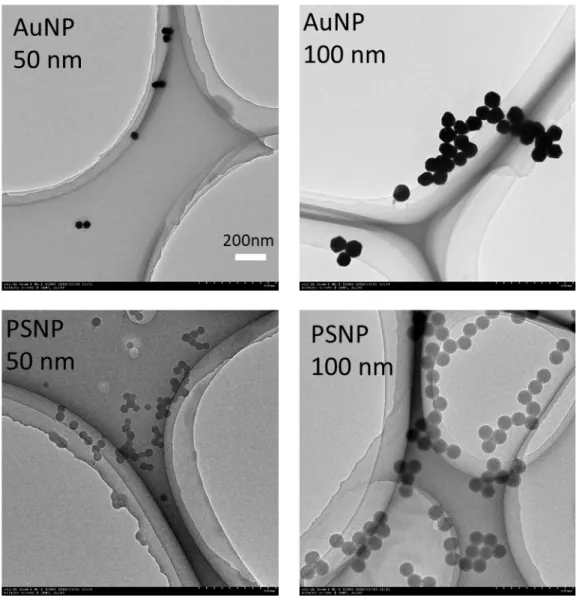
We checked the actual sizes of Au and PS nanoparticles by TEM and the results are shown
28
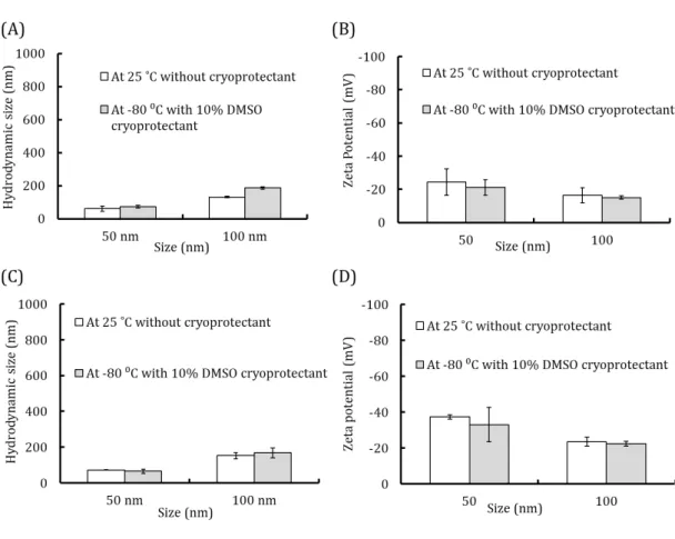
in Figure S1. Both Au and PS nanoparticles had uniform sizes of 50 nm and 100 nm. Our
8
main aim was to use the freeze concentration procedure for the internalisation of Au and
1
PS nanoparticles of various sizes. However, the colloidal stability of these nanoparticles
2
is a major challenge in frozen conditions.31 At the time of freezing, the nanoparticle 3
concentration increases due to solute exclusion from ice crystal formation. This stress
4
could affect the ionic strength, size, and surface potential of nanoparticles.32 A variety of 5
cryoprotectants have been used to shield nanoparticles during freezing.33 Therefore, we 6
investigated the aggregation behaviour of Au and PS nanoparticles in response to freeze
7
concentration and determined suitable conditions for minimising irreversible
8
aggregation. We used DMSO, a well-established cryoprotectant for cell storage.34 We 9
evaluated the size and surface charge of particles in non-frozen and frozen conditions. Au
10
and PS nanoparticles (2 × 10¹⁰ particles/mL) were dispersed in 1 mL of milli-Q water and 11
the hydrodynamic sizes and zeta potential were determined by the DLS technique. As
12
shown in Figure S2A, the hydrodynamic sizes of Au nanoparticles (2 × 10¹⁰ particles/mL) 13
were around 60 nm and 130 nm, consistent with the expectation at room temperature in
14
the absence of a cryoprotectant for 50- and 100-nm Au nanoparticles. When Au
15
nanoparticles were frozen in the presence of 10% DMSO, their sizes did not change
16
substantially. We also evaluated the influence of freezing on the surface charge of 50- and
17
100-nm Au nanoparticles. As expected, the zeta potential was negative due to the efficient
18
distribution of citrate groups in Au nanoparticles, even during freezing. The zeta potential
19
was -24.3 ± 8.0 mV for 50-nm Au nanoparticles at room temperature, but was -16.4 ± 4.6
20
mV for 100-nm Au nanoparticles. When nanoparticles were frozen at -80°C in the
21
presence of 10% DMSO as a cryoprotectant, the surface potential was nearly equal to that
22
in normal conditions. These results suggested that freezing and cryoprotectants do not
23
have a substantial influence on zeta potential, and particles retain their anionic property
24
(Figure S2 b).
25
We also characterised the hydrodynamic size and zeta potential of PS nanoparticles
26
(Figure S2C and D). With respect to the size of PS nanoparticles in aqueous medium, the
27
results were similar to those obtained for Au nanoparticles. At room temperature, the
28
hydrodynamic size of polystyrene nanoparticles was approximately 71.6 ± 1.1 nm in the
29
case of 50-nm nanoparticles. The hydrodynamic size of approximately 152.4 ± 16.7 nm
30
was slightly greater than the actual size of 100 nm of polystyrene nanoparticles (Figure
31
S2C). We also evaluated the influence of freezing on the surface potential of PS 32
9
nanoparticles and found that the potential was nearly the same in the presence of 10%
1
DMSO and at room temperature (Figure S2 D).
2
Since the estimates of size and potential were similar and consistent with the TEM results,
3
it is difficult to determine whether the cryoprotectant contributes to aggregation (Figure
4
S2A–D). These results indicated that 10% DMSO is inert and does not affect colloidal 5
stability for both Au and polystyrene nanoparticles.
6
Gold and polystyrene nanoparticle adsorption on L929 cells using the freeze 7
concentration approach 8
Next, we investigated the efficacy of the freeze concentration method for the in vitro
9
delivery of two different materials. We chose L929 fibroblasts as a model cell line to
10
evaluate the effects of different materials with different sizes in a single cell line. L929
11
cell line can be easily cultured in a reproducible manner. Also, this cell lines are widely
12
used for preliminary studies for wide range of biomaterials due to easily proliferation
13
and adherence on the biomaterials surface. In our previous studies, we also chose L929
14
cell line for our research.13,14 To evaluate cell viability, L929 cells were mixed with various 15
concentrations of 50-nm and 100-nm Au and PS nanoparticles that were cryopreserved
16
with 10% DMSO in culture medium without FBS. Cell viability was then calculated using
17
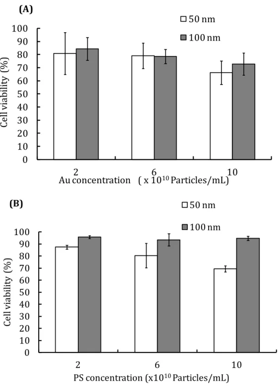
trypan blue staining solution after thawing. As shown in Figure S3, cell viability
18
decreased as the concentration of nanoparticles (Au and PS) increased (Figure S3A–B).
19
A low concentration of nanoparticles for both sizes resulted in a cell viability of around
20
80%. We used small concentrations of Au and PS nanoparticles for in vitro delivery.
21
Interestingly, we observed that cell viability was lower for small nanoparticles than for
22
large nanoparticles. This result is in agreement with previous studies indicating that
23
smaller nanoparticles are generally more toxic.35 Likewise, as shown in Figure S3 B, cell 24
viability for 100-nm PS beads was significantly greater than that for 50-nm PS beads at
25
every concentration, suggesting that cytotoxicity in greater for the latter than the former.
26
This result was consistent with the findings of Jeong et al., who found that microbead
27
toxicity is size-dependent and smaller microbeads are more toxic.36 It is worth pointing 28
out that although our nanoparticles had negative charges of different values, size was the
29
predominant determinant of cytotoxicity.
10
We then used the freeze concentration methodology to investigate adsorption behaviour
1
on cell membranes. A brief illustration of the protocol is shown in Scheme 1. First, we
2
determined the effect of freezing on the adsorption efficacy of Au nanoparticles. The Au
3
nanoparticles did not require modification with fluorescent dyes owing to their intrinsic
4
optical properties (plasmon scattering). Scattering images can be easily observed for Au
5
nanoparticles at a laser wavelength of 559 nm for excitation. Many studies have evaluated
6
nanoparticles inside cells by utilising plasmon scattering properties.37 Scattered Au 7
nanoparticles of both sizes (50 and 100 nm) were efficiently visualised by confocal
8
microscopy after freeze concentration, as shown in Figure 1. The non-frozen conditions
9
did not influence the absorption of Au nanoparticles around the cell membrane for both
10
50 and 100 nm (Figure 1A and C). However, Au internalisation increased when using the
11
freezing approach (Figure 1B and C).
12
We also quantified the mean scattering intensity of Au nanoparticles for Au nanoparticles
13
of 50 and 100 nm by confocal microscopy. We found greater intensities for frozen Au
14
nanoparticles than non-frozen nanoparticles, indicating increased adsorption on the cell
15
surface (Figure 1E).
16
Furthermore, the adsorption of PS nanoparticles of both 50 and 100 nm was greater in
17
frozen than in non-frozen conditions (Figure 2A–D). Quantification by confocal
18
microscopy revealed that the freezing technique increases the penetration of PS
19
nanoparticles (Figure 2E).
20
When cells and nanoparticles are subjected to an ultra-cold environment, water in the
21
system is converted into ice crystals, leading to an increased concentration of
22
nanoparticles in the extracellular solution. The increased concentration of nanoparticles
23
leads to increased interactions with the cell membrane in the frozen system, thereby
24
promoting the efficient adsorption of particles. Although negatively charged
25
nanoparticles are expected to be less efficiently adsorbed by a negatively charged cell
26
membrane, the cellular adsorption of negatively charged NPs was high, possibly due to
27
strong, localised, and nonspecific interactions with the plasma membrane.38 Taken 28
together, these results indicate that the freeze-concentration method enhances the
29
association of both Au and PS nanoparticles on cells, while maintaining a high cell
30
viability.
11
Intracellular delivery of gold and polystyrene nanoparticles using L929 cells by 1
freeze concentration 2
The adsorption of nanoparticles on the cell membrane was enhanced using the freeze
3
concentration approach. Usually, the internalisation of nanoparticles in cells is very
4
important for achieving a therapeutic effect. To investigate this, a similar protocol was
5
applied to that summarised in Scheme 1. The confocal microscopic images for the
6
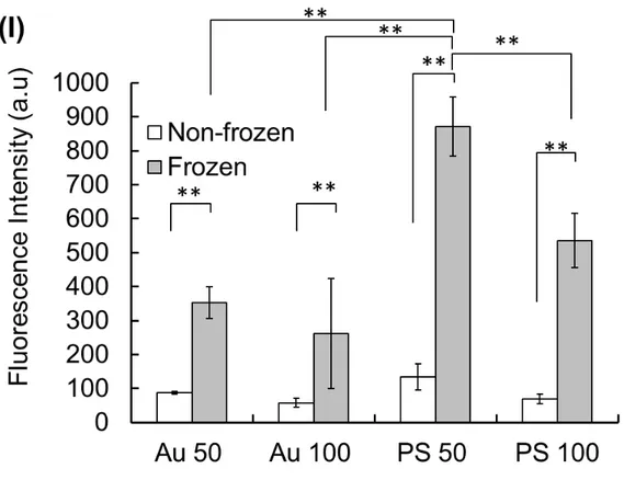
internalisation of Au and PS nanoparticles are shown in Figure 3A–H. The results were
7
quite similar to those shown in Figure 1 and 2. Both Au and PS nanoparticles were
8
efficiently internalised into cells (Figure 3B, D, F, H). However, the non-freezing
9
approach did not induce internalisation for similar conditions (Figure 3A, C, E, G).
10
Moreover, greater internalisation was observed for smaller Au and PS particles than for
11
large particles. Figure 3I summarises the quantitative analysis of intensity values
12
obtained by confocal microscopy for PS and Au nanoparticles at 50 and 100 nm.
13
Interestingly, we found higher intensities using 50-nm PS nanoparticles than using other
14
materials for both the non-frozen and frozen systems. Various studies have reported that
15
small particles are efficiently taken up by cells, in general.17,39,40 16
Nanoparticle size is an important determinant of cellular internalisation. Chithrani et al.
17
showed that 50-nm transferrin-coated Au nanoparticles exhibit greater uptake than that
18
of 100 nm particles. They attributed this to the “wrapping effect,” which induces more
19
efficient cell internalisation. Additionally, they described the roles of ligand-receptor
20
interactions as well as receptor diffusion kinetics in cellular internalisation. The so called
21
“Wrapping effect” indicated how the cellular membrane encloses nanoparticles. Two
22
Factors are associated with the term “Wrapping effect” wherein it dictates the free energy
23
for ligand-receptor interaction and other is the receptor diffusion kinetics onto wrapping
24
sites around the cellular membranes. 41 25
It should be noted that PS nanoparticles have increased interactions with the plasma
26
membrane compared with Au nanoparticles. The increased internalisation of PS
27
nanoparticles might be explained by the difference in affinity towards the cell membrane.
28
Endocytosis mechanisms 29
12
A better understanding of the interactions of functional nanoparticles with biological
1
cells is needed not only for the development of new methods for imaging and therapy,
2
but also to improve our knowledge of the fundamental cellular mechanisms and
3
cytotoxicity of nanomaterials. Therefore, we evaluated the effects of the freeze
4
concentration method on endocytic pathways. Various endocytic inhibitors, such as
5
chlorpromazine (for clathrin-mediated endocytosis), EIPA (for macropinocytosis), and
6
filipin (for caveolae mediated endocytosis), were added to elucidate the endocytic
7
pathway. Before evaluating the endocytic pathways, it is crucial to investigate the effects
8
of these nanoparticles and inhibitors on cell viability, as these inhibitors are cytotoxic at
9
particular concentrations. Hence, we evaluated cell viability using trypan-blue exclusion
10
assays after freezing. The optimal concentration of inhibitors with respect to cell viability
11
(Figure 4) was used to elucidate the endocytic pathway. Before freezing, the cells are
12
100% alive whereas after freezing the cells viability are slightly decrease even after
13
adding cryoprotectant. We examined the endocytic mechanism for two different
14
materials of different sizes and frozen as well as non-frozen conditions by fluorescent
15
confocal microscopy. Quantitative results were obtained from confocal images, which are
16
shown in the supporting information (Figure S4A–D). For both nanoparticle types,
17
intensity was quantified by confocal microscopy (Figure 5A–D). Figure 5A and B show
18
the scattering intensity of Au nanoparticles in the presence of inhibitors for frozen and
19
non-frozen conditions. A high scattering intensity was observed for L929 cells used as a
20
positive control, without inhibitors. When particular inhibitors were added, a change in
21
the scattering intensity was observed. A decreased intensity of Au nanoparticles in the
22
presence of inhibitors implies that the particular inhibitor affected the uptake and
23
restricted the entry of nanoparticles into the cell membrane. We observed a lower
24
scattering intensity for 50 and 100 nm Au nanoparticles in the presence of
25
chlorpromazine; this outcome suggested that chlorpromazine inhibits endocytosis.
26
Accordingly, Au particles of 50 and 100 nm primarily enter cells via clathrin-mediated
27
endocytosis (Figure 5A). Interestingly, using the freeze concentration approach, the
50-28
nm Au nanoparticles exhibited a decreased scattering intensity when the filipin inhibitor
29
was applied, indicating that freezing induces a change in the uptake pathway to
caveolae-30
mediated endocytosis (Figure 5B). Au nanoparticles of 100 nm showed a similar trend
31
with respect to the alteration of pathways. For 100-nm particles, in non-frozen conditions,
13
clathrin-type endocytosis was dominant (Figure 5A). However, frozen conditions
1
resulted in a shift to the caveolae as well as macropinocytosis pathways, even for 100-nm
2
particles (Figure 5B).
3
The endocytic mechanism for PS-based nanomaterials differed somewhat from that for
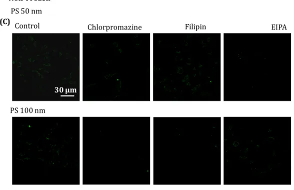
4
Au nanoparticles. The 50-nm PS nanoparticles tended to exhibit uptake by
5
macropinocytosis pathways, as evidenced by the decreased fluorescence intensity in the
6
presence of the EIPA inhibitor (Figure 5C). When the freezing approach was applied, the
7
pathway changed to clathrin-mediated endocytosis (Figure 5D). PS nanoparticles of 100
8
nm were associated with both clathrin- and caveolae-dependent endocytosis in normal
9
conditions (Figure 5C). In particular, the freeze concentration-mediated internalisation
10
of PS nanoparticles occurred only by clathrin-mediated endocytosis (Figure 5D).
11
Previous reports have suggested that endocytic mechanisms are usually dependent on
12
size and cell type.42 However, these are not the only factors responsible for specific 13
uptake. In one study, Mironava et al. reported that 45-nm Au NPs penetrated cells via
14
clathrin-mediated endocytosis,43 consistent with the results of our study. However, the 15
frozen system changes the pathway for 50-nm Au nanoparticles. An increase in cell stress
16
caused by the freeze process could explain the change in the endocytic pathway.
17
Understanding the detailed determinants of the endocytic mechanism can clarify the
18
importance of freeze concentration-induced changes in the endocytic pathway. The same
19
trend was observed for particles of other sizes.
20
An “electroporation” method has previously been used to promote nanoparticle uptake
21
into cells by applying external electrical impulses, resulting in transient membrane pores
22
through which nanoparticles can easily pass.44 Another approach, “microinjection”, is a 23
valuable tool to deliver molecules inside cells. These previous methods adopt a
non-24
endocytic pathway that can induce significant tension, thereby disrupting the lipid
25
bilayers and nanoscale hole formation around the cell membrane.44, 45 These strategies 26
result in substantial cell loss, deformation of the cell membrane, and the non-specific
27
entry of materials, and accordingly they are not practical for applications in biomedical
28
engineering.
14
In our study, a new “freeze concentration” method was developed that does not involve
1
the direct entry of materials inside cells, but rather promotes the interaction between the
2
cell membrane and materials. Compared with previously described physical approaches,
3
this freezing strategy has several benefits, including its high reliability, low cost, and
4
simplicity, using an external force to induce cell membrane interactions without toxicity.
5
Regardless, further detailed investigations are needed to elucidate the underlying
6
mechanism by which freezing induces nanoparticle uptake via different pathways.
7
Studies of nanoparticle internalisation by cells can provide a basis for understanding
8
their ultimate sub-cellular fate and localisation. A comprehensive knowledge of how
9
particle size and freezing conditions affect the interactions between cells and appropriate
10
combinations of nanomaterials using the freeze concentration approach is crucial for
11
delivery applications. In future studies, we will apply the approach to various cell lines,
12
such as HeLa cells, SK-Mel-28 melanoma cells, and RAW 264.7 macrophages, to evaluate
13
the endocytic entry of different materials using the freeze concentration approach.
14
Conclusion 15
Our results demonstrated that the freeze concentration strategy enables the efficient
16
internalisation of Au and PS nanoparticles into fibroblast cells. DLS and zeta potential
17
measurements showed that the colloidal stability of these nanomaterials is retained in
18
the presence of 10% DMSO (a cryoprotectant) after freezing. Further, 50-nm PS
19
nanoparticles showed high intracellular uptake efficacy, while 50-nm Au nanoparticles
20
showed the lowest efficacy. We also investigated the endocytic uptake mechanism using
21
frozen and non-frozen systems with a model fibroblast cell line. Interestingly, Au
22
nanoparticles of both 50 and 100 nm exhibited clathrin-dependent endocytosis in
non-23
frozen conditions, but freezing resulting in a change to the caveolae-dependent and
24
macropinocytosis pathways. Other PS nanoparticles utilised a different endocytosis
25
pathway depending on size and frozen conditions. Our results emphasise the importance
26
of the physical “freezing” approach for nanomaterials of various sizes to guide endocytic
27
uptake for efficient delivery systems. We expect that the precise information about the
28
endocytic uptake of nanoparticles will facilitate the design of materials with increased
29
cellular uptake and the use of the freeze concentration approach for extensive biomedical
30
applications.
15 Conflicts of interest
1
There are no conflicts of interest to declare.
2
Acknowledgements 3
This study was supported in part by a Grant-in-Aid, KAKENHI (16K12895) for scientific
4
research from the Japan Society for the Promotion of Science.
5 6
References
7
1. M. K. Yu, J. Park and S. Jon, Thernostics, 2012, 2, 3-44.
8
2. S.Huo, S. Jin, X. Ma, X. Xue, K. Yang, A. Kumar, P.C. Wang, J. Zhang, Z.Hu and X.J. Liang,
9
ACS Nano, 2014, 8(6), 5852-5862.
10
3. K. Saralidze, L.H. Koole and M.L.W. Knetsch, Materials, 2010, 3, 3537-3564.
11
4. X. Zhang, Q. Guo and D. Cui, Sensors (Basel), 2009, 9(2), 1033-1053.
12
5. Y. Zhou, S. Kundu, J.D. Cirillo and H. Liang, J Nanobiotechnology, 2012, 10, 1-9.
13
6. Y. Xing, J. Zhao, P.S. Conti and K. Chen, Thernostics, 2014, 4(3), 290-306.
14
7. F.Y. Kong, J.W. Zhang, R.F. Li, Z.X. Wang, W.J. Wang and W. Wang, Molecules, 2017,
15
22, 1-13. 16
8. K. Kenesei, K. Murali, A. Czeh, J. Piella, V. Puntes and E. Madarasz, J
17
Nanobiotechnology, 2016, 14, 1-14.
18
9. Y. Zu, S. Huang, W.C. Liao and S. Wang, J Biomed Nanotechnol, 2014, 10(6), 982-992.
19
10. I. Lentacker, S.C. DeSmedt and N.N. Sanders, Soft matter, 2009, 5, 2161-2170.
20
11. S. J. Beebe, N.M. Sain and W. Ren, Cells, 2013, 2(1), 136-162.
21
12. W.S. Meaking, J. Edgerton, C.W. Wharton and R.A. Meldrum, Biochim. Biophys. Acta,
22
1995, 1264, 357-362.
23
13. S. Ahmed, F. Hayashi, T. Nagashima and K. Matsumura, Biomaterials, 2014, 35,
6508-24
6518.
25
14. S. Ahmed, S. Fujita and K. Matsumura, Nanoscale, 2016, 8, 15888-15901.
26
15. S. Ahmed, S. Fujita and K. Matsumura, Adv. Healthc. Mater., 2017, 6, 1700207.
27
16. S. Ahmed, N.K. Tadashi, T. Watanabe, T. Hohsaka and K. Matsumura, ACS Biomater.
28
Sci. Eng., 2017, 3(8), 1677-1689.
16
17. N. Oh and J.H. Park, Int. J Nanomedicine 2014, 9, 51-63.
1
18. G. Sahay, D.Y. Alakhova and A.V. Kabanov, J Control Release, 2010, 145(3), 182-195.
2
19. S.Xu, B.Z. Olenyuk, C.T. Okamoto and S.F. Hamm- Alvarez, Adv. Drug Deliv. Rev., 2013,
3
65(1), 121-138. 4
20. S. Kumari, S. MG and S. Mayor, Cell Res., 2010, 20, 256-275.
5
21. M.O. Oyewumi, A. Kumar and Z. Cui, Expert Rev Vac., 2010, 9(9), 1095-1107.
6
22. A. El-sayed and H. Harashima, Mol Ther., 2013, 21(6), 1118-1130.
7
23. B. Alberts, A. Johnson and J. Lewis, Molecular Biology of the Cell. 4th edition. Garland 8
Science, New York, 2002.
9
24. T.P Welliver, S. L. Chang, J. L. Linderman and J.A. Swanson, J Cell Sci. 2011, 124(23),
10
4106-4114.
11
25. X. Zhao, Y. Wu, D. Gallego-Perez, K.J. Kwak, C. Gupta, X. Quyang and L.J. Lee, Anal
12
Chem., 2015, 87(6), 3208-3215.
13
26. P.E. Boukany, Y. Wu, X. Zhao, K.J. Kwak, P.J. Glazer, K. Leong and L.J. Lee, Adv. Healthc
14
Mater., 2013, 3, 682-689.
15
27. P. Krystosiak, W. Tomaszewski and E. Megiel, J Colloid Interface Sci. 2017, 498, 9-21.
16
28. M. Gozdziewska, G. Cichowicz, K. Markowska, K. Zawada and E. Megiel, RSc Adv. 2015,
17
5, 58403-58415. 18
29. W. Zauner, N.A. Farrow and A.M. Haines, J Control Release 2001, 71(1), 39-51.
19
30. P.Jani, G.W. Halbert, J. Langridge and A.T. Florence, J Pharm Pharmacol. 1990, 42,
20
821-826.
21
31. M.J. Choi, S. Briancon, J. Andrieu, S.G. Min and H. Fessi, Drying Technol., 2004, 22,
22
335-346.
23
32. C. Vauthier, B. Cabane and D. Lanbarre, Eur J Pharm Biopharm, 2008, 69(2),
466-24
475.
25
33. A.M. Alkilany, S.R. Abulateefeh, K.K. Mills, A.I.B. Yaseen, M.A. Hamaly, H.S. Alkhatib,
26
K.M. Aiedeh and J.W. Stone, Langmuir, 2014, 30, 13799-13808.
27
34. G. Chen, A. Yue, Z. Ruan, Y. Yin, R. Wang, Y. Ren and L. Zhu, Stem cells Int., 2016, 7,
1-28
7.
29
35. L. Yildirimer, N.T.K. Thanh, M. Oizidou and A.M. Seifalian, Nano today, 2011, 6,
585-30
607.
17
36. C.B. Jeong, E.J. Won, H.M. Kang, M.C. Lee, D. Hwang and U.K. Hwang, Environ Sci
1
Technol., 2016, 50(16), 8849-8857
2
37. M. Takahashi, P. Mohan, K. Mukai, Y. Takada, T. Matsumoto, K. Matsumura, M.
3
Takakura, H. Arai, T. Taguchi and S. Maenosono, ACS Omega, 2017, 2, 4929- 4937.
4
38. C. Billotey, C. Wilhelm, M. Devaud, J.C. Bacri, J. Bittoun and F. Gazeau, Magn. Reson.
5
Med., 2003, 49, 646-654.
6
39. S. Zhang, J. Li, G. Lykotrafitis, G. Bao and S. Suresh, Adv Mater., 2009 , 21,419–424
7
40. L. Shang, K. Nienhaus and G.U. Nienhaus, J. Nanobiotechnology, 2014, 12, 1-48.
8
41. B.D. Chithrani and W.C. Chan, Nano Lett. 2007, 7(6), 1542-1550.
9
42. J. Giudice, E.A. Jares-Erijman and F.C. Leskow, Bioconjugate Chem, 2013, 24(3),
431-10
442.
11
43. T. Mironava, M. Hadjiargyrou, M. Simon, V. Jurukovski and M.H. Rafailovich,
12
Nanotoxicology, 2010, 4,120–137.
13
44. C.P Jen, Y.H. Chen, C.S. Fan, C.S. Yeh, Y.C. Lin, D.B. Shieh, C.L. Wu, D.H. Chen and C.H.
14
Chou, Langmuir 2004, 20, 1369-1374.
15
45. G. Liu, D. Li, M. K. Pasumarthy, T. H. Kowalczyk, C. R. Gedeon, S. L. Hyatt, J. M. Payne,
16
T. J. Miller, P. Brunovskis and T. L. Fink, J Biol Chem. 2003, 278, 32578–32586.
17 18
Figures 19
20
Scheme-1 Protocol representation of nanoparticles adsorption and internalisation into 21
fibroblast L929 cells by freeze concentration approach
18 1
19 1
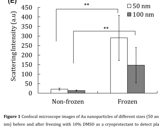
Figure 1 Confocal microscope images of Au nanoparticles of different sizes (50 and 100 2
nm) before and after freezing with 10% DMSO as a cryoprotectant to detect plasmon
3
scattering from the Au nanoparticles of 50 nm and 100 nm. (A, B) Non-frozen and frozen
4
50 nm Au. (C, D) Non-frozen and frozen 100 nm Au. Scale bar: 30 µm (E) Mean scattering
5
intensity of Au nanoparticles determined by confocal microscopy. Results are expressed
6
as means ± SD. **p < 0.01
20 1
21 1
Figure 2 Confocal microscope images of PS nanoparticles of different sizes (50 and 100 2
nm) before and after freezing with 10% DMSO as a cryoprotectant. (A, B) Non-frozen and
3
frozen 50-nm PS nanoparticles. (C, D) Non-frozen and frozen 100-nm PS nanoparticles.
4
Scale bar: 30 µm (E) Mean fluorescence intensity of PS nanoparticles investigated by
5
confocal microscopy. Results are expressed as means ± SD. **p < 0.01
22 1
23 1
24 1
Figure 3 Internalisation of Au nanoparticles in L929 cells. (A, C) Without the freeze-2
concentration of Au nanoparticles of 50 and 100 nm (B, D) With the freeze concentration
3
of Au nanoparticles at -80°C using 10% DMSO as a cryoprotectant. Internalisation of PS
4
nanoparticles in L929 cells. (E, G) Without the freeze-concentration of PS nanoparticles
5
of 50 and 100 nm. (F, H) With the freeze concentration of PS nanoparticles at -80°C using
6
10% DMSO as a cryoprotectant. Scale bar: 30 µm. (I) Mean intensities of scattering and
7
fluorescence images of Au and PS nanoparticles were obtained by confocal microscopy.
8
Results are expressed as means ± SD. **p < 0.01
25 1
Figure 4 Quantification of cell viability determined by trypan blue-exclusion assays after 2
the addition of endocytic inhibitors to Au and PS nanoparticles of 50 nm and 100 nm after
3
freezing. Results are expressed as means ± SD.
4 5
26 1
27 1
28
Figure 5 Quantitative analysis of the fluorescence/scattering intensity by confocal 1
microscopy during endocytic uptake via clathrin-mediated endocytosis (ME), caveolae
2
ME, and macropinocytosis ME, following treatment with various inhibitors. (A)
Non-3
frozen Au 50 nm (B) Frozen Au 50 nm (C) Non-frozen PS 50 nm (D) Frozen PS 100 nm.
4
Results are expressed as means ± SD. **p < 0.01, *p < 0.05 vs. without inhibitors for
50-5
nm nanoparticles and ##p < 0.01, #p < 0.05 vs. without inhibitors for 100-nm
6
nanoparticles.
Electronic supplementary information (ESI) for
Comparative analysis of the cellular entry of polystyrene and gold nanoparticles using the freeze concentration method
Sana Ahmed, Koyo Okuma, and Kazuaki Matsumura*
School of Materials Science, Japan Advanced Institute of Science and Technology, Nomi, Ishikawa 923-1292, Japan
Figure S2 Hydrodynamic diameter and Zeta potential of nanoparticles at room
temperature (25°C) with no cryoprotectant (White bars) and after freezing with 10% DMSO as a cryoprotectant at -80°C (Grey bars). Au nanoparticle (A) hydrodynamic diameter and (B) Zeta potential. PS nanoparticle (C) hydrodynamic diameter and (D) Zeta potential.
Figure S3 Cell viability using nanoparticles at various concentrations after freezing
with 10% DMSO as a cryoprotectant, as determined by trypan blue exclusion assays, for (A) Au and (B) PS.
0
10
20
30
40
50
60
70
80
90
100
2
6
10
Ce
ll
vi
ab
ili
ty
(%
)
Au concentration ( x 10
10Particles/mL)
50 nm
100 nm
(A)
0 10 20 30 40 50 60 70 80 90 100 2 6 10 Ce ll v ia bi lity (% ) PS concentration (x1010 Particles/mL) 50 nm 100 nm (B)Frozen
Control Chlorpromazine Filipin EIPA
Au 100 nm Au 50 nm (B)
Non-Frozen Au 50 nm
Control Chlorpromazine Filipin EIPA
Au 100 nm (A)
Figure S4 Confocal images of non-frozen and frozen endocytic uptake of 50- and
100-nm gold nanoparticles (A, B) or 50- and 100-nm polystyrene beads (C, D). For freezing, the samples were pre-incubated with inhibitors in the presence of 10% DMSO as a cryoprotectant. After thawing, the cells were seeded and incubated for at least 10 h. Scale bar: 30 µm.
Non-Frozen
Control Chlorpromazine Filipin EIPA
PS 50 nm PS 100 nm (C) 30 µm Frozen PS 50 nm
Control Chlorpromazine Filipin EIPA
PS 100 nm