はじめに
近年,脳血管内治療の登場により,脳梗塞の急性期治療は 劇的に変化した.特に灌流域が広範な内頸動脈,中大脳動脈等 が閉塞する急性主幹動脈閉塞症(large vessel occlusion; LVO)に 対する脳血管内治療の有効性が五つの大規模臨床試験で確立
された1).しかし,脳血管内治療は頭蓋内出血等の合併症の
リスクがある侵襲的な治療であり,神経症状が軽症の症例は Multicenter Randomized Clinical Trial of Endovascular Treatment for Acute Ischemic Stroke in the Netherlands(MRCLEAN)2),
Extending the Time for Thrombolysis in Emergency Neurological Deficits-Intra-Arterial(EXTEND-IA)3)試験以外の大規模臨床
試験には含まれていない.その結果,上記の五つの大規模臨 床試験に含まれた症例の National Institutes of Health Stroke Scale(NIHSS)の中央値は 17 で,中等症から重症の症例が 主に対象となっている.また,五つの大規模臨床試験のメタ 解析である,Highly Effective Reperfusion evaluated in Multiple Endovascular Stroke trials(HERMES)collaboration のデータ では NIHSS 10 以下の症例の subgroup に対する血管内治療の
統計学的な優位性は示されていない1).そのため,American
Heart Association(AHA)guideline では,NIHSS 6 以上の症例 に対して level 1a で血管内治療が推奨されている4).同様に,
rt-PA静注療法の大規模臨床試験にも軽症例はほとんど含ま
れておらず5),本邦における rt-PA 静注療法適正治療指針第
二版で,軽症例は慎重投与とされている6).
一方,軽症脳梗塞が必ずしも良好な転帰を辿るとは限らず, 神経症状増悪(early neurological deterioration; END)により,
最終的に転帰不良となる症例が存在する7)~9).また,LVO を 有する症例が,良好な側副血行により軽微な神経症状で来院 する場合もある10).しかし,軽症脳梗塞は上記の大規模臨床 試験で適応基準に含まれていないため,rt-PA 静注療法や血管 内治療等の再開通療法の適応,治療の施行時期について一定 の見解が得られておらず,治療方針の決定に難渋する場合が 少なくない.軽症脳梗塞の END の病態および再開通療法の 意義を中心に最近の知見を整理し,自験例を交えながら考え てみたい. 軽症脳梗塞
軽症脳梗塞は,NIHSS 3~4 以下を minor stroke,NIHSS 5~
7以下を mild stroke とする等,文献によって様々に定義され ている7)11)~13).集団ベース研究や全国登録研究によれば,脳 梗塞の約 30~50%が NIHSS 3 以下の軽症例とされており,軽 症脳梗塞は比較的よくみられる14)15).軽症脳梗塞は必ずしも 良好な経過を辿るとは限らず,発症時の NIHSS 5 以下の症例 の約 1/4 の症例で END が認められ,約 1/3 の症例が 90 日後 の時点で後遺症を認めたと報告されている7).「軽症」や「症 状の急速改善」は,rt-PA 静注療法が行われない代表的な理由 とされ,時間的には rt-PA 静注療法を受けられる症例の 29~ 43%が,実際は治療を受けていないとされる16)17).しかし, 「軽症」あるいは「症状の急速改善」を理由に rt-PA 静注療法 を受けなかった症例の約 27%が,その後自力歩行困難や自宅 退院困難であったとされており,最終的に転帰不良となる症 例が混在している18).また軽症脳梗塞は,中等症以上の脳梗
総 説
軽症脳 塞の再開通療法
高下 純平
1)*
要旨: 軽症脳梗塞は,神経症状の増悪により転帰不良となる症例が少なからず存在し,特に主幹動脈閉塞を有 する例でその危険性が高い.しかし,軽症脳梗塞に対する再開通療法の有効性は不明である.神経症状増悪の病態 は,側副血行の破綻等,様々なメカニズムが提唱されているが不明な場合も多い.内頸動脈閉塞を有するが,側副 血行を介して同側の中大脳動脈が描出される中大脳動脈開存型内頸動脈閉塞症は,血栓の遠位への移動により側 副血行が遮断され,劇的に神経症状が増悪する場合があり注意を要する.最近の知見を整理し,主幹動脈閉塞を伴 う軽症脳梗塞を中心に,神経症状増悪の病態や再開通療法について概説した. (臨床神経 2019;59:84-92) Key words: 急性期脳梗塞,側副血行,神経症状増悪,血栓溶解療法,機械的血栓回収術 *Corresponding author: 済生会福岡総合病院脳神経内科〔〒 810-0001 福岡県福岡市中央区天神 1-3-46〕 1)済生会福岡総合病院脳神経内科(Received November 26, 2018; Accepted December 19, 2018; Published online in J-STAGE on January 31, 2019) doi: 10.5692/clinicalneurol.cn-001255
塞と比較して発症から画像撮像,rt-PA 静注療法まで時間を要 することが多い点も問題視されている13).発症時の NIHSS が 低値であることは必ずしも最終的な転帰良好を意味しない, ということを踏まえて初期診療を行わなければならない. 主幹動脈閉塞を伴う軽症脳梗塞 LVOを有する症例は一般的に NIHSS が高値である19).一方,
NIHSS 0~4 の 18%,NIHSS 5~8 の 39%に LVO が認められ19),
LVO症例の約半数が NIHSS 10 未満とする報告もあり,LVO
の診断における NIHSS の感度は必ずしも高くない10).LVO
を有する軽症脳梗塞では特に転帰不良の可能性が高く8)9)20),
そのオッズは約 7 倍とする報告もある8).他の報告では,LVO
のない症例の END が 3%であったのに対し,LVO を有する例 では 38%に END を認めている9).LVO の有無は END を予測
する重要な因子であり,軽症脳梗塞であっても頭蓋内血管の
精査を省略すべきではない20).
LVOを有する軽症脳梗塞の自然経過に関する報告は少な
い.Mokin らは,LVO を伴う NIHSS 7 以下の脳梗塞例のうち, 再開通療法を受けていない 204 例の転帰を検討したところ, 退院時に自力歩行可能な症例は 62%で,転帰は多様であっ
た21).Lima らは,再開通療法を受けていない LVO を有する
脳梗塞 126 例の自然経過を報告している.56 例(44%)の NIHSS 10以下の軽症例のうち,6 ヶ月後の転帰良好(modified Rankin scale(mRS)0~2)の割合は 75%であった22).Heldner
らの報告では,LVO を有する NIHSS 5 以下の軽症脳梗塞のう ち,内科治療群 47 例(rt-PA 静注療法を除く)の 3 ヶ月後の 転帰良好(mRS 0~2)の割合は 67%であった19)(Table 1). このように,LVO を伴う軽症脳梗塞の転帰は必ずしも良好と はいえず,再開通療法による転帰の改善が期待される. 主幹動脈閉塞を伴う軽症脳梗塞の病型 脳梗塞の発症機序は,軽症脳梗塞の転帰に関与する重要な 因子である23).動脈硬化性病変を背景とした LVO を有する症例 では,慢性かつ緩徐進行性の経過から,心原性脳塞栓症による LVOの症例と比較して側副血行が豊富とされ,臨床経過に違
いが生じる可能性がある24)25).また,carotid stump syndrome26),
vertebral artery stump syndrome27)のように,慢性完全閉塞を
背景に形成された血栓が同側の頭蓋内に塞栓症を引き起こす 病態も存在する.つまり急性閉塞から慢性完全閉塞まで,あ らゆる時期の LVO が脳梗塞の原因となり得る.頭部 MRI T2*
画像28)29),定量 MRA30),頭部 CTA 等31)複数の modality を活
用することで,閉塞機序,側副血行の状態等の診断精度が向 上する可能性がある.しかし,標的血管が閉塞した状態で閉 塞機序を正確に診断することはしばしば困難で,今後の重要 な研究課題と考える.後述する LVO を有する軽症脳梗塞の ENDの病態や再開通療法の有効性についての検討は,閉塞血 管の機序や閉塞時期の診断に制約がある現状において,主幹 動脈の急性かつ塞栓性閉塞を想定して行ったものであること を断っておく. 主幹動脈閉塞を伴う軽症脳梗塞の神経症状増悪 脳梗塞の神経症状増悪の背景には,症候性頭蓋内出血,脳 浮腫,脳梗塞の早期再発,痙攣等,様々な病態が挙げられる32). しかし,END を呈する症例の半数以上は原因が明らかでない END(unexplained END)とされる33).再開通療法を受けてい
ない症例において,END は 15~37%に生じる32).一般的に, NIHSS 4点以上の加点を伴う神経症状増悪を END と定義す ることが多い32).LVO を伴う軽症脳梗塞では特に END を生じ やすく34),上記に加えて,側副血行の破綻(collateral failure)等 の血行力学的な病態や35),血栓の伸展(thrombus extension)36), 血栓の遠位への移動(thrombus migration)37)等の血栓関連の病 態が END の原因として挙げられている.これらは,再開通 療法によって解決できる可能性のある重要な病態と考えら れ,さらに詳しく述べてみたい. LVOにおいて,側副血行は脳梗塞の重症度や梗塞巣増大の 速度と密接な関連が見られる.LVO を有する脳梗塞は,Willis 動脈輪,脳軟膜動脈吻合,外頸動脈系と内頸動脈系の吻合が 側副血行として機能する38).しかし,LVO が残存している場 合,側副血行は経時的に減少し,神経症状が増悪する場合が Table 1 Summary of the clinical course of patients with mild stroke with large vessel occlusion without reperfusion therapy Study Patient
Number
Inclusion
criteria Occlusion site
Baseline NIHSS t-PA mRS 0–2 at final assessment Mortality at final assessment
Mokin et al 204 NIHSS ≤ 7 ICA, M1/2, ACA, BA, P1/2 2† 0 62% 5%*
Lima et al 56 NIHSS ≤ 9 ICA, M1/2 NA 0 75% 5.4%
Heldner et al 47 NIHSS ≤ 5 ICA, M1 3† 0 67% 6.7%
Haussen et al 88 NIHSS ≤ 5 ICA, M1/2, ACA, BA 3† 9% 73% NA
*Data include death or hospice.
†Data are presented as medians.
NIHSS, National Institutes of Health Stroke Scale; t-PA, tissue plasminogen activator; mRS, modified Rankin Scale; ICA, internal carotid artery; M1, M1 segment of the middle cerebral artery; M2, M2 segment of the middle cerebral artery; ACA, anterior cerebral artery; BA, basilar artery; P1, P1 segment of the posterior cerebral artery; P2, P1 segment of the posterior cerebral artery.
ある39).この collateral failure を生じる原因として,側副血行
路の血栓形成,静脈,正常脳組織への盗血40)41),自律神経障
害による血圧の変動42),一過性の頭蓋内圧亢進43)等が指摘さ
れているが不明な点も多い.Seners らは unexplained END を 認めるLVOにおいて,血栓の存在を示すT2* susceptibility vessel
signが有意に拡大していることを示し,凝固亢進,血流鬱滞
による元の血栓の進展(thrombus extension)が END につなが る可能性を推察している36).この thrombus extension は,側 副血行の血流の遅い例でよく認められ,転帰不良と関連して いたという報告もある44).Thrombus migration は,塞栓性梗 塞に特徴的な病態で中大脳動脈閉塞症の約 1/3 に認められる とされ,完全再開通や神経症状の改善が得られる割合が低下 する可能性が指摘されている37).しかし,thrombus migration による END については,いくつかの症例報告が存在するのみ で45)46)まとまった報告はない.Caplan は,塞栓性機序の脳梗 塞の 20%が,発症から 24~48 時間の間に塞栓子の遠位への 移動に関連した END を呈すると報告している47).このよう に,LVO を伴う脳梗塞の END の病態も様々である.また,側 副血行を評価可能な血管造影や灌流画像を用いても,持続的 に脳血流の変化を観察することはできず,END を事前に予測 することは難しい. LVOを有する軽症脳梗塞では,上記の病態によって豊富な 側副血行が破綻し,END を呈する場合がある.しかし理論的 には,側副血行が豊富な例は神経症状増悪の速度は緩徐で48)
治療開始可能時間(therapeutic time window)が長いため49)50),
ENDを早期発見し,迅速に再開通療法を行うことができれば, 神経症状の改善が得られる可能性はあると考える.そのため 来院時に神経症状が軽症であっても,LVO が判明した場合な るべく早く側副血行の評価と病型診断に必要な情報収集を行 い,END を呈した場合の追加治療に備えなければならない. 軽症脳梗塞の再開通療法 1.軽症脳梗塞に対する rt-PA 静注療法 NIHSS 5以下の軽症脳梗塞症例は,大部分が rt-PA 静注療 法のランダム化比較試験から除外されており,その有効性は 証明されていない5).ランダム化比較試験のメタ解析では, rt-PA静注療法は NIHSS 0~4 の軽症例において,3~6 ヶ月後 の転帰良好(mRS0~1)の割合を増加させた(rt-PA 静注療 法群:69% vs. コントロール群:59%)51).軽症脳梗塞に対 する rt-PA 静注療法に関する観察研究のメタ解析では,rt-PA 静注療法は,転帰良好(mRS0~1)の割合を増加させたもの の(rt-PA 静注療法群:75% vs. 非投与群:68%),症候性頭 蓋内出血の増加(rt-PA 静注療法群:1.9% vs. 非投与群:0%) が指摘されている52).NIHSS の点数だけでなく,その内容が 半盲,重度の失語,空間失認,麻痺等,重度の障害につなが るもの(disabling deficits)であるかを検討することも重要で ある53).NIHSS 0~5 で,日常生活や仕事上の障害につなが
らない神経症状(minor nondisabling neurologic deficits)を呈 する軽症脳梗塞例に対する rt-PA 静注療法の有効性を検討し たランダム化比較試験(PRISMS 試験)では,rt-PA 静注療法 はアスピリン 325 mg 投与と比較して 90 日後の転帰を改善し なかった54).rt-PA 静注療法後の症候性頭蓋内出血のリスク となる因子を確認することも意思決定に役立つかもしれな い55).軽症脳梗塞例も rt-PA 静注療法により転帰の改善が得 られる可能性がある一方,出血性合併症のリスクは増加する ことを患者や家族に十分説明し,治療を決定すべきであろう. 2.主幹動脈閉塞を有する軽症脳梗塞の再開通療法 近年,安全性の高い血管内治療デバイスを用いた機械的血 栓回収術(mechanical thrombectomy; MT)が登場したことに より,軽症脳梗塞に対しても有効性が期待されており,最近 の知見を紹介する. 1)rt-PA 静注療法を含まない保存的治療と再開通療法(rt-PA 静注療法または MT)
Urraらは,LVO を有する NIHSS 5 以下の軽症脳梗塞 88 例の うち,再開通療法を受けなかった 47 例(53%)と,再開通療 法(rt-PA 静注療法または MT)を受けた 41 例(47%)の転帰 を後方視的に解析した56).再開通療法群で高い再開通率が得 られ(再開通療法群:79% vs. 保存的治療群:11%,P < 0.001), 発症から 3 ヶ月以内の神経症状増悪(NIHSS 1 以上の増悪)の 割合が少なかった(再開通療法群:15% vs. 保存的治療群: 41%,P < 0.001).症候性頭蓋内出血の頻度には差がなかっ た(再開通療法群:4.9% vs. 保存的治療群:0%,P = 0.134). この報告を含むメタ解析では,再開通療法は,rt-PA 静注療法 を含まない保存的治療と比較して死亡率,症候性頭蓋内出血 を増加させることなく,90 日後の転帰良好(mRS 0~2)の 割合を有意に改善させた(オッズ比 9.27,95%信頼区間 1.71 ~50.29,P = 0.04)57).このように,LVO を有する軽症脳梗塞 において,再開通療法は rt-PA 静注療法を含まない保存的治 療と比較して転帰を改善する可能性が報告されている. 2)rt-PA 静注療法を含む内科治療と MT 近年 LVO を有する軽症脳梗塞に対する MT の治療成績が報 告されている(Table 2).これらの報告は,主に NIHSS 8 以 下の症例を対象に MT を行われており,90 日後の mRS 0~2 の割合は 64~97%であった58)~64).症候性頭蓋内出血は 1.4~ 12%で,HERMES collaboration のデータ(症候性頭蓋内出血: 4.4%,parenchymal hemorrhage type 2: 5.1%)と同等の報告 が多い1).Haussen らは,NIHSS 5 以下の LVO 合併脳梗塞に
ついて 88 例の内科治療群(rt-PA 静注療法施行率 9.1%)と 30例の MT 群(rt-PA 静注療法施行率 40.0%)の治療成績を, 背景因子をマッチング後に比較した.結果は MT が独立して
NIHSSの改善,3~6 ヶ月後の ADL 自立(内科治療群:69%
vs. MT群:93%,P = 0.04)と関連していることを示した62).
一方 Sarraj らは,NIHSS 5 以下の LVO 合併脳梗塞に対する治 療成績を MT 群 124 例(rt-PA 静注療法施行率 31.4%)と内 科治療群 90 例(rt-PA 静注療法施行率 32.2%)の間で,傾向
スコアマッチングを行い比較したところ,MT は有意な治療 効果を示さなかった(オッズ比 1.17,95%信頼区間 0.54~ 2.52,P = 0.69)65).LVO を伴う軽症脳梗塞に対して,rt-PA 静 注療法を含む内科治療と MT のどちらが有効であるか,コン センサスは得られていないのが現状である. 3)MT の施行時期 LVOを有する軽症脳梗塞に対して MT を行う場合,症状増 悪前に緊急で治療を行うべきか(immediate MT),あるいは 内科治療を先行させ,END を認めた場合に追加治療として行 うべきか(rescue MT),最適な治療介入時期についても議論 されている.
Kimらは,NIHSS 5 以下の LVO を有する軽症脳梗塞 92 例 について,END(NIHSS 1 点以上の増悪)を認め rescue MT を行った 21 例と,保存的治療群 71 例の治療成績を比較した. その結果,rescue MT 群で有意に転帰良好が多く(rescue MT 群:52% vs. 保存的治療群:23%,P = 0.013),3 ヶ月後の転 帰良好と関連していたことから(オッズ比 10.9,95%信頼区間 3.06~38.84,P < 0.001)66),rescue MT の有効性が示唆された.
Haussenらは,LVO を有する NIHSS 5 以下の脳梗塞例につい て,immediate MT 群 10 例,内科治療先行群 22 例の転帰を, intention-to-treat解析を用いて比較した67).内科治療先行群 では抗血小板療法,十分な補液,頭位の flat position 等の対策 が行われたものの,9 例(41%)で END(NIHSS 2 点以上の 増悪)を認め,rescue MT が追加された.その結果,NIHSS は immediate MT 群で有意に改善していた(immediate MT 群: 2.5 vs. 内科治療先行群:0,P < 0.05).90 日後の転帰良好 (mRS 0~2)の割合は immediate MT 群で良好な傾向があった (immediate MT 群:100% vs. 内科治療先行群:77%,P = 0.15). 近年,LVO 合併軽症脳梗塞例に対する,MT 施行時期に 関する多施設共同研究が報告されている.Dargazanli らは, NIHSS 7以下の LVO 合併軽症脳梗塞例について immediate
MTを行った 170 例と,内科治療先行群 131 例の転帰を傾向 スコアマッチング後に比較した12).内科治療先行群では,24 例(18%)に END(NIHSS 4 点以上の増悪)を認め,rescue MTが追加された.その結果,3 ヶ月後の mRS 0~2 の割合 (immediate MT 群:77% vs. 内科治療先行群:78%,P = 0.84) は両群で差を認めなかった.一方,Nagel らは,NIHSS 5 以 下の LVO を有する脳梗塞例について immediate MT を行った 80例と,内科治療先行群 220 例の転帰を比較し,immediate MTは転帰良好と関連していた(オッズ比 3.1,95%信頼区間 1.4~6.9)68). LVOを伴う軽症脳梗塞に対する rescue MT の有効性が報告 されたことにより,内科治療や,最適な MT の介入時期を含 めた総合的な治療戦略が注目されている.現時点では,来院 後直ちに MT を行う方法と,内科治療を先行し症状増悪時に rescue MTを追加する方法のどちらが有効であるかは明らか でなく,今後ランダム化比較試験に期待したい.しかし, rescue MTのように,発症時間に関わらず常に MT を組み合 わせて治療方針を考える時代となった今,LVO を伴う軽症脳 梗塞に対しては,END を念頭に置いた厳重な観察,MT を含 めた治療方針の説明,rescue MT を施行可能な施設への搬送 等,臨機応変な対応が求められるのではないかと考える. 中大脳動脈が開存している内頸動脈閉塞 最後に,LVO の中でも軽症脳梗塞を呈する場合が多いにも 関わらず,劇的な神経症状増悪を呈することがある,中大脳 動脈の開存した内頸動脈閉塞症について筆者らの検討を紹介 したい.内頸動脈閉塞は,発症機序や閉塞部位が多様で69),
Table 2 Summary of the clinical course of patients with mild stroke with large vessel occlusion who underwent endovascular therapy Study Patient
Number
Inclusion
criteria Target vessel
Baseline NIHSS t-PA Successful reperfusion mRS 0–2 at 90 days Symptomatic intracranial hemorrhage Mortality at 90 days
Bowen et al 72 NIHSS ≤ 8 ICA, M1/2, VA, BA 6.3† 39% 93% 72% 5.6%* 9.7%
Dargazanli et al 138 NIHSS ≤ 7 ICA, M1/2 5‡ 58% 81% 78% 1.4% 5.1%
Pfaff et al 33 NIHSS ≤ 8 ICA, M1/2 5‡ 67% 79% 64% 6.1% 9.1%
Bhogal et al 41 NIHSS ≤ 5 M1 NA NA 88% 75% 4.9% 7.5%
Haussen et al 30 NIHSS ≤ 5 ICA, M1/2, ACA, BA 4‡ 40% 93% 97% 6.7%* 0%
Uraa et al 34 NIHSS ≤ 5 ICA, M1/2, posterior 4‡ 47% 91% 76% 12% 12%
Shang et al 93 NIHSS ≤ 8 ICA, M1/2 7‡ 28% 90% 73% 8.6% 5.4%
*Data include any parenchymal hemorrhage.
†Data are presented as means. ‡Data are presented as medians.
NIHSS, National Institutes of Health Stroke Scale; t-PA, tissue plasminogen activator; mRS, modified Rankin Scale; ICA, internal carotid artery; M1, M1 segment of the middle cerebral artery; M2, M2 segment of the middle cerebral artery; ACA, anterior cerebral artery; BA, basilar artery; P1, P1 segment of the posterior cerebral artery; P2, P1 segment of the posterior cerebral artery.
軟膜動脈吻合(leptomeningeal anastomosis),Willis 動脈輪,外 頸動脈系を介した側副血行のバリエーションも豊富なため,
多様な経過,転帰を呈する70).我々は,内頸動脈が閉塞して
いるにも関わらず,側副血行を介して同側の中大脳動脈が灌 流されている症例を,中大脳動脈開存型の内頸動脈閉塞症 (Internal carotid artery occlusion with patent middle cerebral
artery; ICO with patent MCA)と呼び,同側の中大脳動脈が閉 塞している内頸動脈閉塞症(ICO with occluded MCA)と区別し ている71).ICO with patent MCA は,Willis 動脈輪を介した豊
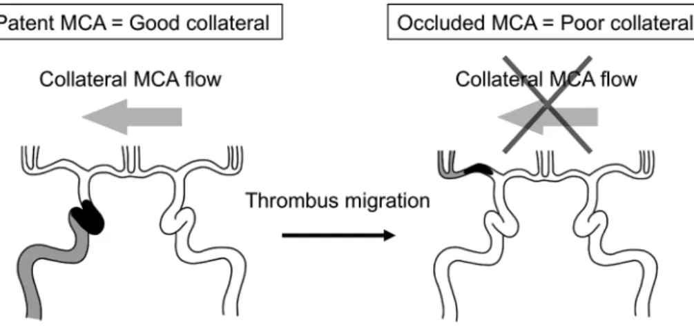
富な側副血行により,発症時神経症状が軽症の場合がある72). しかし,内頸動脈に存在する血栓が内頸動脈先端部や中大脳 動脈に移動することによって,Willis 動脈輪を介した側副血 行の灌流が突然途絶え,急激に神経症状が増悪する場合があ る(Fig. 1).つまり,側副血行が豊富な緩徐進行型の脳梗塞 から,側副血行の乏しい急速進行型の脳梗塞に突然変化する ため,最初の側副血行の状態からは予測困難な経過を呈し得 る.また,thrombus migration による神経症状増悪は,治療前 だけでなく rt-PA 静注療法後や血管内治療中にも生じ得るた め,再開通療法の適応および施行時期の決定に難渋する.
筆者らはこれらの ICO with patent MCA の臨床経過,再開 通療法後の転帰を検討した.2006 年 1 月から 2015 年 2 月に かけて,小倉記念病院脳卒中センターに入院した発症から
7日以内の虚血性脳卒中のうち,内頸動脈閉塞を認める症例
を後方視的に解析した.77 例の ICO のうち,MRA または血 管造影で,側副血行を介して同側の中大脳動脈が描出される ICO with patent MCA 34例を対象とした.これらの症例を,入 院時 NIHSS 10 未満の軽症は 17 例で,NIHSS 10 以上の重症 群は 17 例であった.軽症群のうち,3 例(18%)に緊急血管 内治療が行われ,14 例(82%)はまず保存的に加療された. 保存的加療を受けた 14 例のうち 7 例(47%)で神経症状の増 悪(NIHSS 4 点以上の増悪)が見られ,そのうち 6 例が血管 内治療を受けた.重症群のうち,12 例(71%)で血管内治療 が行われ,5 例(29%)が保存的に加療された.軽症群と,重 症群の血管内治療の時間経過,経過中の血栓移動,90 日後の 転帰良好(mRS 0~2)の割合を比較した.経過中の血栓移動 は,閉塞部位が同側の内頸動脈先端部や中大脳動脈近位部に 変化した場合と定義した. 背景因子は,入院時の NIHSS が有意に軽症であった [軽症 群:2(Interquartile range(IQR)1~4.5)vs. 重症群:18(IQR 12.5~23),P < 0.01].病型や治療内容には差がなかった.血 管内治療例において,有効再開通率(TICI grade 2b~3)に差 はなかった(軽症群:56% vs. 重症群:67%,P = 0.67).発 症から再開通までの時間は,まず保存的に加療された症例が 多く含まれているため,軽症群で有意に延長していた[軽症 群:673 分(IQR 403~1,860)vs. 重症群:331 分(IQR 185~ 484),P = 0.04].しかし,90 日後の転帰良好の割合は重症群 で低い傾向があった(軽症群:53% vs. 重症群:24%,P = 0.16).一方,経過中(治療前,血管内治療中)の thrombus migrationは,軽症群で有意に 90 日後の転帰良好の割合を低
下させたものの(thrombus migration なし:80% vs. thrombus migrationあり:14%,P = 0.02,Fisher の正確検定),重症群 では転帰に対して有意な影響を与えなかった(thrombus migrationなし:0% vs. thrombus migration あり:29%,P = 0.54,Fisher の正確検定).
以上の結果から ICO with patent MCA において,側副血行 の豊富な軽症例の転帰は,thrombus migration による急激な側 副血行の遮断に大きな影響を受ける可能性が示された.一方, 側副血行が不十分な重症例は therapeutic time window が短く, 再開通までの時間が転帰に対して大きな影響を与えると考え た.また解剖学的な側副血行の参入経路だけでなく,機能的 な側面から見た側副血行の多寡も重要で,側副血行が豊富な 軽症の ICO with patent MCA の症例(ICO with functionally patent MCA)に対しては,特に血栓移動の防止に主眼を置い た治療戦略が必要な可能性を示した.LVO を有する軽症脳梗 塞例に対して血管内治療を行う場合,therapeutic time window と,thrombus migration,出血性合併症等,治療による症状増 悪のリスクを考えながら,周到に治療を計画しなければなら ない.
Fig. 1 Schematic of internal carotid artery (ICA) occlusion with a patent middle cerebral artery (MCA) and thrombus migration. The migrated thrombus interrupts the collateral MCA flow. Consequently, acute ischemic stroke with a good collateral circulation changes to stroke with poor collateral circulation. NIHSS, National Institutes of Health Stroke Scale.
おわりに 本稿では,LVO を有する軽症脳梗塞の END の病態,再開 通療法について概説した.軽症脳梗塞は,rt-PA 静注療法,MT 等の科学的根拠を持った超急性期の治療が登場した現在にお いて,未だ課題が残る領域である.軽症脳梗塞は,発症時の 神経症状が軽微でも,END により最終的な転帰は不良な場合 があり,特に LVO を有する例では,END に備えて迅速に側 副血行の状態,脳梗塞の発症機序についての精査を行わなけ ればならない.しかし,LVO を有する軽症脳梗塞は,特に END を生じる危険性が高く,collateral failure 等,未だ不明な点の 多い様々な機序によって END を呈する.現在我々が行える血 管造影や灌流画像では,脳血流の状態を持続的に評価するこ とはできず,END を正確に予測することは困難である.END の病態をさらに明らかにするため,持続的に脳血流の状態を 観察できる検査手段の開発が望まれる.軽症脳梗塞に対して も rt-PA 静注療法,MT 等の再開通療法が有効な可能性はあ るが,出血性合併症のリスクもある.そのため,現時点で報 告されているデータを参考に,本人,家族への十分な病状説 明を行い,治療方針を決定する他ないのが現状と考える.ま た,血管内治療の最適な介入時期は不明であるが,END を認 めた場合の rescue MT は,保存的加療と比較して有効な可能 性がある.ICO with patent MCA では thrombus migration に よって側副血行の状態が劇的に変化するため,血管内治療を 行う場合,側副血行の状態によっては thrombus migration を 防止に主眼を置いた治療戦略が必要かもしれない.今後,再 開通療法の有効性や最適な施行時期を明らかにするために, ランダム化比較試験による検証が待たれる. 謝辞:本稿の執筆にあたり,御高閲頂いた済生会福岡総合病院脳 神経内科山田猛先生,倉敷中央病院脳神経外科石橋良太先生,藤田医 科大学脳卒中科中原一郎先生,松本省二先生,九州大学大学院医学研 究院神経内科学吉良潤一先生に深謝致します. ※著者に本論文に関連し,開示すべき COI 状態にある企業,組織, 団体はいずれも有りません. 文 献
1) Goyal M, Menon BK, van Zwam WH, et al. Endovascular thrombectomy after large-vessel ischaemic stroke: a meta-analysis of individual patient data from five randomised trials. Lancet 2016;387:1723-1731.
2) Goyal M, Demchuk AM, Menon BK, et al. Randomized assess-ment of rapid endovascular treatassess-ment of ischemic stroke. N Engl J Med 2015;372:1019-1030.
3) Campbell BC, Mitchell PJ, Kleinig TJ, et al. Endovascular therapy for ischemic stroke with perfusion-imaging selection. N Engl J Med 2015;372:1009-1018.
4) Powers WJ, Rabinstein AA, Ackerson T, et al. 2018 guidelines for the early management of patients with acute ischemic stroke: a guideline for healthcare professionals from the American Heart Association/American Stroke Association.
Stroke 2018;49:e46-e110.
5) Grotta JC, Welch KM, Fagan SC, et al. Clinical deterioration following improvement in the NINDS rt-PA stroke trial. Stroke 2001;32:661-668.
6) 日本脳卒中学会脳卒中医療向上・社会保険委員会 rt-PA (アルテプラーゼ)静注療法指針部会編.rt-PA(アルテプラー
ゼ)静注療法適正治療指針第二版.脳卒中 2017;39:43-86. 7) Khatri P, Conaway MR, Johnston KC. Ninety-day outcome rates
of a prospective cohort of consecutive patients with mild ischemic stroke. Stroke 2012;43:560-562.
8) Nedeltchev K, Schwegler B, Haefeli T, et al. Outcome of stroke with mild or rapidly improving symptoms. Stroke 2007;38:2531-2535.
9) Rajajee V, Kidwell C, Starkman S, et al. Early MRI and outcomes of untreated patients with mild or improving ischemic stroke. Neurology 2006;67:980-984.
10) Maas MB, Furie KL, Lev MH, et al. National Institutes of Health Stroke Scale score is poorly predictive of proximal occlusion in acute cerebral ischemia. Stroke 2009;40:2988-2993. 11) Fischer U, Baumgartner A, Arnold M, et al. What is a minor
stroke? Stroke 2010;41:661-666.
12) Dargazanli C, Arquizan C, Gory B, et al. Mechanical throm-bectomy for minor and mild stroke patients harboring large vessel occlusion in the anterior circulation: a multicenter cohort study. Stroke 2017;48:3274-3281.
13) Laurencin C, Philippeau F, Blanc-Lasserre K, et al. Throm-bolysis for acute minor stroke: outcome and barriers to manage-ment. Results from the RESUVAL stroke network. Cerebrovasc Dis 2015;40:3-9.
14) Reeves M, Khoury J, Alwell K, et al. Distribution of National Institutes of Health stroke scale in the Cincinnati/Northern Kentucky Stroke Study. Stroke 2013;44:3211-3213.
15) Ferrari J, Knoflach M, Keichi S, et al. Early clinical worsening in patients with TIA or minor stroke: the Austrian Stroke Unit Registry. Neurology 2010;74:136-141.
16) Kleindorfer D, Kissela B, Schneider A, et al. Eligibility for recombinant tissue plasminogen activator in acute ischemic stroke: a population-based study. Stroke 2004;35:e27-e29. 17) California Acute Stroke Pilot Registry (CASPR) Investigators.
Prioritizing interventions to improve rates of thrombolysis for ischemic stroke. Neurology 2005;64:654-659.
18) Romano JG, Smith EE, Liang L, et al. Distinct short-term outcomes in patients with mild versus rapidly improving stroke not treated with thrombolytics. Stroke 2016;47:1278-1285. 19) Heldner MR, Zubler C, Mattle HP, et al. National Institutes of
Health stroke scale score and vessel occlusion in 2152 patients with acute ischemic stroke. Stroke 2013;44:1153-1157. 20) Kim JT, Park MS, Chang J, et al. Proximal arterial occlusion in
acute ischemic stroke with low NIHSS scores should not be considered as mild stroke. PloS One 2013;8:e70996.
21) Mokin M, Masud MW, Dumont TM, et al. Outcomes in patients with acute ischemic stroke from proximal intracranial vessel occlusion and NIHSS score below 8. J Neurointerv Surg 2014; 6:413-417.
22) Lima FO, Furie KL, Silva GS, et al. Prognosis of untreated strokes due to anterior circulation proximal intracranial arterial
occlusions detected by use of computed tomography angiography. JAMA Neurol 2014;71:151-157.
23) Hao Z, Liu M, Wang D, et al. Etiologic subtype predicts outcome in mild stroke: prospective data from a hospital stroke registry. BMC Neurol 2013;13:154.
24) Rebello LC, Bouslama M, Haussen DC, et al. Stroke etiology and collaterals: atheroembolic strokes have greater collateral recruitment than cardioembolic strokes. Eur J Neurol 2017;24: 762-767.
25) Lee PH. Isolated middle cerebral artery disease: clinical and neuroradiological features depending on the pathogenesis. J Neurol Neurosurg Psychiatry 2004;75:727-732.
26) Lakshminarayan R, Scott PM, Robinson GJ, et al. Carotid stump syndrome: Pathophysiology and endovascular treatment options. Cardiovasc Intervent Radiol 2011;34(Suppl 2):48-52.
27) Kawano H, Inatomi Y, Hirano T, et al. Vertebral artery stump syndrome in acute ischemic stroke. J Neurol Sci 2013;324: 74-79.
28) Kim SK, Yoon W, Heo TW, et al. Negative susceptibility vessel sign and underlying intracranial atherosclerotic stenosis in acute middle cerebral artery occlusion. AJNR Am J Neuroradiol 2015;36:1266-1271.
29) Bourcier R, Volpi S, Guyomarch B, et al. Susceptibility vessel sign on MRI predicts favorable clinical outcome in patients with anterior circulation acute stroke treated with mechanical throm-bectomy. AJNR Am J Neuroradiol 2015;36:2346-2353.
30) Bae YJ, Jung C, Kim JH, et al. Quantitative magnetic resonance angiography in internal carotid artery occlusion with primary collateral pathway. J Stroke 2015;17:320-326.
31) Baek JH, Kim BM, Yoo J, et al. Predictive value of computed tomography angiography-determined occlusion type in stent retriever thrombectomy. Stroke 2017;48:2746-2752.
32) Seners P, Turc G, Oppenheim C, et al. Incidence, causes and predictors of neurological deterioration occurring within 24 h following acute ischaemic stroke: a systematic review with pathophysiological implications. J Neurol Neurosurg Psychiatry 2015;86:87-94.
33) Seners P, Turc G, Tisserand M, et al. Unexplained early neuro-logical deterioration after intravenous thrombolysis: incidence, predictors, and associated factors. Stroke 2014;45:2004-2009. 34) Thanvi B, Treadwell S, Robinson T. Early neurological
deterioration in acute ischaemic stroke: predictors, mechanisms and management. Postgrad Med J 2008;84:412-417.
35) Alexandrov AV, Felberg RA, Demchuk AM, et al. Deterioration following spontaneous improvement: sonographic findings in patients with acutely resolving symptoms of cerebral ischemia. Stroke 2000;31:915-919.
36) Seners P, Hurford R, Tisserand M, et al. Is unexplained early neurological deterioration after intravenous thrombolysis associated with thrombus extension? Stroke 2017;48:348-352.
37) Kaesmacher J, Maegerlein C, Kaesmacher M, et al. Thrombus migration in the middle cerebral artery: incidence, imaging signs, and impact on success of endovascular thrombectomy. J Am Heart Assoc, 2017;6:pii: e005149.
38) Shuaib A, Butcher K, Mohammad AA, et al. Collateral blood vessels in acute ischaemic stroke: a potential therapeutic target.
Lancet Neurol 2011;10:909-921.
39) Campbell BC, Christensen S, Tress BM, et al. Failure of collateral blood flow is associated with infarct growth in ischemic stroke. J Cereb Blood Flow Metab 2013;33:1168-1172.
40) Pranevicius O, Pranevicius M, Pranevicius H, et al. Transition to collateral flow after arterial occlusion predisposes to cerebral venous steal. Stroke 2012;43:575-579.
41) Alexandrov AV, Sharma VK, Lao AY, et al. Reversed robin hood syndrome in acute ischemic stroke patients. Stroke 2007;38: 3045-3048.
42) Palamarchuk I, Kimpinski K, Lippert C, et al. Nocturnal deterioration after ischemic stroke and autonomic dysfunction: hypothesis and implications. Cerebrovasc Dis 2013;36:454-461. 43) Beard DJ, McLeod DD, Logan CL, et al. Intracranial pressure
elevation reduces flow through collateral vessels and the penetrating arterioles they supply. A possible explanation for ‘collateral failure’ and infarct expansion after ischemic stroke. J Cereb Blood Flow Metab 2015;35:861-872.
44) Zhang R, Zhou Y, Yan S, et al. Slow collateral flow is associated with thrombus extension in patients with acute large-artery occlusion. AJNR Am J Neuroradiol 2018;39:1088-1092.
45) Watanabe M, Mori T, Imai K, et al. Distal migration of a floating carotid thrombus in a patient using oral contraceptives: a case report. J Med Case Reports 2009;3:8389.
46) Yoon JH, Shin YS, Lim YC, et al. Distal migration of thrombus during intra-arterial thrombolysis. Eur Neurol 2010;63:62-63. 47) Caplan LR. Worsening in ischemic stroke patients: is it time for
a new strategy? Stroke 2002;33:1443-1445.
48) Hwang YH, Kang DH, Kim YW, et al. Impact of time-to-reperfusion on outcome in patients with poor collaterals. AJNR Am J Neuroradiol 2015;36:495-500.
49) de Havenon A, Southerland AM. In large vessel occlusive stroke, time is brain... But collaterals are time. Neurology 2018;90: 153-154.
50) Kim BM, Baek JH, Heo JH, et al. Collateral status affects the onset-to-reperfusion time window for good outcome. J Neurol Neurosurg Psychiatry 2018;89:903-909.
51) Emberson J, Lees KR, Lyden P, et al. Effect of treatment delay, age, and stroke severity on the effects of intravenous throm-bolysis with alteplase for acute ischaemic stroke: a meta-analysis of individual patient data from randomised trials. Lancet 2014; 384:1929-1935.
52) You S, Saxena A, Wang X, et al. Efficacy and safety of intravenous recombinant tissue plasminogen activator in mild ischaemic stroke: a meta-analysis. Stroke Vasc Neurol 2018;3: 22-27.
53) Levine SR, Khatri P, Broderick JP, et al. Review, historical context, and clarifications of the NINDS rt-PA stroke trials exclusion criteria: Part 1: rapidly improving stroke symptoms. Stroke 2013;44:2500-2505.
54) Khatri P, Kleindorfer DO, Devlin T, et al. Effect of alteplase vs aspirin on functional outcome for patients with acute ischemic stroke and minor nondisabling neurologic deficits: the PRISMS randomized clinical trial. JAMA 2018;320:156-166.
55) Cappellari M, Turcato G, Forlivesi S, et al. Starting-SICH nomogram to predict symptomatic intracerebral hemorrhage
after intravenous thrombolysis for stroke. Stroke 2018;49: 397-404.
56) Urra X, Arino H, Llull L, et al. The outcome of patients with mild stroke improves after treatment with systemic thrombolysis. PloS One 2013;8:e59420.
57) Griessenauer CJ, Medin C, Maingard J, et al. Endovascular mechanical thrombectomy in large-vessel occlusion ischemic stroke presenting with low National Institutes of Health stroke scale: systematic review and meta-analysis. World Neurosurg 2018;110:263-269.
58) Pfaff J, Herweh C, Pham M, et al. Mechanical thrombectomy in patients with acute ischemic stroke and lower NIHSS scores: recanalization rates, periprocedural complications, and clinical outcome. AJNR Am J Neuroradiol 2016;37:2066-2071.
59) Dargazanli C, Consoli A, Gory B, et al. Is reperfusion useful in ischaemic stroke patients presenting with a low National Institutes of Health stroke scale and a proximal large vessel occlusion of the anterior circulation? Cerebrovasc Dis 2017;43: 305-312.
60) Bowen MT, Rebello LC, Bouslama M, et al. Clinical and imaging outcomes of endovascular thrapy in patients with acute large vessel occlusion stroke and mild clinical symptoms. Interv Neurol 2018;7:91-98.
61) Bhogal P, Bücke P, Ganslandt O, et al. Mechanical thrombectomy in patients with M1 occlusion and NIHSS score ≤5: a single-centre experience. Stroke Vasc Neurol 2016;1:165-171.
62) Haussen DC, Lima FO, Bouslama M, et al. Thrombectomy versus medical management for large vessel occlusion strokes with minimal symptoms: an analysis from STOPStroke and GESTOR cohorts. J Neurointerv Surg 2018;10:325-329. 63) Urra X, San Roman L, Gil F, et al. Medical and endovascular
treatment of patients with large vessel occlusion presenting with mild symptoms: an observational multicenter study.
Cerebrovasc Dis 2014;38:418-424.
64) Shang X, Lin M, Zhang S, et al. Clinical outcomes of endovascular treatment within 24 hours in patients with mild ischemic stroke and perfusion imaging selection. AJNR Am J Neuroradiol 2018; 39:1083-1087.
65) Sarraj A, Hassan A, Savitz SI, et al. Endovascular thrombectomy for mild strokes: how low should we go? Stroke 2018;49: 2398-2405.
66) Kim JT, Heo SH, Yoon W, et al. Clinical outcomes of patients with acute minor stroke receiving rescue IA therapy following early neurological deterioration. J Neurointerv Surg 2016;8: 461-465.
67) Haussen DC, Bouslama M, Grossberg JA, et al. Too good to intervene? Thrombectomy for large vessel occlusion strokes with minimal symptoms: an intention-to-treat analysis. J Neurointerv Surg 2017;9:917-921.
68) Nagel S, Bouslama M, Krause LU, et al. Mechanical thrombectomy in patients with milder strokes and large vessel occlusions. Stroke 2018;49:2391-2397.
69) Kwak JH, Zhao L, Kim JK, et al. The outcome and efficacy of recanalization in patients with acute internal carotid artery occlusion. AJNR Am J Neuroradiol 2014;35:747-753.
70) Liebeskind DS, Flint AC, Budzik RF, et al. Carotid I’s, L’s and T’s: collaterals shape the outcome of intracranial carotid occlusion in acute ischemic stroke. J Neurointerv Surg 2015;7: 402-407.
71) 田中悠二郎,中原一郎.頭頸部主幹動脈閉塞に対する急性期 血行再建:血栓回収デバイスの治療成績.脳と循環 2013;18: 31-41.
72) Kim JT, Park MS, Choi KH, et al. Clinical implications of collateral middle cerebral artery flow in acute ischaemic stroke with internal carotid artery occlusion. Eur J Neurol 2011;18: 1384-1390.
Abstract
Reperfusion therapy in patients with minor or mild ischemic stroke
Junpei Koge, M.D.
1)1)Division of Neurology, Saiseikai Fukuoka General Hospital