56:352 はじめに 脳梗塞に比べて脊髄梗塞は稀な疾患で,その診断は必ずし も容易ではないことがある1).急性期脊髄梗塞の診断はその 臨床的な経過,神経症状に加えて脊髄 MRI の有用性が報告さ れているが2)~5),発症時から慢性期まで MRI の画像的変化を 経時的に検討した報告は少ない.今回,脊髄梗塞症例において MRIの経時的変化を観察し得た 1 例を経験したので報告する. 症 例 症例:80 歳女性 主訴:下半身の疼痛,両下肢の脱力,排尿障害 既往歴:高血圧,脂質代謝異常症で治療中. 現病歴:2015 年 2 月某日,突然両下腿背側に痛みが出現 し,5 分後に腰部まで広がった.30 分後に両下肢は脱力し, 尿意が消失した.臀部から両下腿の触覚が低下し,床に足を ついても感覚がわからなかった.壁伝いにトイレに行ったが, 自力で排尿できなかった.翌日,症状の改善なく,近医を受 診.脊髄 MRI で腰髄髄内に異常信号病変を認め,原因精査の ため当科に紹介入院した. 入院時身体所見:血圧 150/79 mmHg,体温 37.1°C,脈拍 82回 / 分・整,胸腹部に異常所見はなかった. 入院時神経学的所見:意識清明.脳神経に異常はなかった. 運動系では上肢筋力は正常だが,両下肢は徒手筋力検査にて 腸腰筋が 2 程度であり,それ以外の下肢筋力はすべて消失し ていた.筋緊張は両下肢で低下し,腱反射は左側の大腿四頭 筋腱のみ保たれていたが右側では減弱し,下腿三頭筋腱は両 側で消失していた.Babinski 徴候はみられなかった.感覚系 では第 10 胸髄髄節レベル以下で両側性に温痛覚低下があり, 遠位は障害が著しかった.下肢関節位置覚は保たれていた. 自力で排尿,排便はできなかった. 入院時検査所見:血液検査で血算に異常はなく,LDL コレ ステロール 157 mg/dl(正常 65~163),HDL コレステロール 59 mg/dl(正常 48~103)であった.空腹時血糖 89 mg/dl, HbA1c 6.1%.凝固系は活性化部分トロンボプラスチン時間 30.5 sec(正常 24~38),フィブリノーゲン 463 mg/dl(正常 200~400),D-dimer 0.9 μg/ml(正常≦ 1.0)であった.各種 抗核抗体,腫瘍マーカーは陰性であった. 脊髄 MRI で第 1~2 腰椎椎体レベルの髄内に T2強調画像で 高信号域を認めた(Fig. 1).同部位に拡散強調画像(diffusion weighted image; DWI)で信号上昇があり,みかけの拡散係数 (apparent diffusion coefficient; ADC)は低下していた.脂肪抑
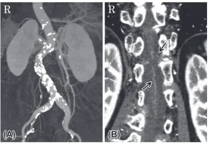
制 T2強調画像で椎体に異常信号はみられなかった.造影 MRI で髄内に造影効果は認めなかった.造影胸腹部 CT で腹部大 動脈に解離はなかったが,腹部大動脈は石灰化が多く,動脈 硬化が著しかった.Adamkiewicz 動脈は左第 12 肋間動脈から 分岐していたが,比較的細く,一部に連続性が不明瞭な部分 があった(Fig. 2).前脊髄動脈は概ね描出されていた.
短 報
脊髄 塞急性期の MRI の経時的変化:症例報告と文献的考察
竹下 翔
1)緒方 利安
1)米良 英和
1)津川 潤
1)深江 治郎
1)坪井 義夫
1)*
要旨: 症例は 80 歳女性.突然腰部から両下腿に激痛が出現し,約 30 分後に両下肢脱力,感覚低下,尿意消失 を呈した.神経所見では完全対麻痺,第 1 腰髄髄節レベル以下の解離性感覚障害,膀胱直腸障害を認めた.第 2 病 日の腰部 MRI の T2強調画像及び拡散強調画像(diffusion weighted image; DWI)で,第 1∼2 腰椎椎体レベルの 髄内に強い高信号域を認め,見かけの拡散係数(apparent diffusion coefficient; ADC)は低下しており脊髄梗塞と 診断した.経時的に MRI を撮像し,DWI の信号強度は時間と共に減弱し,約 1 か月後の ADC 値は高値であった.T2強調画像で浮腫性変化は一旦増強したが,最終的には縮小した.本症例は脊髄梗塞の MRI 画像変化を経時的に
捉えた点で貴重な症例と思われた. (臨床神経 2016;56:352-355)
Key words: 脊髄梗塞,前脊髄動脈症候群,MRI,拡散強調画像,経時的変化
*Corresponding author: 福岡大学医学部神経内科学教室〔〒 814-0180 福岡市城南区七隈 7-45-1〕
1)福岡大学医学部神経内科学教室
(Received December 25, 2015; Accepted February 18, 2016; Published online in J-STAGE on April 19, 2016) doi: 10.5692/clinicalneurol.cn-000858
脊髄梗塞急性期の MRI の経時的変化 56:353
経 過
本症例は突然発症の前脊髄動脈症候群を呈し,発症翌日の
MRIで脊髄内に DWI で高信号,ADC 値が低値を示す病変を認
め,造影胸腹部 CT で腹部大動脈に強い動脈硬化を認めたこと より脊髄梗塞と診断した.急性期にヘパリン 1×10⁴単位 / 日及 びエダラボン 60 mg/ 日の投与を開始した.第 3 病日に徒手筋 力検査で右下肢は 3 程度,左下肢は 4 程度まで改善した.表 在感覚は両下肢近位部の改善がみられたが,第 4 腰髄髄節以 下の改善は乏しく,右優位に障害が残った.同日の脊髄 MRI Fig. 1 Time course of MRI findings on days 1, 3, 10, 28 and 67.
Sagittal and axial T2-weighted images (T2WIs) of the lumbar spinal cord. Sagittal and axial T2WIs revealed high signal in the spinal cord at vertebral level L1 to L2 on days 1 and 3. The largest abnormal lesion is seen on T2WI, and its boundary appears vague on day 10. Its intensity became more obvious and its boundary became clearer on day 67 and this was associated with spinal cord atrophy. Axial finding of L2 segment and sagittal finding of the lumbar spinal cord on diffusion weighted imaging (DWI). DWI was not acquired on day 1. DWI revealed the brightest intensity on day 3. The DWI signal attenuated on days 10 and 28. Hyperintensity areas of sagittal DWI correspond with sagittal T2WI. DWI hypersignal intensity no longer existed on day 67. Scanning parameters for axial DWI were as follows; on day 3 (TR: 7,372.8 ms, TE: 73.3 ms, thickness: 7.0 mm, b = 800), on day 10 (TR: 5,608 ms, TE: 73.4 ms, thickness: 5.0 mm, b = 800), on day 28 (TR: 3,000 ms, TE: 67.1 ms, thickness: 5.0 mm, b = 1,000), on day 67 (TR: 3,200 ms, TE: 80 ms, thickness: 5.0 mm, b = 1,000). Scanning parameters for sagittal DWI were as follows; on day 3 (TR: 3,000 ms, TE: 68.5 ms, thickness: 4.5 mm, b = 800), on day 10 (TR: 3,000 ms, TE: 73.6 ms, thickness: 4.5 mm, b = 800), on day 28 (TR: 1,644.4 ms, TE: 51.3 ms, thickness: 5.0 mm, b = 800), on day 67 (TR: 3,200 ms, TE: 85 ms, thickness: 5.0 mm, b = 1,000). Axial findings of L2 segment on apparent diffusion coefficient (ADC). ADC was not acquired on day 1. ADC values were lowered on day 3 (0.55 ± 0.16 × 10⁻³
mm2/s) compared with the spinal cord in adjacent non-ischemic regions (1.15 ± 0.24 × 10⁻³ mm2
/s). Except for partial area, ADC values were still lowered on day 10 (0.91 ± 0.23 × 10⁻³
mm2
/s). On day 28, ADC values of the lesion were increased (1.53 ± 0.34 × 10⁻³
mm2/s), indicating pseudo-normalization. On day 67, ADC values remained high (1.59 ± 0.23 × 10⁻³ mm2
/s). The Gadolinium-enhanced T1-weighted images and STIR are shown below ADC imaging. There was no enhanced lesion in the spinal cord on T1-weighted axial images with Gd. STIR and T2-weighted images showed no pathological area in any vertebral body.
臨床神経学 56 巻 5 号(2016:5) 56:354 は第 1~2 腰椎椎体レベルの髄内に T2強調画像で高信号域の 拡大を認め,同部位は DWI で高信号,ADC 値が低値のまま であった.その後,下肢の運動・感覚機能は徐々に改善した が,膀胱直腸障害は残存した.第 10 病日の脊髄 MRI は T2強 調画像で髄内信号はより上昇し,浮腫性変化は増悪していた (Fig. 1).DWI の信号はやや低下しており,ADC 値も前回よ りわずかに上昇していた.第 22 病日には軽介助下で歩行器歩 行が可能となり,膀胱直腸障害に関しては導尿の継続が必要 だが排便できるようになった.第 28 病日の脊髄 MRI は T2強 調画像で髄内信号が辺縁明瞭になりさらに上昇し,脊髄の浮 腫性変化は軽減していた.DWI の信号は低下し,ADC 値は上 昇していた.第 29 病日にリハビリテーション目的で転院し た.第 67 病日に転院先で脊髄 MRI が行われ,T2強調画像で 髄内の高信号域は縮小し,脊髄は全体的に萎縮していた.DWI の信号はさらに低下しており,ADC 値は高値のままであった. 考 察 近年脊髄梗塞の診断における MRI の有用性が認知され,広 く用いられるようになり,画像的特徴の報告も多い.前脊髄 動脈症候群を呈した場合に脊髄 MRI で脊髄前角が両側性に 高信号を呈することが知られ,“snake eyes sign”や“owlʼs eye(s) sign” と呼ばれるが必ずしも特異性が高くない6)7).Weidauer ら は脊髄梗塞の形状から責任病巣を前脊髄動脈,脊髄中心動脈, 後脊髄動脈,Adamkiewicz 動脈,脊髄前角における分水嶺領 域の五つに分類している7).本症例は前脊髄動脈領域の梗塞に 分類される一方で,障害部位である腰仙髄領域が Adamkiewicz 動脈の分水嶺領域とされている中部胸髄と並んで血行力学的 に虚血を起こしやすいと考えられている8).本症例は強い腹 部大動脈の動脈硬化を背景に,Adamkiewicz 動脈から潅流さ れる前脊髄動脈の支配領域の最も遠位部に位置する部分が梗 塞になったとも考えられた.中枢神経虚血急性期の MRI でみ られる経時的変化は脳梗塞でよく検討されており,一般に発 症後 3 時間程度で DWI が高信号を示し,ADC 値が低下,3~ 4週間で DWI,ADC の信号強度が逆転すると言われている9). T2強調画像に関しては 24 時間~1 週間以内に高信号を呈し, その後浮腫により周囲の不鮮明化が生じた後に辺縁が明瞭 となる.脊髄梗塞の MRI でみられる経時的変化は脳梗塞と 同様であるとの報告もあるが5)10)発症後 3 か月経過しても ADC値が低下しなかった例や10) DWIが見かけ上等信号となる pseudonormalizationが脳梗塞より早期に起こった4)という報 告もある.本症例は発症後早期に T2WI,DWI で高信号,ADC 値の低下を示し,発症後約 1 か月で DWI,ADC の信号が逆転 している点で脳梗塞の MRI 経過と類似していた.脊髄梗塞の MRI所見は,急性期から慢性期まで経時的に大きく変化する ため,その時期に応じた画像理解がないと他疾患と見誤る可 能性がある.特に脱髄や脊髄炎は T2強調画像で高信号を示す ため,拡散強調画像でも高信号を呈する,いわゆる T2-shine
throughが MRI で脊髄梗塞を鑑別する際に問題となり,ADC
と併せた評価が重要である.また,発症急性期に拡散強調画 像で著明な高信号を示すこと,亜急性期に造影効果があるこ とや椎体梗塞を合併することもあり,脊髄梗塞の特徴とされ ている11)12).脊髄梗塞における画像の経時的変化を追えた報 告は稀で,本症例はその点で貴重な症例と考えられた.今後 は脊髄梗塞の病型別の画像経過の特徴や,画像所見と重症度 や予後との関連について検討していく必要がある. 本報告の要旨は,第 210 回日本神経学会九州地方会で発表し,会長 推薦演題に選ばれた. 11 22
Fig. 2 Contrast CT of the spine.
(a) Abdominal aorta accompanied by remarkable calcification. (b) Adamkiewicz artery originated from 12th intercostal artery and not visualized everything (arrow 1). The anterior spinal artery was visualized from lower thoracic cord to upper lumbar cord. We could not found any arteriovenous malformation (arrow 2).
脊髄梗塞急性期の MRI の経時的変化 56:355 謝辞:稿を終えるにあたり,貴重な画像の提供をいただいた福岡リ ハビリテーション病院神経内科 金森祐治先生に深謝致します. ※本論文に関連し,開示すべき COI 状態にある企業,組織,団体 はいずれも有りません. 文 献
1) Sandson TA, Friedman JH. Spinal cord infarction. Report of 8 cases and review of the literature. Medicine 1989;68:282-292. 2) Fujikawa A, Tsuchiya K, Takeuchi S, et al. Diffusion-weighted
MRI imaging in acute spinal cord ischemia. Eur Radiol 2004;14: 2076-2078.
3) Loher TJ, Bassetti CL, Lovblad KO, et al. Diffusion weighted MRI in acute spinal cord ischemia. Neuroradiology 2003;45:557-561.
4) Küler W, Weller M, Klose U, et al. Diffusion-weighted MRI of spinal cord infarction high resolution imaging and time course of diffusion abnormality. J Neurol 2004;251:818-824.
5) Benjamin KT, Foster E, Kam A, et al. Diffusion weighted imaging with trace diffusion weighted imaging, the apparent diffusion coefficient and exponential images in the diagnosis of spinal cord infarction. J Clin Neurosci 2013;20:1630-1632.
6) Weidauer S, Nichtweiß M, Berkefeld J. Spinal cord infarction. In: Hattingen E, Weidauer S, Setzer M, editors. Disease of the Spinal Cord-Novel Imaging, Diagnosis and Treatment. 1st ed. Heidelberg: Springer 2015. p. 435-452.
7) Weidauer S, Nichtweiß M, Hattingen E, et al. Spinal cord ischemia: aetiology, clinical syndromes and imaging features. Neuroradiology 2015;57:241-257.
8) Duggal N, Lach B. Selective vulnerability of the lumbosacral spinal cord after cardiac arrest and hypotension. Stroke 2002; 33:116-121.
9) Burdette H, Ricci E, Nicola P, et al. Cerebral Infarction: time course of signal intensity changes on diffusion-weighted MR images. AJR Am J Roentgenol 1998;171:791-795.
10) Fujikawa A, Tsuchiya K, Koppera P, et al. Case report: spinal cord infarction demonstrated on diffusion-weighted MR imaging with a single-shot fast spin-echo sequence. J Comput Assist Tomog 2003;27:415-419.
11) Hirono H, Yamadori A, Komiyama M, et al. MRI of spontaneous spinal cord infarction: serial changes in gadolinium-DTPA enhancement. Neuroradiology 1992;34:95-97.
12) Yuh WT, Marsh EE 3rd, Wang AK, et al. MR imaging of spinal cord and vertebral body infarction. AJNR Am J Neuroradiology 1992;13:145-154.
Abstract
Time course of diffusion weighted image and apparent diffusion coefficient
in acute spinal cord infarction: A case report and review of the literature
Sho Takeshita, M.D.
1), Toshiyasu Ogata, M.D.
1), Hidekazu Mera, M.D.
1),
Jun Tsugawa, MD.
1), Jiro Fukae, M.D.
1)and Yoshio Tsuboi, M.D.
1)1)Department of Neurology, Faculty of Medicine, Fukuoka University