IRUCAA@TDC : Lymphoepithelial cysts of oral mucosa: two cases in different regions.
7
0
0
全文
(2) Bull Tokyo Dent Coll (2012) 53(1): 17–22. Case Report. Lymphoepithelial Cysts of Oral Mucosa: Two Cases in Different Regions Roberta Targa Stramandinoli-Zanicotti, Lúcia Fátima de Castro Ávila, Ana Claudia Santos de Azevedo Izidoro, Fábio Alves Izidoro* and Juliana Lucena Schussel** Department of Stomatology, School of Post Graduate Dentistry, General Hospital of Curitiba, Brazil * Department of Radiology, School of Post Graduate Dentistry, General Hospital of Curitiba, Paraná, Brazil ** Department of Oral and Maxillofacial, Erasto Gaertner Hospital, Curitiba, Brazil Received 13 July, 2011/Accepted for publication 11 October, 2011. Abstract Lymphoepithelial cyst of the oral cavity is a rare cystic lesion that presents as an asymptomatic, well-circumscribed, yellowish small submucosal nodule covered by normal overlying mucosa, usually located in the floor of the mouth or in the ventral or posterolateral surface of the tongue. Histopathological examination reveals a cyst lined by a stratified squamous epithelium surrounded by lymphoid tissue. In this paper, we report two cases of oral lymphoepithelial cyst, one on the anterior floor of the mouth and another on the posterior lateral tongue. In both cases, the patients were women aged approximately 50 years and the lesions were treated by local surgical excision, with no evidence of recurrence after 2 years of follow-up. Key words:. Lymphoepithelial cyst — Ectopic lymphoid tissue — Oral lymphoepithelial cyst — Oral cysts — Benign oral lesion. Introduction. y ellow-white discoloration. Occasional cysts are transparent. Usually the OLC is located on the floor of the mouth or on the ventral or posterolateral surface of the tongue. Approximately half of all intraoral cases are located on the floor of the mouth6). Histopathologically, it presents as a central cystic lesion lined with stratified squamous epithelium with desquamated keratin in the lumen. Surrounding the cyst lining there are variable amounts of lymphoid tissue and a. Oral lymphoepithelial cyst (OLC) is a relatively rare lesion that develops within lymphoid tissue of unknown etiopathogenesis. These cystic lesions, usually located in the lateral aspect of the neck, were first described by Bhaskar and Bernier as “branchial cyst” in 19594). Clinically, OLCs present as movable, painless submucosal nodules with a yellow or 17.
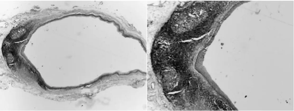
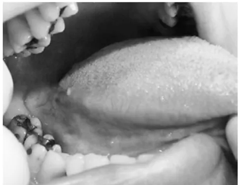
(3) 18. Zanicotti RTS et al.. fibrous connective tissue capsule3). Two cases with OLC are reported in this paper.. Case Reports 1. Case 1 A 57-year-old woman presented with a nodule on the floor of her mouth. Her personal and family medical histories were unremarkable. Intraoral examination revealed a submucosal nodule 2 mm in diameter. The nodule was non-elastic, movable, and soft, appearing as a yellow discoloration on the left side of the floor of the mouth, adjacent to the. Fig. 1 Submucosal nodule, 2 mm in diameter, with yellow discoloration, on left side of floor of mouth adjacent to lingual frenum. lingual frenum (Fig. 1). The mucosal surface was intact and the patient complained of no pain or discomfort. The clinical diagnosis was lipoma or OLC. The lesion was totally excised under local anesthesia. Histopathological examination revealed a cystic lesion lined with thin parakeratinized stratified squa mous epithelium; lymphoid tissue exhibiting germinal centers was observed on the cyst wall (Fig. 2). The diagnosis was lymphoepithelial cyst. No complications were encountered in the postoperative period of two years. 2. Case 2 A 55-year-old woman visited a dental clinic for a routine examination. On physical examination, an elastic, hard, submucosal nodule, yellow in color and 15 mm in diameter was observed on the posterior edge of the right side of the tongue (Fig. 3). The overlying mucosa was normal and the patient complained of no discomfort. The clinical diagnosis was lipoma or OLC. Under local anesthesia, the nodular lesion was excised together with surrounding normal tissues and sent for histopathological examination. Micro scopically, the cystic cavity was lined with a thin flattened layer of parakeratinized stratified squamous epithelium, and was surrounded by a well-circumscribed mass of lymphoid tissue, with desquamated cells and lymphocytes present in the lumen (Fig. 4). The. Fig. 2 Cystic lesion lined with thin stratified squamous epithelium; lymphoid tissue exhibiting germinal centers observed on cyst wall.
(4) Report of Two Cases of Oral Lymphoepithelial Cysts. pathological diagnosis was OLC. Three years after surgery, the postoperative course was uneventful.. Discussion and Conclusion Bhaskar and Bernier first recommended the term lymphoepithelial in 1959 in a report of 468 cases of branchial cysts of the neck4,25). In 1962, Gold and Lewittown described an OLC as a case report, which was the first one in the world14,25). A lymphoepithelial cyst may occur in virtually any organ. After the neck and oral cavity, the most frequent sites reported in the literature are the pancreas and thyroid gland. Pancreatic lesions are. Fig. 3 Elastic, hard, submucosal nodule, yellow in color observed on posterior edge of tongue. 19. also believed to be benign epithelial inclusions embedded within the pancreas during embryogenesis30). OLCs are rare cystic lesions lined with stratified squamous epithelium surrounded by lymphoid tissue and have been reported as branchial cysts, branchial cleft cysts, branchiogenic cysts, or pseudocysts3). The pathogenesis of OLCs is uncertain. The two theories that have been proposed are the entrapment and obstruction theories. Several authors have suggested that ectopic foci of glandular epithelium become entrapped within nodal tissue and may proliferate ultimately to form a cyst5,27). However, other authors have suggested that OLCs are the result of obstruction of crypts of otherwise normal oral tonsils17). Clinically, an OLC presents as a small asymptomatic, well-circumscribed, yellowish, elevated, mobile, submucosal tumor of varying duration3). Most of the lesions are located in the floor of the mouth (60%) or on the lateral and ventral surfaces of the tongue (40%)5,25). In our cases, the sites and diameters of the lesions concurred with the literature. Giunta and Cataldo13) and Buchner and Hansen7) also observed that these cysts may vary in diameter from 2 to 15 mm. OLCs occurring on the parotid gland are often associated with immunocompromised patients and lymphoproliferative disorders20). Table 1. Fig. 4 Cystic lesion lined with thin keratinized stratified squamous epithelium and surrounded by wellcircumscribed mass of lymphoid tissue with desquamated cells and lymphocytes presents in lumen.
(5) 20. Zanicotti RTS et al.. shows cases reported in the literature, since the first report by Gold and Lewittown (1962)14), excluding cases related to immunocompromised patients and salivary glands. The OLC is covered with intact mucosa, clinically interpreted most often as mucocele or lipoma. Although most of the cases. reported are from male patients27), our two cases were from women, in their fifth decade of life. The actual prevalence of OLCs is not known, because many of these cystic lesions are not diagnosed. The cysts are often misdiagnosed as mucous cyst, other cystic lesions or even as a lipoma, and many times they are. Table 1 Oral lymphoepithelial cyst cases reported in literature. Publication. Cases. Gold & Lewittown, 196214) 1 8). Location. Gender. Age. Floor of mouth. M. 32. Calman, 1963 1. Floor of mouth. M. 40. Vickers & Von Der Muhl, 196627) 1. Floor of mouth. M. 30. Bhaskar & Colonel, 19665) 24 . Floor of mouth (n=15); Tongue (n=8); Palate (n=1). M=17/F=7. 40 (15–65). Young & Claman, 196729) 1. Floor of mouth. M. 42. 17). Floor of mouth (n=7); Tongue (n=2) Palate (n=4). M=12/F=1. 32 (17–48). Acevedo & Nelson, 19711) 9. Floor of mouth. M=9. 33 (20–46). Merchant, 197222) 1. Floor of mouth. M. 21. Giunta & Cataldo, 1973 21 . Floor of mouth (n=17); Palate (n=2) Buccal mucosa (n=1); Retromolar area (n=1). M=9/F=12. 36 (7–65) . Buchner & Hansen, 19807) 38 . Floor of mouth (n=19); Tongue (n=14); Palate (n=4); Retromolar area (n=1). M=23/F=15. 43 (14–81). Toto et al., 198226) 6 . Floor of mouth (n=2); Tongue (n=3) Palate (n=1). M=6/F=6. 43 (25–60). Knapp, 1970 13 . 13). Sakoda et al., 198324) 1. Tongue. F. 19. 24. Floor of mouth. M=9/F=15. 43 (12–74). Iwase et al., 198515) 1. Floor of mouth. M. 34. Chaudhry et al., 19849). 21). McDonnell, 1990 1. Floor of mouth. M 5. Kumara et al., 199518) 1. Floor of mouth. M. 29. 2). Ahn et al., 1996 1. Floor of mouth. F. 56. Tongue. M. 30. Suzuki et al., 2000 1. Flaitz, 200011) 1. Tongue. M 4. Flaitz & Davis, 200412) 1. Tongue. M. 72. Tongue. F. 27. 25). 19). López-Jornet, 2007 1 16). Khelemsky & Mandel, 2010 1. Floor of mouth. M. 34. Nonaka et al., 201123) 10 . Floor of mouth (n=2); Tongue (n=4) Palate (n=1); Oropharynx (n=2). M=2/F=8. 38 (16–60). Yang et al., 201128) 120 . Floor of mouth (n=46); Tongue (n=60); Palate (n=6); Buccal mucosa (n=3); Others (n=5). M=37/F=83. 44 (2–75) .
(6) Report of Two Cases of Oral Lymphoepithelial Cysts. not examined microscopically, and incidence may vary among different institutions. Differential diagnosis is important10) for definitive therapy. Several lesions appear as submucosal nodules in the floor of the mouth. Lipomas are usually located in the buccal mucosa, and rarely affect the floor of the mouth. It can also have a yellowish appearance but is usually larger than an LC. Some salivary lesions must also be considered as LCs in differential diagnosis. Mucoceles are the most frequent benign salivary gland in the oral cavity and can have the same clinical aspects as an LC. Fluid drainage and size fluctuation facilitates differential diagnosis between the two lesions. Salivary stones or sialolithiasis can also present as a submucosal mass in the floor of the mouth, and can induce pain depending on duct obstruction and salivary gland swelling. An LC is usually encapsulated and small in size, facilitating surgical excision in an ambulatorial environment. There are no LC recurrences reported in the literature, and recovery after surgical removal should be fast and without recurrence.. References 1) Acevedo A, Nelson JF (1971) Lymphoepithelial cysts of the oral cavity: report of nine cases. Oral Surg Oral Med Oral Pathol Oral Radiol Endod 31:632–635. 2) Ahn SK, Won JH, Lee SH, Choi EH, Choi SI (1996) Lymphoepithelial cyst associated with epithelial inclusion cyst. Am J Dermatopathol 18:424–426. 3) Bernier J, Bhaskar S (1958) Lymphoepithelial lesions of salivary glands: histogenesis and classification based on 186 cases. Cancer 11: 1156–1179. 4) Bhaskar SN, Bernier JL (1959) Histogenesis of branchial cysts: A report of 468 cases. Am J Pathol 35:407–423. 5) Bhaskar SN, Colonel L (1966) Lymphoepithelial cysts of the oral cavity. Oral Surg Oral Med Oral Pathol 21:120–128. 6) Bouquot J, Nikai H (2001) Lesions of the oral cavity. Diagnostic surgical pathology of the head and neck, Gnepp D ed., pp.141–221, WB. 21. Saunders, Philadelphia. 7) Buchner A, Hansen LS (1980) Lymphoepithelial cysts of the oral cavity. Oral Surg 50:441– 449. 8) Calman HI (1963) Sublingual branchiogenic cyst-report of a case. J Oral Surg 16:333–338. 9) Chaudhry AP, Yamane GM, Scharlorck SE, SunderRaj M, Jain R (1984) A clinicopathological study of intraoral lymphoepithelial cysts. J Oral Med 39:79–84. 10) Epivatianos A, Zaraboukas T, Antoniade D (2005) Coexistence of lymphoepithelial and dermoid cysts on the floor of the mouth: report of a case. Oral Dis 11:330–333. 11) Flaitz CM (2000) Oral lymphoepithelial cyst in a young child. Pediatr Dent 22:422–423. 12) Flaitz CM, Davis SE (2004) Oral and maxillofacial pathology case of the mouth: oral lymphoepithelial cyst. Tex Dent J 121:630–631. 13) Giunta J, Cataldo E (1973) Lymphoepithelial cysts of the oral mucosa. Oral Surg 35:77–84. 14) Gold C, Lewittown NJ (1962) Branchial cleft cyst located in the floor of the mouth. Oral Surg Oral Med Oral Pathol 15:1118–1120. 15) Iwase T, Teratani K, Saito A, Funatsu K, Sato M, Kiuchi K, Umemura S (1985) Immunohistochemical and lectin histochemical studies on the lymphoepithelial cyst of the oral cavity and neck. J Nihon Univ Sch Dent 27:28–34. 16) Khelemsky R, Mandel L (2010) Lymphoepithelial cyst of mouth floor. J Oral Maxillofac Surg 68:3055–3057. 17) Knapp MJ (1970) Pathology of oral tonsils. Oral Surg 29:295–304. 18) Kumara GR, Gillgrass TJ, Bridgman JB (1995) A lymphoepithelial cyst (branchial cyst) in the floor of the mouth. N Z Dent J 91:14–15. 19) López-Jornet P (2007) Oral lymphoepithelial cyst. Ann Dermatol Venereol 134:588. 20) Marsot-Dupuch K, Quillard J, Meyohas MC (2004) Head and neck lesions in the immunocompromised host. Eur Radiol 3:E155–E167. 21) McDonnell D (1990) Spontaneous regression of a yellow sublingual swelling: a case report. Pediatr Dent 12:388–389. 22) Merchant NE (1972) Lymphoepithelial cyst of the floor of the mouth: a case report. Br Dent J 132:271–272. 23) Nonaka CF, Henriques AC, de Matos FR, de Souza LB, Pinto LP (2011) Nonodontogenic cysts of the oral and maxillofacial region: demographic profile in a Brazilian population over a 40-year period. Eur Arch Otorhinolaryngol 268:917–922. 24) Sakoda S, Kodama Y, Shiba R (1983) Lymphoepithelial cyst of oral cavity: report of a case and review of the literature. Int J Oral Surg 12:127–131..
(7) 22. Zanicotti RTS et al.. 25) Suzuki H, Baba S, Hashimoto K (2000) Lymphoepithelial cyst in the sublingual region: Report of a case and review of literature. Oral Med Pathol 5:105–108. 26) Toto PD, Wortel JP, Joseph G (1982) Lymphoepithelial cysts and associated immunoglobulins. Oral Surg Oral Med Oral Pathol Oral Radiol Endod 54:59–65. 27) Vickers RA, Von Der Muhl OH (1966) An investigation concerning inducibility of lymphoepithelial cysts in hamsters by autogenous epithelial transplantation. J Dent Res 45:1029– 1032. 28) Yang X, Ow A, Zhang C, Wang L, Yang W, Hu Y, Zhong L (2011) Clinical analysis of 120 cases of intraoral lymphoepithelial cyst. Oral Surg Oral Med Oral Pathol Oral Radiol Endod.. [Epub ahead of print] 29) Young WG, Claman SM (1967) A lymphoepithelial cyst of the oral cavity. Oral Surg Oral Med Oral Pathol Oral Radiol Endod 23:62–70. 30) Younus S, Bleibel W, Bleibel H, Hernady N (2007) Lymphoepithelial cyst of the pancreas. Dig Dis Sci 52:3136–3139. Reprint requests to: Dr. Juliana Lucena Schussel Department of Oral and Maxillofacial, Erasto Gaertner Hospital, Rua João Guariza, 422 Curitiba, Brazil Tel & Fax: +55-41-3256-2409 E-mail: juliana.schussel24@gmail.com.
(8)
図

関連したドキュメント
If condition (2) holds then no line intersects all the segments AB, BC, DE, EA (if such line exists then it also intersects the segment CD by condition (2) which is impossible due
This equation encompasses many important integral and functional equations that arise in nonlinear analysis and its applications, in particular integral equations (1.1), (1.2),
We show that a discrete fixed point theorem of Eilenberg is equivalent to the restriction of the contraction principle to the class of non-Archimedean bounded metric spaces.. We
In this case, the extension from a local solution u to a solution in an arbitrary interval [0, T ] is carried out by keeping control of the norm ku(T )k sN with the use of
Here general is with respect to the real analytic Zariski topology and the dimension of a fiber is well-defined since the fiber is covered by a countable union of real analytic
Applications of msets in Logic Programming languages is found to over- come “computational inefficiency” inherent in otherwise situation, especially in solving a sweep of
Shi, “The essential norm of a composition operator on the Bloch space in polydiscs,” Chinese Journal of Contemporary Mathematics, vol. Chen, “Weighted composition operators from Fp,
[2])) and will not be repeated here. As had been mentioned there, the only feasible way in which the problem of a system of charged particles and, in particular, of ionic solutions
