REVIEW
Ectopic fat deposition and global cardiometabolic risk :
New paradigm in cardiovascular medicine
Michio Shimabukuro
1,2, Chisayo Kozuka
3, Shin-ichiro Taira
3, Koichi Yabiku
3,
Munkhbaatar Dagvasumberel
5, Masayoshi Ishida
2, Sachiko Matsumoto
2,
Shusuke Yagi
2, Daiju Fukuda
1,2, Ken Yamakawa
4, Moritake Higa
4, Takeshi Soeki
2,
Hisashi Yoshida
5, Hiroaki Masuzaki
3, and Masataka Sata
2 1Department of Cardio-Diabetes Medicine,2
Department of Cardiovascular Medicine, the University of Tokushima Graduate School of Health Biosciences, Tokushima, Japan ;3
Division of Endocrinology, Diabetes and Metabolism, Hematology, Rheumatology, Second Department of Internal Medicine, University of the Ryukyus School of Medicine Graduate School of Medicine, Okinawa, Japan ;4
Diabetes and Lifestyle-Related Disease Center, Tomishiro Central Hospital, Okinawa, Japan ;5
Cardiovascular Division, Shonan Hospital, Okinawa, Japan
Abstract : The obesity epidemic is a global public health concern that increases the likeli-hood of morbidity and mortality of metabolic and cardiovascular disease (CVD) and threat-ens to reduce life expectancy around the world. The concept of the metabolic syndrome (MetS) takes into account that visceral fat plays an essential role in the development of metabolic and cardiovascular diseases. However, MetS cannot be used to assess global CVD risk but is at best one more modifiable CVD risk factor. Thus, global cardiometabolic risk (the global risk of cardiovascular disease resulting from traditional risk factors combined with the additional contribution of the metabolic syndrome and/or insulin resistance) should be considered individually. There is solid evidence supporting the notion that ex-cess abdominal fat is predictive of insulin resistance and the presence of related metabolic abnormalities currently referred to as MetS. Despite the fact that abdominal obesity is a highly prevalent feature of MetS, the mechanisms by which abdominal obesity is causally related to MetS are not fully elucidated. Besides visceral fat accumulation, ectopic lipid deposition, especially in liver and skeletal muscle, has been implicated in the pathophysiol-ogy of diabetes, insulin resistance and obesity-related disorders. Also, ectopic fat deposi-tion could be deteriorated in the heart components such as (1) circulatory and locally re-cruited fat, (2) intra- and extra-myocellular fat, (3) perivascular fat, and (4) pericardial fat. In this review, the contribution of ectopic lipid deposition to global cardiometabolic risk is reviewed and also discussed are potential underlying mechanisms including adipocy-tokine, insulin resistance and lipotoxicity. J. Med. Invest. 60 : 1-14, February, 2013
Keywords : obesity, insulin resistance, cardiovascular disease, diabetes mellitus
Abbreviations : CVD : cardiovascular disease ; T2DM : type 2 dia-betes mellitus ; BMI : body mass index ; AMI : acute myocardial infarction ; CHD : coronary heart diseases ; FFA : free fatty acids ; ROS : reactive oxygen species ; eNOS : endothelial nitric oxide synthase ; NO : nitric oxide ; TG : triglycerides ; lipoprotein -cholesterol ; HDL- C : high- density lipoprotein - -cholesterol.
Received for publication January 4, 2013 ; accepted February 14, 2013.
Address correspondence and reprint requests to Michio Shimabukuro, MD, Departments of Cardio - Diabetes Medicine, the University of Tokushima Graduate School of Health Bi-osciences, 3 - 18 - 15 Kuramoto, Tokushima 770 - 8503, Japan and Fax : + 81 - 88 - 633 - 7894.
Glucose intolerance Hypertension
Dyslipidemia
Visceral fat obesity
Glucose intolerance/Type 2 diabetes Visceral fat obesity
High fat diet + Physical inactivity
Metabolic syndrome
Enhanced FFA eflux
Pancreatic fat deposition Tissue fat deposition
Whole body Insulin resistance Abnormal insulin secretion Cardiovascular diseases Cardiac fat deposition
INTRODUCTION
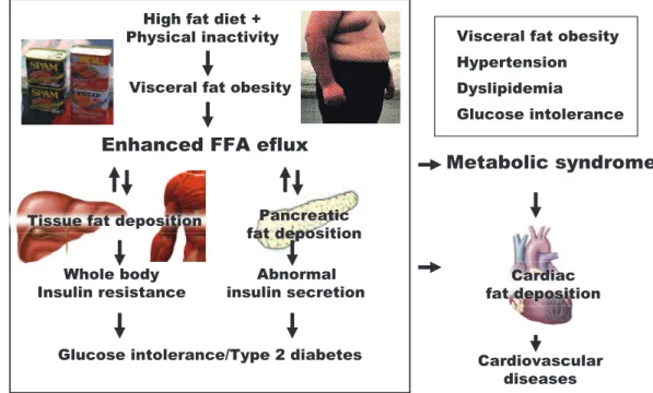
The obesity epidemic is a global public health concern that increases the likelihood of morbidity and mortality of metabolic and cardiovascular dis-ease (CVD) and threatens to reduce life expectancy around the world (1-2). The concept of the meta-bolic syndrome (MetS) takes into account that vis-ceral fat plays an essential role in the development of metabolic and cardiovascular diseases (3-5). MetS is merely a modifiable CVD risk factor as well as traditional risk factors such as LDL-cholesterol, diabetes, hypertension and smoking, but accounts for a great proportion of mechanisms under the obesity endemic era. Thus, global cardiometabolic risk (the global risk of cardiovascular disease re-sulting from traditional risk factors combined with the additional contribution of the metabolic syn-drome and/or insulin resistance) should be consid-ered individually (6-8). Despite the fact that abdomi-nal obesity is a highly prevalent feature of MetS, ectopic fat deposition, especially in liver and skele-tal muscle, has been implicated in the pathophysi-ology of diabetes, insulin resistance and obesity-related disorders (Fig. 1) (6-8). Also, ectopic fat deposition in the cardiovascular components is now recognized as a new cardiometabolic risk marker, as it is associated with increased insulin resistance, visceral fat and, in general, with the metabolic syn-drome (6, 9-11). In this review, the contribution of ectopic fat deposition to global cardiometabolic risk
and its potential underlying mechanisms are re-viewed.
1. CONTRIBUTION OF ECTOPIC FAT
DESPOSION TO CARDIOVASCULAR
DIS-EASE
The amount of adipose tissue, as well as its dis-tribution, is of special importance in the pathogene-sis of insulin repathogene-sistance and type 2 diabetes. Visceral adipose tissue is metabolically highly active (3-5) and its major role in the pathogenesis of insulin resistance is widely accepted. Simultaneously, lipids in ectopic (non-adipose) tissues such as liver and skeletal muscle are of increasing interest. Liver fat is elevated in insulin-resistant subjects (12, 13) and, furthermore, is strongly correlated with the amount of visceral fat in a prediabetic population (14). In addition, high intramyocellular fat is a marker of insulin resistance (15, 16). Thus, determination of fat in the visceral depot and in the ectopic tissues liver and muscle is thought to be a predictor for subjects who are insulin resistant and have a high risk of type 2 diabetes and cardiovascular dieases (Fig 2). Recenlty, we evaluated the relationship be-tween hepatic and muscular lipid deposition and vis-ceral fat accumulation in middle-aged Japanese men with MetS and found that visceral fat accumulation is accompanied by excess lipid deposition in skele-tal muscle (iliopsoas muscle, back muscle, rectus
Normal lean Whole-body adiposity Blood circulation Ectopic fat deposition Insulin resistance Subcutaneous fat obesity Lipids leptin, adiponectin no leptin, no adiponectin Lipids Visceral fat obesity Generalized lipodystrophy fat fat no fat fat Vascular failure fat fat fat fat Lipids leptin, adiponectin Lipids leptin, adiponectin
abdominis muscle) and the liver (17). Interstingly, changes in lipid contents by atnti-diabetic medica-tions were different in the tissues (visceral fat" liver fat"muscle fat), suggesting that ectopic fat desposion is regulated differently in these tissues. Contribution of ectopic fat desposion to cardiovas-cular disease are discussed below.
1.1. Coronary heart diseases
In the INTERHEART study (18), Yusuf et al. re-ported the effect of various measures of adiposity on rates of acute myocardial infarction (AMI) by comparing 12,461 AMI cases and 14,637 standard-ized-controls of variable ethnicity from 52 countries. BMI showed a modest association with AMI (un-adjusted odds ratio [OR] 1.44 for the top quintile vs. the bottom quintile), but this association was lost after adjustment for other risk factors. Mean-while, adjusted OR for quintile of waist-to-hip ratio was consecutively higher than that of the previous one (OR 1.15 ; 1.39 ; 1.90 ; and 2.52, respectively).
Targher et al. assessed prospectively whether Nonalcoholic fatty liver disease NAFLD, a typical phenotype of ectopic fat deposition, predicts future CVD events including nonfatal coronary heart dis-ease (myocardial infarction and coronary revascu-larization procedures), ischemic stroke, or cardio-vascular death, among type 2 diabetic individuals (19). After adjustment for age, sex, smoking his-tory, diabetes duration, HbA1c, LDL cholesterol,
liver enzymes, and use of medications, the presence of NAFLD was significantly associated with an in-creased CVD risk (odds ratio 1.84, P!0.001). Ad-ditional adjustment for the metabolic syndrome ap-preciably attenuated, but did not abolish, this asso-ciation (1.53, P=0.02).
One form of ectopic fat deposition, epicardial adi-pose tissue (EAT), is correlated with various car-diovascular risk factors, independent of abdominal visceral adiposity, body mass index (BMI), hyper-tension, and diabetes mellitus (9-11). Two popu-lation-based studies, the Multi-Ethnic Study of Atherosclerosis and the Framingham Heart Study, showed that EATV is an independent risk predictor for cardiovascular disease (20, 21). EAT is shown to be metabolically active and the source of pro-atherogenic mediators and adipocytokines. We (22) and others (23) showed that proinflammatory cytoki-nes and adipocytokicytoki-nes are expressed and secreted at a higher level in the adipose tissue of individuals with coronary artery disease (CAD) than in indi-viduals without CAD. Abdominal fat distribution is dissimilar between men and women : Visceral fat obesity is the dominant form in men, while subcu-taneous fat obesity is the dominant form in women (24, 25). We evaluated gender disparities in EATV and its impact on coronary atherosclerosis by us-ing multi-detector computed tomography (MDCT) (Fig. 3). EATV/body surface area (BSA) was higher among men in the CAD group than in the non-CAD Fig 2. Ectopic fat deposition causes metabolic derrangements and cardiovascular diseases
Visceral fat obesity rather than subcutaneous fat obesity leads to lipid (free fatty acid) spreading to end - organs, causing ectopic fat deposition and resultant insulin resistance via lipotoxicity mechanism(s)). Vascular failure is a consequence of insulin resistance/ glucose intolerance. Generalized lipodystrophy, genetic or acquired disorders without capacity to store fat in adipose tissue, shows lipotoxic damage such as severe insulin resistance and vascular failure.
Cardiovasc Diabetol, 2012
non-CAD CAD
EATV/body surface area
cm
3
/m
2
Men
Women
group (62!13 vs. 33!10 cm3/m2, p!0.0001), but
did not differ significantly among women in the 2 groups (49!18 vs. 42!9 cm3/m2, not significant)
(26). Multivariate logistic analysis showed that EATV/BSA was the single predictor for"50% coro-nary luminal narrowing in men (p!0.0001). Thus, increased EATV might be strongly associated with coronary atherosclerosis only in men (26).
1.2. Cardiac dysfunction and heart failure
Echocardiographical measures of left ventricu-lar (LV) structure and LV function were altered in metabolic syndrome (27). Patients with metabolic syndrome appeared more likely to show LV diastolic dysfunction, independently of LV mass (27). Thus, Veglobal, an index of global LV relaxation function, decreased progressively from absent group (0 of metabolic syndrome component), pre-metabolic syndrome group (1-2 component), to metabolic syn-drome group ("=3 component), even after adjust-ment for LV mass (27). In patients hospitalized for CHF, 30%-40% present only with LV diastolic dys-function but not with LV systolic dysdys-function (28). The presence of metabolic syndrome provides im-portant risk information beyond that of established risk factors also for congestive heart failure (CHF). In a community-based sample of middle-aged men, BMI and/or metabolic syndrome was a significant
predictor of CHF, independent of established CHF risk factors (hypertension, T2DM, LV hypertrophy and smoking) (29). There is strong evidence for lipotoxic mechanisms in rodents showing that lipid accumulation in the heart leads to heart failure. 1H-MRS has been adapted to quantify lipid content in cardiac muscle of human subjects (30), showing that triglyceride (TG) was detectable in the myocardium of healthy human subjects even in those who are very lean. In overweight subjects myocardial TG content was elevated and was accompanied by in-creased left ventricular mass and a subtle reduc-tion of septal wall thickening, which represents mild systolic dysfunction (30). Myocardial fat was found to be higher in obese than in lean subjects and myocardial fat correlated with FFA levels, epicardial fat, and waist-to-hip ratio (31). Epicardial fat was positively associated with peripheral vascu-lar resistance and negatively with the cardiac in-dex. Combined, the cardiac accumulation of TG is related to FFA exposure, generalized ectopic fat excess, and peripheral vascular resistance and that these changes precede left ventricular overload and hypertrophy (32).
1.3. Cerebrovascular diseases
In the Atherosclerosis Risk in Communities (ARIC) study, which included 14,448 men and Fig 3. Men with coronary atherosclerotic disease (CAD) showed an increase in epicardial adipose tissue (EAT) volume
women, Ohira et al. determined contribution of risk factors on ischemic stroke subtype (33). In addi-tion to tradiaddi-tional risk factors such as hypertension, current smoking and T2DM, waist-to-hip ratio was associated with increased risk for nonlacunar and cardioembolic stroke, but not with lacunar stroke. The population-attributable fraction for hyperten-sion was approximately 35% for all ischemic stroke subtypes. The each population-attributable fraction for T2DM, current smoking, and waist-to-hip ratio were 26.3%, 22.0% and -5.6% for lacunar ; 11.3%, 11.4%, and 9.7% for nonlacunar stroke ; 16.4%, 20.7% and 2.9% for cardioembolic stroke. In Japanese popu-lation of the Hisayama Study, the multivariate-ad-justed incidence of non-ischemic and ischemic stroke appeared higher in subjects with metabolic syndrome in men (hazard ration [HR] 1.68, p=0.06 and 2.54, p=0.02) and women (1.78, p=0.01 and 0.99, p=0.91), as compared with those without meta-bolic syndrome (34).
1.4. Atrial fibrilation, arrhythmic events and sudden death
In the Paris Prospective Study I, which investi-gated mortality of 6,678 middle-aged men, sagittal abdominal diameter, substituted for waist circum-ference, and the presence of metabolic syndrome were associated with an increase in!1 hour sud-den death (multivariate adjusted HR, 2.26 and 2.02), so as with non-sudden death from AMI (HR 1.69 and 1.60) (35). Sudden death could be coming from arrhythmic events including lethal ventricular fib-rillation, since patients with metabolic syndrome had significantly higher values of corrected QT interval (QTc) and QT dispersion (QTd) on electrocardio-gram, which reflects myocardial refractoriness and electrical instability (36). In the Multicenter Auto-matic Defibrillator Implantation Trial II (MADIT-II) study (37), obesity (BMI"=30 kg/m2) was a risk factor for sustained ventricular tachyarrhythmia in patients after myocardial infarction with severe left ventricular dysfunction (LV ejection fraction! 30%).
In analysis of Japanese 592 hospitalized patients without obvious structural heart diseases (38), the metabolic syndrome was a significant risk factor for paroxysmal atrial fibrillation/flutter, independently of left atrial diameter ("44 mm) or age ("70 years) (OR 2.8, p!0.01). In a community - based cohort in Japan ("28,000 subjects), Watanabe et al. dem-onstrated an apparent correlation between the presence of metabolic syndrome and increased
susceptibility to atrial fibrillation (39). Among the metabolic syndrome components, obesity (BMI"= 25) (age- and sex-adjusted HR, 1.64), as well as ele-vated blood pressure (systolic pressure "=130 mmHg and/or diastolic pressure"= 85 mmHg) (HR 1.69), low HDL-cholesterol (HR 1.52), and high fasting plasma glucose ("=110 mg/dL) (HR 1.44), showed an increased risk for atrial fibrillation (39). The association between the metabolic syndrome and atrial fibrillation remained significant in subjects without treated hypertension or T2DM (HR 1.78). Obesity and metabolic syndrome were also shown to be independent risk factors for atrial fibrillation after coronary artery bypass graft surgery (40). Whether the increased atrial fibrillation risk in meta-bolic syndrome is due to the syndrome as a whole or simply the sum of the risks of its individual components is currently equivocal. A recent report showed that EAT volume measured by MDCT was highly associated with AF, independent of traditional risk factors including left atrial (LA) enlargement (41). A large sample from the Framingham Heart Study (n=3,217) has shown that pericardial fat vol-ume was associated with AF even after adjustment for risk factors, including body mass index (42). Nakanishi et al. also demonstrated that the peri-atrial EAT volume predicted future AF events more accurately than total EAT volume during follow-up of 3.3!1.0 years (43). These results suggest the potential role of peri-atrial EAT in the development of AF such as local and direct effects on LA struc-tures, generation of inflammatory cytokines, and modulation of the intrinsic autonomic nervous sys-tem.
1.5. Peripheral arterial disease
There are few studies examining the relationship between obesity and peripheral arterial disease (PAD). Whether obesity is a risk factor for devel-opment of PAD remains controversial (44, 45). The discrepancy may be due to the higher prevalence of PAD in elderly males and in smokers ; elderly males show a weaker relationship between obesity and CVD, and smokers tend to have lower BMI than non-smokers (46). A recent prospective cohort study revealed a positive relationship between waist-to-hip ratio, not BMI, and PAD prevalence (47). Mechanisms by which obesity causes PAD (if any), could be different to CHD, two do have different risk profiles ; e.g. cigarette smoking is more strongly associated with development of PAD than CHD (48). Studies should be done to clarify effects of
obesity on onset of PAD, complications such as rupture of aortic aneurysm and severity of limb ischemia.
1.6. Venous thromboembolism
Numerous studies have shown a clear relation-ship between obesity and the risk of idiopathic ve-nous thromboembolism (deep vein thrombosis and pulmonary embolism), independent of other tradi-tional risk factors (48, 49). In a study from Sweden, men with a waist circumference"=100 cm had a 4-fold higher risk of venous thromboembolism than with!100 cm (49). Obese patients have chronically raised intra-abdominal pressure and decreased blood velocity in the common femoral vein. Inac-tivity, poor gait, as well as other co-morbidity may collectively impair venous return from the lower limbs. Alternatively, obesity, in particular visceral obesity, may have prothrombotic propensity via mechanisms including : actions of adipocytokines, increased activity of the coagulation cascade and decreased activity of the fibrinolytic cascade, inflam-mation, oxidative stress, endothelial dysfunction, and disturbances in lipids and glucose homeostasis (50).
2. MECHANISTIC LINK BETWEEN ECTOPIC
FAT DESPOSION AND CARDIOVASCULAR
DISEASE
2.1. Adipocytokine and insulin resistance
There is solid evidence supporting the notion that excess abdominal fat is predictive of insulin resis-tance and of the presence of related metabolic ab-normalities currently referred to as the metabolic syndrome (3-8). Despite the fact that abdominal obesity is a highly prevalent feature of the metabolic syndrome, the mechanisms by which abdominal obesity is causally related to the metabolic syndrome are not fully elucidated. When categorized by whole-body distribution of adiposity, insulin sensitivity is well explained by ectopic fat deposition in insulin-sensitive non-adipose tissue (Fig. 1, 3). Obese sub-jects constantly deliver more lipids and dysregulated adipocytokine than normal lean subjects ; visceral fat obese can produce more pro-atherogenic adi-pocytokine including free fatty acid (FFA) than subcutaneous fat obese. Adipose tissue is not only an energy storage tissue, but also a metabolically active organ secreting hormones, cytokines and growth factors, collectively called as adipocytokine (adipokine), that act in an autocrine, paracrine or
endocrine manner (3-8). It is believed that anti-atherosclerotic adipocytokine such as leptin and adiponectin and pro-atherosclerotic cytokines such as interleukin-6 (IL-6) and tumor necrosis factorα (TNFα) cooperatively regulate metabolic and car-diovascular homeostasis at local and remote site (Table) (3-8, 51). Obesity and atherosclerotic proc-ess at least partly share an inflammatory etiology (52, 53), which hypothetically causes imbalance in the interaction between nitric oxide (NO) and re-active oxygen species (ROS) and result in a pro-atherogenic vascular bed (54).
Comorbidity of hypertension, glucose intolerance and dyslipidemia individually may cause cardiac dysfunction directly via impaired relaxation of LV and/or via vascular failure (endothelial dysfunction) (55). The excess production of ROS may elicit tis-sue damage in the heart, as shown in experimen-tally induced-heart failure model (56). ROS could be involved in the pathophysiology of human CHF (57). Systemic oxidative stress is enhanced in obese animals and in humans with visceral obesity (58) or the metabolic syndrome (59). ROS derived from visceral fat could affect LV geometry and LV function.
2.2. Lipotoxicity
Obesity is associated with lipid accumulation not only in adipose tissue, but also in non-adipose tissues (60). The latter is known as ectopic fat depo-sition and lipotoxicity, which is theorized to produce obesity comorbidities such as insulin resistance, T2DM and cardiovascular disease (Fig. 3). We first described this concept in pancreaticβ cells (β cell-lipotoxicity) (61-63) and expand this to other tissues including the heart (64, 65). As obesity develops, insulin secretion increases parallel to insulin resis-tance in order to maintain normal glucose homeo-stasis. Patients predisposed to diabetes, however, fail to compensate for greater insulin requirements, and develop T2DM (60).
The remarkable hyperlipolytic activity of the vis-ceral adipose tissue, over the subcutaneous adipose tissue, contributes to exposure the liver, skeletal muscle and even the cardiovascular system to ex-cess FFA. This impairs insulin-dependent metabolic process, and leads to hyperinsulinemia, glucose in-tolerance (an increase in hepatic glucose production and decreases in skeletal and hepatic glucose up-take), hypertriglyceridemia (an increase in VLDL-apolipoprotein B secretion), low plasma HDL-cho-lesterol level, and cardiovascular disturbance (60).
2.3. Cardiac adiposity and epicardial fat : cardiac lipotoxicity
Mechanisms by which abdominal adiposity im-duces cardiac dysfunction remain unknown (27-32). From evidence described here, one of authors (MS) previously proposed an idea that ectopic fat deposition in the heart is causally related to car-diac performance and structural remodeling at multi-ple component by (A) circulatory and locally-re-cruited fat (66), (B) intra- and extra-myocellular fat (64, 65), (C) perivascular fat (67-69), and (D) peri-cardial fat (6-11), collectively to be called ”cardiac lipotoxicity”.
2.3.1. Circulatory and locally-recruited fat (Vascu-lar lipotoxicity)
An oversupply of FFA to the bloodstream from visceral adipose tissues may disturb vascular homeo-stasis. We found a ”vascular lipotoxicity” phenome-non that FFA does directly activate vascular ROS production and leads endothelial dysfunction in obese model (66). Circulating FFA can acutely in-crease vascular ROS signals and chronically enhance vascular expression of NADPH oxidase multisubunit complex, which is the major source of superoxide anion in the vasculature (70, 71). Two general mechanisms underlying activation of NADPH oxi-dase are either an acute increase in oxioxi-dase com-plex formation secondary to post-translational modi-fication of regulatory subunits or mitochondrial un-coupling (72, 73), or a chronic increase in the ex-pression and abundance of component subunits (70, 71). The locally produced ROS (66) and fat-de-rived ROS (58) simultaneously react with NO, gen-erate peroxynitrite, and finally impair cGMP-de-pendent vasodilatation.
2.3.2. Intra- and extra-myocellular fat
Contents of intra- and extra-myocellular fat are shown to be closely related to visceral fat adiopos-ity in animal model (64, 65) and in human (69). Fat accumulation of the myocardium is associated with LV hypertrophy and dysfunction that ultimately pro-gresses to lipotoxic heart disease (64). Myocardial fat accumulation and lipotoxic cardiomyopathy in this model can be prevented by an insulin sensitizer treatment initiated at an early age (60). In human, Szczepaniak et al. showed that the contractile func-tion of myocardium measured by MRS was nega-tively correlated with myocardial TG levels (69), and also found that elevated myocardial TG levels
developed LV concentric hypertrophy. This obser-vation suggests that high myocardial TG levels her-ald contractile dysfunction in humans.
2.3.3. Perivascular fat
Perivascular fat is another candidate of mediator for obesity-associated cardiovascular risk. It is de-fined as the accumulation of fat around vascular structures, mostly in the proximity of all blood ves-sels and around the coronaries and the aorta. Con-ventionally, perivascular fat was considered to act largely as a structural support for vasculature. Re-cent experimental data from ex-vivo epicardial adi-pose tissue and arteries suggest that periadventitial fat can modulate vascular responsiveness to vasoac-tive agents (55, 56) (68-70). Perivascular fat can secrete a variety of cytokines and chemokines and could contribute to the pathogenesis and/or pro-gression of obesity-induced atherosclerosis.
2.3.4. Pericardial fat
Accumulation of excess pericardial fat, which shares the capacity to secrete cytokines with vis-ceral fat might be related to cardiac remodeling (6-11). Its quantity is well correlated with the mass of visceral adipose tissue. Human pericardial adipose tissue has a considerable secretory activity ; epicar-dial adipose tissue from patients undergoing elec-tive coronary aortic bypass grafting contained more mRNA and protein for IL-1β, IL-6, monocyte chemoattractant protein 1 (MCP-1) and TNFα com-pared to subcutaneous adipose tissue (22, 23). In pericardial tissue, the cytokine concentrations were correlated well with an accumulation of inflamma-tory cells, such as T-lymphocytes, macrophages and mast cells. Adiponectin might also play a role in pericardial adipose tissue, as patients with ad-vanced coronary heart disease have lower level of epicardial adiponectin. A recent clinical study showed that pericardial adipose tissue represents a novel indicator of cardiovascular risk (20, 21).
3. MANAGEMENT STRATEGY FOR GLOBAL
CARDIO-METABOLIC RISK
3.1. Individual risk factors and global cardio-meta-bolic risk
Although remarkable progress has been made in the management of traditional CVD risk fac-tors such as hypertension, elevated LDL-choles-terol, smoking, and diabetes mellitus, approach to
Macroangiopathy CVD Visceral Obesity + dyslipidemia + hyper tension + Hyper glycemia
Micro
angiopathy
Type 2 diabetes
5%/10 yrs AMI CVD 17%/10 yrsMetS•IGT•PPHG
8-10%/10 e s e b o -n o N diabetes (No insulin resistance) athy + dyslipidemiayslipidemia dyslipidemia + hyper tension + Hyper glycemi gMetS•I
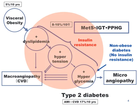
8-10%/10 Insulin resistanceabdominal obesity/metabolic syndrome have not been reached to a worldwide consunsus (7, 8, 74). The American Diabetes Association (ADA) and the European Association for the Study of Diabetes (EASD) as well as the International Chair on Car-diometabolic Risk have emphasized the critical im-portance of first using global risk calculators such as the Framingham risk score, the PROCAM algo-rithm or the European SCORE (74, 75). However, there is evidence to suggest that the risk assess-ment algorithms may not accurately estimate the global CVD risk in patients with visceral obesity/ metabolic syndrome. Thus, it is essential to de-velop a risk assessment model of global CVD in the presence of traditional risk factors and emerg-ing markers found in individuals with visceral obe-sity. Després et al. defined this model as global cardio-metabolic risk (76). Under this model, car-diometabolic risk is the global risk of CVD result-ing from the presence of traditional risk factors combined with the possible additional contribution of the metabolic syndrome. Namely, the metabolic syndrome cannot be used to assess global CVD risk but is at best one more modifiable CVD risk factor. To modify this model for clinical setting, we propose cardiometabolic risk cascade causing
cardiovasuclar disease (Fig. 4).
It is also claimed that the Framingham risk score does not properly assess lifetime risk particularly among young adults or adolescents with abdomi-nal obesity (77), which are not considered at a risk of CVD by current algorithms because of young age. Surrogate markers to detect abnormalities in ROS and renin-angiotensin-aldosterone system (RAAS), vascular failure (endothelial dysfunction), neurohumoral stress and arrhythmogenicity could have additional powers in long-term assessment of global CVD risk.
3.2. Visceral fat management
Obesity management goals should encompass re-duction in total cardiovascular morbidity and mor-tality. Losing 5% to 10% of body weight reduces the traditional CV risks (78). Increasing physical activ-ity, in combination with a diet that emphasizes fresh fruits and vegetables, whole grains, and low-fat dairy products, can help patients reduce weight and obe-sity comorbidity.
Less is known about the long-term effect of weight loss on the development of T2DM and CVD out-comes in the form of death, myocardial infarction, and stroke. A trial to answer the most important
Fig 4. Cardiometabolic risk cascade causing cardiovascular disease
question whether the improvements in cardiovas-cular risk factors by managed weight loss will be associated with reduction in long-term cardiovas-cular events was investigated (The Look AHEAD (Action for Health in Diabetes) trial) (79). The study enrolled 5,145 people with type 2 diabetes and a BMI greater than 25, randomizing half to a life-style intervention and half to a general program of diabetes support and education (80). Although those in the intervention group kept off 5% of their initial body weight at 4 years, there was no difference be-tween them and the standard care group in the rate of myocardial infarction, stroke, hospitalizations for angina, and cardiovascular death -- the primary out-come. Despite no reduction in cardiovascular events in those in the intense intervention arm, they did experience other health benefits. Patients in this group saw improvements in sleep apnea and mobil-ity, as well as quality of life. In addition, their dia-betes medications were reduced. In addition, at 1
and 4 years, both diabetes control (glucose, HbA1c) and most cardiovascular disease risk factors (blood pressure, HDL cholesterol, triglycerides) were more favorable in the lifestyle intervention than in the con-trol group with the exception of LDL cholesterol, which was not different between groups at year 1. At year 4, those in the intensive lifestyle interven-tion group continued to have more favorable dia-betes control and CVD risk factor reduction, with the exception of LDL-C in which there were slightly greater reductions in the standard care group. Par-ticipants in the lifestyle intervention group main-tained greater improvements in fitness at both years 1 and 4.
According to AHA/ACCF Secondary Prevention and Risk Reduction Therapy for Patients With Coro-nary and Other Atherosclerotic Vascular Disease : 2011 Update : Intervention Recommendations With Class of Recommendation and Level of Evidence (Fig. 5) (81), weight management is recognized as
Fig 5. Body weight management : AHA/ACCF Secondary Prevention and Risk Reduction Therapy for Patients With Coronary and Other Atherosclerotic Vascular Disease : 2011 Update : Intervention Recommendations With Class of Recommendation and Level of Evidence
Class I (Procedures/Treatment should be per-formed/administered), but estimated of certainty of treatment effect is named as Level B or Levels C. When indicated, exercise, caloric intake, and formal behavioral programs are recommended to main-tain/achieve a body mass index between 18.5 and 24.9 kg/m2 (Level of Evidence : B), or to achieve waist circumference (!89 cm) in women and (!102 cm) in men or to reduce body weight by approxi-mately 5% to 10% from baseline.
CONCLUSION
The concept of the metabolic syndrome takes into account the central role that visceral fat plays essential role in the development of metabolic and cardiovascular diseases, and indicates how waist circumference measurement is useful in aiding pa-tient identification in a clinical setting. The meta-bolic syndrome cannot be used to assess global CVD risk but is at best one more modifiable CVD risk factor. Thus, global cardiometabolic risk should be considered individually. Increased visceral fat is associated with a shift in the normal balance of the adipocytokines resulting in pro-inflammatory and pro-atherosclerotic state. Although evidence for therapeutic efficacy in the treatment of abdominal adiposity and clustering of cardiometabolic risks are limited, this should be a promising challenge to reduce the highly contagious state around the world.
ACKNOWLEDGEMENTS
This work was supported by Grants-in-Aid for Scientific Research from the Japanese Society for the Promotion of Science, the Ministry of Education, Culture, Sports, Science and the Technology and Ministry of Health, Labour and Welfare. Authors sincerely thanks to every collaborators especially to Dr Roger H. Unger, Chris B. Newgard, and J Dennis McGarry for superb mentorship ; Drs Yoichiro Hirata, Minoru Tabata, Hirotsugu Kurobe, Hirofumi Izaki, Hiroomi Kanayama, and Shuichiro Takanashi for crucial collaborations ; Mss Hiromi Sato, Etsuko Uematsu, Junko Yata, Horoko Goda for secretary dedication.
REFERENCE
1. Olshansky SJ, Passaro DJ, Hershow RC, Layden J, Carnes BA : A potential decline in life expec-tancy in the United States in the 21st century. N Engl J Med 352 : 1138-1145, 2005
2. Hossain P, Kawar B, E1 Nahas M : Obesity and diabetes in the developing world-a growing challenge. N Engl J Med 356 : 213-215, 2007 3. Matsuzawa Y, Funahashi T, Nakamura T :
Mo-lecular mechanism of metabolic syndrome X : Contribution of adipocytokines adipocyte-de-rived bioactive substances. Ann NY Acad Sci 18 : 146-154, 1999
4. Montague CT, O’Rahilly S : The perils of port-liness : Causes and consequences of visceral adiposity. Diabetes 49 : 883-888, 2000
5. Eckel RH, Grundy SM, Zimmet PZ : The meta-bolic syndrome. Lancet 365 : 1415-1428, 2005 6. Shimabukuro M : Cardiac adiposity and global
cardiometabolic risk : new concept and clini-cal implication. Circ J 73 : 27-34, 2009
7. Després JP : Body fat distribution and risk of cardiovascular disease : an update. Circulation 126 : 1301-1313, 2012
8. Cardiometabolic Risk Working Group : Execu-tive Committee, Leiter LA, Fitchett DH, Gilbert RE, Gupta M, Mancini GB, McFarlane PA, Ross R, Teoh H, Verma S, Anand S, Camelon K, Chow CM, Cox JL, Després JP, Genest J, Harris SB, Lau DC, Lewanczuk R, Liu PP, Lonn EM, McPherson R, Poirier P, Qaadri S, Rabasa-Lhoret R, Rabkin SW, Sharma AM, Steele AW, Stone JA, Tardif JC, Tobe S, Ur E : Cardiometabolic risk in Canada : a detailed analysis and position paper by the cardiome-tabolic risk working group. Can J Cardiol 27 : e1-e33, 2011
9. Cherian S, Lopaschuk GD, Carvalho E : Cellu-lar cross-talk between epicardial adipose tissue and myocardium in relation to the pathogenesis of cardiovascular disease. Am J Physiol Endo-crinol Metab 303 : E937-949, 2012
10. Iacobellis G, Bianco AC : Epicardial adipose tis-sue : emerging physiological, pathophysiologi-cal and clinipathophysiologi-cal features. Trends Endocrinol Metab 22 : 450-457, 2011
11. Sacks HS, Fain JN : Human epicardial fat : what is new and what is missing? Clin Exp Pharma-col Physiol 38 : 879-887, 2011
12. Utzschneider KM, Kahn SE : Review : The role of insulin resistance in nonalcoholic fatty liver
disease. J Clin Endocrinol Metab 91 : 4753-4761, 2006
13. Thamer C, Machann J, Stefan N, Haap M, Schafer S, Brenner S, Kantartzis K, Claussen C, Schick F, Haring H, Fritsche A : High vis-ceral fat mass and high liver fat are associated with resistance to lifestyle intervention. Obe-sity (Silver Spring) 15 : 531-538, 2007
14. Thamer C, Machann J, Haap M, Stefan N, Heller E, Schnödt B, Stumvoll M, Claussen C, Fritsche A, Schick F, Haring H : Intrahepatic lipids are predicted by visceral adipose tissue mass in healthy subjects. Diabetes Care 27 : 2726-2729, 2004
15. Krssak M, Falk Petersen K, Dresner A, DiPietro L, Vogel SM, Rothman DL, Roden M, Shulman GI : Intramyocellular lipid concentra-tions are correlated with insulin sensitivity in humans : a 1H NMR spectroscopy study. Dia-betologia 42 : 113-116, 1999
16. Kelley DE, Goodpaster BH, Storlien L : Muscle triglyceride and insulin resistance. Annu Rev Nutr 22 : 325-346, 2002
17. Taira S, Shimabukuro M, Higa M, Yabiku K, Kozuka C, Ueda R, Sunagawa S, Kawamoto E, Nakayama Y, Nakamura H, Iha T, Tomoyose T, Ikema T, Yamakawa K, Masuzaki M : Lipid deposition in various sites of skeletal muscle and liver shows correlation with visceral fat accumulation in male patients with metabolic syndrome. Intern Med. in press
18. Yusuf S, Hawken S, Ounpuu S, Dans T, Avezum A, Lanas F, et al. : Effect of potentially modifi-able risk factors associated with myocardial infarction in 52 countries (the INTERHEART study) : case-control study. Lancet 364 : 937-952, 2004
19. Targher G, Bertolini L, Poli F, Rodella S, Scala L, Tessari R, Zenari L, Falezza G : Nonalcoholic fatty liver disease and risk of future cardiovas-cular events among type 2 diabetic patients. Diabetes 54 : 3541-3546, 2005
20. Rosito GA, Massaro JM, Hoffmann U, Ruberg FL, Mahabadi AA, Vasan RS, O’Donnell CJ, Fox CS : Pericardial fat, visceral abdominal fat, cardiovascular disease risk factors, and vascular calcification in a community-based sample : the Framingham Heart Study. Circulation 117 : 605-613, 2008
21. Ding J, Hsu FC, Harris TB, Liu Y, Kritchevsky SB, Szklo M, Ouyang P, Espeland MA, Lohman KK, Criqui MH, Allison M, Bluemke
DA, Carr JJ : The association of pericardial fat with incident coronary heart disease. The Multi-Ethnic Study of Atherosclerosis (MESA). Am J Clin Nutr 90 : 499-504, 2009
22. Hirata Y, Tabata M, Kurobe H, Motoki T, Akaike M, Nishio C, Higashida M, Mikasa H, Nakaya Y, Takanashi S, Igarashi T, Kitagawa T, Sata M : Coronary atherosclerosis is associ-ated with macrophage polarization in epicar-dial adipose tissue. J Am Coll Cardiol 58 : 248-255, 2011
23. Kremen J, Dolinkova M, Krajickova J, Blaha J, Anderlova K, Lacinova Z, et al. : Increased subcutaneous and epicardial adipose tissue pro-duction of proinflammatory cytokines in cardiac surgery patients : possible role in postoperative insulin resistance. J Clin Endocrinol Metab 91 : 4620-4627, 2006
24. Kotani K, Tokunaga K, Fujioka S, et al. : Sexual dimorphism of age-related changes in whole-body fat distribution in the obese. Int J Obes Relat Metab Disord 18 : 202-207, 1994
25. Camhi SM, Bray GA, Bouchard C, et al. : The relationship of waist circumference and BMI to visceral, subcutaneous, and total body fat : sex and race differences. Obesity (Silver Spring) 19 : 402-408, 2011
26. Dagvasumberel M, Shimabukuro M, Nishiuchi T, Ueno J, Takao S, Hirata Y, Kurobe H, Soeki T, Iwase T, Kusunose K, Niki T, Tamaguchi K, Taketani Y, Yagi S, Tomita N, Yamada H, Wakatsuki T, Harada M, Kitagawa T, Sata M : Gender disparities in the association between epicardial adipose tissue volume and coronary atherosclerosis : A 3-dimensional cardiac com-puted tomography imaging study in Japanese subjects. Cardiovasc Diabetol 11 : 106, 2012 27. de las Fuentes L, Brown AL, Mathews SJ,
Waggoner AD, Soto PF, Gropler RJ, et al. : Metabolic syndrome is associated with abnor-mal left ventricular diastolic function independ-ent of left vindepend-entricular mass. Eur Heart J 28 : 553-559, 2007
28. Redfield MM, Jacobsen SJ, Burnett JC Jr, Mahoney DW, Bailey KR, Rodeheffer RJ : Bur-den of systolic and diastolic ventricular dysfunc-tion in the community : appreciating the scope of the heart failure epidemic. JAMA 289 : 194-202, 2003
29. Ingelsson E, Arnlov J, Lind L, Sundstrom J : Metabolic syndrome and risk for heart failure in middle-aged men. Heart 92 : 1409-1413, 2006
30. Szczepaniak LS, Dobbins RL, Metzger GJ, Sartoni-D’Ambrosia G, Arbique D, Vongpatanasin W, et al. : Myocardial triglycerides and systolic function in humans : in vivo evaluation by lo-calized proton spectroscopy and cardiac imag-ing. Magn Reson Med 49 : 417-423, 2003 31. Reingold JS, McGavock JM, Kaka S, Tillery T,
Victor RG, Szczepaniak LS, et al. : Determina-tion of triglyceride in the human myocardium by magnetic resonance spectroscopy : repro-ducibility and sensitivity of the method. Am J Physiol Endocrinol Metab 289 : E935-939, 2005 32. Kankaanpaa M, Lehto HR, Parkka JP, Komu M, Viljanen A, Ferrannini E, et al. : Myocardial triglyceride content and epicardial fat mass in human obesity : relationship to left ventricu-lar function and serum free fatty acid levels. J Clin Endocrinol Metab 91 : 4689-4695, 2006 33. Ohira T, Shahar E, Chambless LE, Rosamond
WD, Mosley TH Jr, Folsom AR : Risk factors for ischemic stroke subtypes : the Atheroscle-rosis Risk in Communities study. Stroke 37 : 2493-2498, 2006
34. Ninomiya T, Kubo M, Doi Y, Yonemoto K, Tanizaki Y, Rahman M, et al. : Impact of meta-bolic syndrome on the development of cardio-vascular disease in a general Japanese popula-tion : the Hisayama study. Stroke 38 : 2063-2069, 2007
35. Empana JP, Duciemetiere P, Balkau B, Jouven X : Contribution of the metabolic syndrome to sudden death risk in asymptomatic men : the Paris Prospective Study I. Eur Heart J 28 : 1149-1154, 2007
36. Pietrasik G, Goldenberg I, McNitt S, Moss AJ, Zareba W : Obesity as a risk factor for sustained ventricular tachyarrhythmias in MADIT II pa-tients. J Cardiovasc Electrophysiol 18 : 181-184, 2007
37. Soydinc S, Davutoglu V, Akcay M : Uncompli-cated metabolic syndrome is associated with prolonged electrocardiographic QTc interval and QTc dispersion. Ann Noninvasive Electro-cardiol 11 : 313-317, 2006
38. Umetani K, Kodama Y, Nakamura T, Mende A, Kitta Y, Kawabata K, et al. : High prevalence of paroxysmal atrial fibrillation and/or atrial flut-ter in metabolic syndrome. Circ J 71 : 252-255, 2007
39. Watanabe H, Tanabe N, Watanabe T, Darbar D, Roden DM, Sasaki S, et al. : Metabolic syndrome and risk of development of atrial
fibrillation : the Niigata preventive medicine study. Circulation 117 : 1255-1260, 2008 40. Echahidi N, Mohty D, Pibarot P, Després JP,
O’Hara G, Champagne J, et al. : Obesity and metabolic syndrome are independent risk fac-tors for atrial fibrillation after coronary artery bypass graft surgery. Circulation 116(Suppl) : I213-I219, 2007
41. Al Chekakie MO, Welles CC, Metoyer R, Ibrahim A, Shapira AR, Cytron J, et al. : Peri-cardial fat is independently associated with human atrial fibrillation. J Am Coll Cardiol 56 : 784-788, 2010
42. Thanassoulis G, Massaro JM, O’Donnell CJ, Hoffmann U, Levy D, Ellinor PT, et al. : Peri-cardial fat is associated with prevalent atrial fibrillation : The Framingham Heart Study. Circ Arrhythm Electrophysiol 3 : 345-350, 2010 43. Nakanishi K, Fukuda S, Tanaka A, Otsuka K,
Sakamoto M, Taguchi H, et al. : Peri-atrial epicardial adipose tissue is associated with newonset nonvalvular atrial fibrillation. Circ J 76 : 2748-2754, 2012
44. Bainton D, Sweetnam P, Baker I, Elwood P : Peripheral vascular disease : consequence for survival and association with risk factors in the Speedwell prospective heart study. Br Heart J 72 : 128-132, 1994
45. Smith GD, Shipley MJ, Rose G : Intermittent claudication, heart disease risk factors and mor-tality : the Whitehall Study. Circulation 82 : 1925-1931, 1990
46. Planas A, Clara A, Pou JM, Vidal-Barraquer F, Gasol A, De Moner A, et al. : Relationship of obesity distribution and peripheral arterial oc-clusive disease in elderly men. Int J Obes Relat Metab Disord 25 : 1068-1070, 2001
47. Hansson PO, Eriksson H, Welin L, Svardsudd K, Wilhielmsen L : Smoking and abdominal obe-sity : risk factors for venous thromboembolism among middle-aged men : “the study of men born in 1913.” Arch Intern Med 159 : 1886-1890, 1999
48. Leng GC, Lee AJ, Fowkes FG, Lowe GD, Housley E : The relationship between cigarette smoking and cardiovascular risk factors in pe-ripheral arterial disease compared with ischae-mic heart disease. The Edinburgh Artery Study. Eur Heart J 16 : 1542-1548, 1995
49. Samama MM : An epidemiologic study of risk factors for deep vein thrombosis in medical outpatients : the Sirius study. Arch Intern Med
160 : 3415-3420, 2000
50. Darvall KA, Sam RC, Silverman SH, Bradbury AW, Adam DJ : Obesity and thrombosis. Eur J Vasc Endovasc Surg 33 : 223-233, 2007 51. Kadowaki T, Yamauchi T, Kubota N, Hara K,
Ueki K, Tobe K : Adiponectin and adiponectin receptors in insulin resistance, diabetes, and the metabolic syndrome. J Clin Invest 116 : 1784-1792, 2006
52. Ross R : Atherosclerosisdan inflammatory dis-ease. N Engl J Med 340 : 115-126, 1999 53. Hansson GK : Inflammation, atherosclerosis,
and coronary artery disease. N Engl J Med 352 : 1685-1695, 2005
54. Harrison DG : Cellular and molecular mecha-nisms of endothelial cell dysfunction. J Clin Invest 100 : 2153-2157, 1997
55. Inoue T, Node K : Vascular failure : A new clini-cal entity for vascular disease. J Hypertens 24 : 2121-2130, 2006
56. Seddon M, Looi YH, Shah AM : Oxidative stress and redox signalling in cardiac hypertrophy and heart failure. Heart 93 : 903-907, 2007 57. Ingelsson E, Arnlov J, Sundstrom J, Zethelius
B, Vessby B, Lind L : Novel metabolic risk fac-tors for heart failure. J Am Coll Cardiol 46 : 2054-2060, 2005
58. Furukawa S, Fujita T, Shimabukuro M, Iwaki M, Yamada Y, Nakajima Y, et al. : Increased oxidative stress in obesity and its impact on metabolic syndrome. J Clin Invest 114 : 1752-1761, 2004
59. Fujita K, Nishizawa H, Funahashi T, Shimomura I, Shimabukuro M : Systemic oxidative stress. A potential biomaker for visceral fat obesity and metabolic syndrome. Circ J 70 : 1437-1442, 2006
60. Unger RH : Lipotoxic diseases. Annu Rev Med 53 : 319-336, 2002
61. Shimabukuro M, Ohneda M, Lee Y, Unger RH : Role of nitric oxide in obesity-induced β-cell disease. J Clin Invest 100 : 290-295, 1997 62. Shimabukuro M, Koyama K, Chen G, Wang
MY, Trieu F, Lee Y, et al. : Direct antidiabetic effect of leptin through triglyceride depletion of tissues. Proc Natl Acad Sci USA 94 : 4637-4641, 1997
63. Shimabukuro M, Zhou Y-T, Levi M, Unger RH : Fatty acid-inducedβ-cell apoptosis : A link between obesity and diabetes. Proc Natl Acad Sci USA 95 : 2498-2502, 1998
64. Shimabukuro M, Meng F, Zhou YT, Grayburn
PA, Asad K : Increased extracelluar matrix re-modeling during the development of a specific diabetic cardiomyopathy in the Zucker Fatty Rats. Circulation 98(Supple) : 180, 1998 65. Zhou YT, Grayburn P, Karim A, Shimabukuro
M, Higa M, Baetens D, et al. : Lipotoxic heart disease in obese rats : implications for human obesity. Proc Natl Acad Sci USA 97 : 1784-1789, 2000
66. Chinen I, Shimabukuro M, Yamakawa K, Higa N, Matsuzaki T, Noguchi K, et al. : Vascular lipotoxicity : endothelial dysfunction via fatty acid-induced reactive oxygen species over-production in obese Zucker diabetic fatty rats. Endocrinology 148 : 160-165, 2007
67. Yudkin JS, Eringa E, Stehouwer CD : “Vasocrine” signalling from perivascular fat : a mechanism linking insulin resistance to vas-cular disease. Lancet 365 : 1817-1820, 2005 68. Henrichot E, Juge-Aubry CE, Pernin A, Pache
JC, Velebit V, Dayer JM, et al. : Production of chemokines by perivascular adipose tissue : a role in the pathogenesis of atherosclerosis? Arterioscler Thromb Vasc Biol 25 : 2594-2599, 2005
69. Szczepaniak LS, Victor RG, Orci L, Unger RH : Forgotten but not gone : the rediscovery of fatty heart, the most common unrecognized disease in America. Circ Res 101 : 759-767, 2007 70. Takaoka M, Suzuki H, Shioda S, Sekikawa K,
Saito Y, Nagai R, Sata M : Endovascular injury induces rapid phenotypic changes in perivas-cular adipose tissue. Arterioscler Thromb Vasc Biol 30 : 1576-82, 2010
71. Brandes RP, Kreuzer J : Vascular NADPH oxi-dases : molecular mechanisms of activation. Cardiovasc Res 65 : 16-27, 2005
72. Mueller CF, Laude K, McNally JS, Harrison DG : Redox mechanisms in blood vessels. Ar-terioscler Thromb Vasc Biol 25 : 274-278, 2005 73. Du X, Edelstein D, Obici S, Higham N, Zou MH, Brownlee M : Insulin resistance reduces arterial prostacyclin synthase and eNOS activi-ties by increasing endothelial fatty acid oxida-tion. J Clin Invest 116 : 1071-1080, 2006 74. Kahn R, Buse J, Ferrannini E, Stern M : The
metabolic syndrome : time for a critical ap-praisal : joint statement from the American Dia-betes Association and the European Associa-tion for the Study of Diabetes. Diabetes Care 28 : 2289-2304, 2005
Menotti A, De Backer G, et al. : Estimation of ten-year risk of fatal cardiovascular disease in Europe : the SCORE project. Eur Heart J 24 : 987-1003, 2003
76. Després JP, Lemieux I, Bergeron J, Pibarot P, Mathieu P, Larose E, et al. : Abdominal obe-sity and the metabolic syndrome : contribution to global cardiometabolic risk. Arterioscler Thromb Vasc Biol 28 : 1039-1049, 2008 77. Grundy SM : Metabolic syndrome : a multiplex
cardiovascular risk factor. J Clin Endocrinol Metab 92 : 399-404, 2007
78. Selwyn AP, Weight reduction and cardiovascu-lar and metabolic disease prevention : Clinical trial update. Am J Cardiol 100(suppl) : 33-37, 2007
79. Look AHEAD Research Group : Reduction in weight and cardiovascular disease risk factors in individuals with type 2 diabetes : one-year results of the Look AHEAD trial. Diabetes Care 30 : 1374-1383, 2007
80. Wing RR, Lang W, Wadden TA, Safford M, Knowler WC, Bertoni AG, Hill JO, Brancati FL, Peters A, Wagenknecht L ; Look AHEAD Research Group : Benefits of modest weight loss in improving cardiovascular risk factors in overweight and obese individuals with type 2 diabetes. Diabetes Care 34 : 1481-1486, 2011 81. Smith SC Jr, Benjamin EJ, Bonow RO, Braun LT, Creager MA, Franklin BA, Gibbons RJ, Grundy SM, Hiratzka LF, Jones DW, Lloyd-Jones DM, Minissian M, Mosca L, Peterson ED, Sacco RL, Spertus J, Stein JH, Taubert KA ; World Heart Federation and the Preven-tive Cardiovascular Nurses Association. AHA/ ACCF Secondary Prevention and Risk Reduc-tion Therapy for Patients with Coronary and other Atherosclerotic Vascular Disease : 2011 update : a guideline from the American Heart Association and American College of Cardi-ology Foundation. Circulation 124 : 2458-2473, 2011