http://internmed.jp
【 ORIGINAL ARTICLE 】
Clinical Characteristics and Risk Factors for Rebleeding in
Patients with Obscure Gastrointestinal Bleeding
Yuki Baba, Seiji Kawano, Yoshiyasu Kono, Toshihiro Inokuchi, Hiromitsu Kanzaki,
Masaya Iwamuro, Keita Harada, Sakiko Hiraoka, Yoshiro Kawahara and Hiroyuki Okada
Abstract:
Objective With the advent of capsule endoscopy (CE) and double-balloon endoscopy (DBE), the diagnosis and treatment of obscure gastrointestinal bleeding (OGIB) have markedly progressed. However, rebleeding sometimes occurs and is difficult to diagnose and treat. The aim of the present study was to investigate the clinical features of OGIB and risk factors for rebleeding in our hospital.
Methods A total of 195 patients who underwent CE and/or DBE for OGIB in our hospital from January
2009 to July 2016 were included in the present study. We analyzed 168 cases of small intestinal OGIB, after excluding 27 cases of extra small intestinal bleeding. The clinical characteristics and risk factors related to rebleeding were retrospectively studied.
Results Among the 168 patients who were included in the analysis, 95 patients (56.5%) were male. The mean age was 64.5 years (range, 8 to 87 years). Hypertension (31.0%) was the most frequent comorbidity, followed by chronic kidney disease (19.0%). The final diagnoses were ulcerative lesions (n=50, 29.8%), vas-cular lesions (n=30, 17.9%), tumors (n=7, 4.2%), and diverticula (n=2, 1.2%). The bleeding source was unde-termined in the remaining 79 cases (47.0%). Rebleeding was confirmed in 29 cases (17.3%). In a univariate analysis, chronic kidney disease, vascular lesions, and overt previous bleeding were significantly associated with the risk of rebleeding. A multivariate analysis showed that chronic kidney disease, vascular lesion, and overt previous bleeding were significantly associated with the risk of rebleeding.
Conclusion Patients with OGIB with overt previous bleeding, vascular lesions, and/or chronic kidney dis-ease had a higher risk of rebleeding.
Key words:obscure gastrointestinal bleeding, overt previous bleeding, vascular lesions, chronic kidney disease
(Intern Med 59: 1345-1350, 2020) (DOI: 10.2169/internalmedicine.3628-19)
Introduction
Obscure gastrointestinal bleeding (OGIB) is a form of gastrointestinal bleeding whose source is unknown, despite the performance of upper and lower gastrointestinal endo-scopy. It accounts for 5% of all cases of gastrointestinal bleeding (1). Many of these cases are caused by small intes-tinal lesions (2). OGIB is classified into two subtypes in the American Journal of Gastroenterology Clinical Guideline: i) overt bleeding, manifesting as melena or hematochezia; and
ii) occult bleeding, presenting as a positive fecal occult blood test with or without iron-deficiency anemia (3). Overt bleeding is further divided into two groups: i) overt ongoing bleeding (continuous bleeding); and ii) overt previous bleed-ing (previous bleedbleed-ing). With the development of capsule endoscopy ( CE ) (4) and double-balloon endoscopy (DBE) (5), the diagnosis and treatment of OGIB have mark-edly progressed. However, rebleeding sometimes occurs, and most rebleeding cases are difficult to diagnose and treat. Thus, it is important to identify the characteristics of cases in which rebleeding occurs. The aim of the present study
Department of Gastroenterology and Hepatology, Okayama University Graduate School of Medicine, Dentistry and Pharmaceutical Sciences, Ja-pan
Received: July 7, 2019; Accepted: December 10, 2019; Advance Publication by J-STAGE: February 5, 2020 Correspondence to Dr. Seiji Kawano, skawano@mpd.biglobe.ne.jp
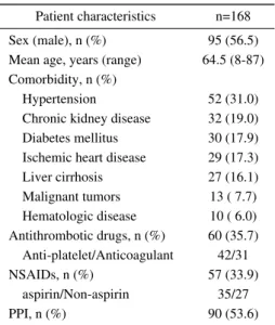
Table 1. Patient Characteristics. Patient characteristics n=168 Sex (male), n (%) 95 (56.5) Mean age, years (range) 64.5 (8-87) Comorbidity, n (%)
Hypertension 52 (31.0)
Chronic kidney disease 32 (19.0) Diabetes mellitus 30 (17.9) Ischemic heart disease 29 (17.3) Liver cirrhosis 27 (16.1) Malignant tumors 13 ( 7.7) Hematologic disease 10 ( 6.0) Antithrombotic drugs, n (%) 60 (35.7) Anti-platelet/Anticoagulant 42/31 NSAIDs, n (%) 57 (33.9) aspirin/Non-aspirin 35/27 PPI, n (%) 90 (53.6)
NSAIDs: nonsteroidal anti-inflammatory drugs, PPI: proton-pump inhibitor
Table 2. The Final Diagnosis of OGIB Cases. Positive group (n=104) Negative group (n=64) Total (n=168) Classified (n=89) Vascular lesions 28 2 30 Ulcerative lesions 49 1 50 Tumors 7 0 7 Diverticulum 2 0 2 Unclassified (n=79) 18 61 79
OGIB: obscure gastrointestinal bleeding
Table 3. The Final Diagnosis of Rebleeding Cases. Positive group (n=21) Negative group (n=8) Total (n=29) Vascular lesions 12 2 14 Ulcerative lesions 8 1 9 Tumors 0 0 0 Diverticula 0 0 0 Unclassified 1 5 6
was to investigate the clinical features of OGIB and risk fac-tors for rebleeding.
Materials and Methods
Study design and patientsThis was a single-center, retrospective study. Among 195 patients who underwent CE and/or DBE for OGIB in our hospital from January 2009 to July 2016, 27 patients in whom the source of bleeding was proven to be outside the small intestine were excluded. We investigated the clinical characteristics of the remaining 168 patients. Among these, 148 underwent CE alone, 8 underwent DBE alone, and 39 underwent both CE and DBE. Total enteroscopy was per-formed in 153 cases. CE was perper-formed with a video cap-sule endoscopy device (PillCam SB2, or SB3; Given Imag-ing, Yokneam, Israel). The images were analyzed with the RAPID Reader 6 software program (Given Imaging) on a RAPID 5 or 6.5 workstation (Given Imaging). DBE was performed with a DBE system (FUJIFILM, Saitama, Japan) employed with a FUJIFILM 450T5, 530T, or EN-580T endoscope. The clinical characteristics and factors re-lated to rebleeding were retrospectively studied. This study was conducted in accordance with Declaration of Helsinki
and was approved by the Okayama University Hospital Eth-ics Committee in June 2018.
Variables
Medical records were retrospectively reviewed and evalu-ated for sex, age, comorbidities, oral medicine, clinical diag-nosis, clinical course of the patient and outcome, and pres-ence of rebleeding. Regarding deceased patients, the obser-vation period was defined as the period until the day of death. Therapeutic intervention was defined as any of the various types of therapeutic intervention that were employed [e.g., endoscopic therapy, interventional radiology (IVR), medication, change of orally administered drugs, and sur-gery].
Types of OGIB bleeding and definition of rebleeding
As mentioned previously, OGIB was divided into three subtypes: overt ongoing bleeding, overt previous bleeding, and occult bleeding. In this study, rebleeding was defined as repeated visible bleeding or progressive anemia requiring re-peated endoscopic examination. When the source of rebleed-ing was not confirmed in the small intestine, upper and lower gastrointestinal endoscopy were repeated in all cases.
Definition of the endoscopic findings and diagnoses
The endoscopic findings were classified as follows: no abnormality, ulcerative lesions (i.e., erosion or ulcer), vascu-lar lesions (i.e., angiectasia, Dieulafoy lesions, or arte-riovenous malformation), tumors, diverticula and others. The final diagnosis was made based on the endoscopic findings at not only the first bleeding episode but also rebleeding episodes. The endoscopic diagnosis was performed and dis-cussed by two expert endoscopists (S.K and Y.B). Only defi-nite findings of bleeding were regarded as positive findings.
Comorbidities and medications
Hypertension, chronic kidney disease (CKD), diabetes mellitus, ischemic heart disease, liver cirrhosis, malignant tumors, and hematologic disease were investigated as comorbidities. In this study, CKD was defined by a cre-atinine level that exceeded the standard laboratory value in our hospital. CKD was further divided into two groups as severe CKD (eGFR<30 mL/min/1.73 m2) or moderate CKD
(eGFR 30 to <60 mL/min/1.73 m2
Table 4. Rebleeding Rates after Therapeutic Intervention.
OGIB cases Vascular lesions
Total, n Rebleeding, n
(% incidence ratio) Total, n
Rebleeding, n (% incidence ratio) Therapeutic intervention 52 11 (21.1) 13 7 (53.8) Hemostasic treatment 24 6 (25.0) 10 5 (50.0) Endoscopic therapy 15 6 (40.0) 9 5 (55.6) Surgery 8 0 0 0 IVR 1 0 1 0
Oral administration change/addition 28 5 (17.9) 3 2 (66.7)
Addition of other drugs 13 2 (15.4) 2 1 (50.0)
NSAIDs change 10 1 (10.0) 0 0
Antithrombotic drug change 5 2 (40.0) 1 1 (100)
IVR: interventional radiology, NSAIDs: nonsteroidal anti-inflammatory drugs, OGIB: obscure gastrointestinal bleeding
Table 5. Clinical Risk Factors for Rebleeding. No rebleeding n=139 Rebleeding n=29 p value Sex (male) 80 15 0.57 Over 70 years 59 13 0.81 Liver cirrhosis 20 7 0.21
Chronic kidney disease 21 11 <0.01
Hemodialysis 6 3 0.23
Antithrombotic drugs 49 11 0.79
NSAIDs 24 3 0.33
Vascular lesions 16 14 <0.01
Overt previous bleeding 65 22 <0.01
Therapeutic intervention 41 11 0.38
NSAIDs: nonsteroidal anti-inflammatory drugs
were classified into antiplatelet drugs (e.g., aspirin and thienopyridine) and anticoagulants (e.g., warfarin and direct oral anticoagulants). Nonsteroidal anti-inflammatory drugs (NSAIDs), including aspirin, cyclooxygenase (COX) inhibi-tors (e.g., loxoprofen and diclofenac), and selective COX2 inhibitors (e.g., celecoxib, etodolac, and meloxicam). Proton-pump inhibitors (PPIs) included omeprazole, esome-prazole, lansoesome-prazole, and rabeprazole. The potassium-competitive acid blocker (P-CAB) vonoprazan was not in-cluded because it was not administered to any of the pa-tients.
Statistical analysis
Sequential data were expressed as the mean and median. Categorical data were compared using the chi-squared test or Fisher’s exact test. Variables with p values of <0.05 in the univariate analysis were included in the multivariate logistic regression models. All statistical analyses were performed using the JMP Pro for Windows software program (version 13). P values of <0.05 were considered to indicate statistical significance.
Results
Patient characteristicsTable 1 shows the clinical characteristics of patients in this study. Among 168 patients, 95 (56.5%) were male, and the mean age was 64.5 years (range, 8-87 years). One-hundred fourteen patients (67.9%) had one or more comor-bidities: hypertension was the most common (31.0%), fol-lowed by CKD (19.0%), diabetes mellitus (17.9%), ischemic heart disease (17.3%), and liver cirrhosis (16.1%). Sixty pa-tients (35.7%) were taking oral antithrombotic drugs. Forty-two patients were using antiplatelet drugs and 31 were using anticoagulant drugs (including combined use). Fifty-seven patients (33.9%) were using NSAIDs, including aspirin (as-pirin, n=35; NSAIDs other than as(as-pirin, n=27). Ninety pa-tients (53.6%) were using PPIs. The median period of vation was 31.5 months. 20 patients died during the obser-vation period. The primary cause of death was progression of comorbidities (n=15). The other causes included sepsis, multiple organ failure, and unknown origin.
Endoscopic findings at the first procedure and final diagnosis
We investigated the endoscopic findings of 168 patients at the first bleeding episode. Endoscopic findings were positive in 104 patients (positive group), while no abnormalities were detected in 64 patients (negative group). The 104 pa-tients in the positive group had ulcerative lesions (erosion and/or ulcer, n=49), vascular lesions (angiectasia, Dieulafoy lesions, and/or arteriovenous malformation, n=28), tumors (n =7), diverticulum (n=2), and unclassified lesions (n=18). In contrast, among the 64 patients in the negative group, vascu-lar lesions (n=2) and ulcerative lesions (n=1) were detected at the second or subsequent bleeding event. Thus, the final diagnoses of 89 patients were as follows: ulcerative lesions (n=50), vascular lesions (n=30), tumors (n=7), and diverticu-lum (n=2) (Table 2). Seventy-nine patients were unclassified or impossible to diagnose.
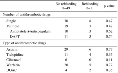
Table 6. Antithrombotic Drugs Administered to Patients with Re-bleeding. No rebleeding n=49 Rebleeding n=11 p value Number of antithrombotic drugs
Single 30 8 0.47
Multiple 19 3 0.47
Antiplatelet+Anticoagulant 10 3 0.62
DAPT 11 3 0.74
Type of antithrombotic drugs
Aspirin 29 6 0.77
Ticlopidine 11 4 0.35
Cilostazol 6 0 0.11
Warfarin 20 5 0.77
DOAC 4 2 0.35
DAPT: dual antiplatelet therapy, DOAC: direct oral anticoagulants
Table 7. The Multivariate Analysis of Risk Factors for Rebleed-ing.
Odds ratio Confidence interval p value Chronic kidney disease 2.83 (1.06-7.58) 0.04 Vascular lesions 6.45 (2.48-16.72) <0.01 Overt previous bleeding 3.68 (1.37-9.92) 0.01
Details of rebleeding cases
Rebleeding was confirmed in 29 patients (17.3%). In the positive group, rebleeding was observed in 21 patients (20.2%), with vascular lesion (n=12) being the most fre-quent diagnosis in cases of rebleeding. There was only one case in which a final diagnosis could not be obtained. In contrast, among the 64 patients in the negative group, rebleeding occurred in 8 patients (12.5%). In three cases, vascular lesions (n=2) and ulcerative lesions (n=1) were identified at the second or subsequent bleeding episode. However, a final diagnosis could not be obtained in 5 cases (62.5%). In the total population of 168 patients, rebleeding occurred in 29 patients (17.3%): vascular lesions (n=14), ul-cerative lesions (n=9), and unclassified lesions (n=6). In this study, vascular lesions were the most frequent cause of rebleeding (Table 3). The median observation period from the first bleeding episode to rebleeding was 4 months.
Detail of rebleeding cases after therapeutic interven-tion
Among 168 patients with OGIB, 52 underwent therapeu-tic intervention such as endoscopic treatment, surgery, and oral administration change at the first event. Rebleeding oc-curred in 11 patients (21.1%). Vascular lesions were the most frequent cause of rebleeding (7 patients). In the 11 pa-tients, 5 patients (55.6%) occurred rebleeding after endo-scopic treatment. On the other hand, no rebleeding occurred after surgery or IVR (Table 4). In the patients who under-went a therapeutic intervention, rebleeding rate was higher
in those with overt previous bleeding (n=9, 81.8%) than in those with occult bleeding (n=2, 18.2%).
Risk factors for rebleeding
We investigated risk factors for rebleeding. In the univari-ate analysis, CKD, vascular lesions, and overt previous bleeding were significantly associated with higher risk of rebleeding (Table 5). Intake of any antiplatelet, anticoagu-lant, or combination use was not a risk factor of rebleeding (Table 6). In the multivariate analysis, CKD, vascular le-sions, and overt previous bleeding were independent risk factors of rebleeding (Table 7).
With respect to the relationship between CKD and rebleeding, the rebleeding rate of patients with normal kid-ney function was 13.2%. In contrast, the rebleeding rate in moderate CKD was 33.3%, and that in severe CKD was 35.2% (Figure). The rebleeding rate in overt bleeding and occult bleeding cases were 22.5% (23 of 102 patients) and 9.1% (6 of 66 patients), respectively. The rebleeding rate in overt bleeding was significantly higher than that in occult bleeding (p=0.02). Additionally, the rebleeding rate in overt previous bleeding and others were 25.6% (22 of 86 patients) and 8.6% (7 of 81 patients), respectively (p<0.01). The rate of overt previous bleeding was significantly higher than that in other bleeding types.
Discussion
We investigated clinical features of patients with OGIB. In this study, we diagnosed OGIB according to the
diagnos-Figure. The rebleeding rates according to the renal function. CKD: chronic kidney disease
tic algorithm of the Japan Gastroenterological Endoscopy Society (6). This algorithm recommends that balloon endo-scopy precede CE in cases when abnormalities are identified on enhanced CT and cases with massive bleeding. Thus, we included cases in which only DBE was performed and in which total enteroscopy was not performed.
The multivariate analysis revealed that vascular lesions, overt previous bleeding, and CKD were independent risk factors for rebleeding. At first, vascular lesions often present as multiple lesions and develop synchronously and/or me-tachronously. Thus, metachronous bleeding can occur natu-rally. In addition, when the bleeding had stopped at the time of endoscopic observation, it was difficult to distinguish the source of bleeding from multiple vascular lesions. Rebleed-ing is likely to occur without appropriate hemostasis for bleeding sources. Furthermore, small vascular lesions may be overlooked during an endoscopic examination. For exam-ple, in the Yano-Yamamoto classification, punctate erythema (<1 mm) without oozing (i.e., type 1a lesions) and punctate lesions (<1 mm) with pulsatile bleeding (i.e., type 2a le-sions) (7) are difficult to detect owing to the small size and subtle appearance. Careful observation is required to detect vascular lesions. Consequently, vascular lesions have been reported in several previous studies as a predictive factor for rebleeding in patients with OGIB (8-10). The results of the present study are consistent with those of previous reports.
In patients with overt previous bleeding, it is often diffi-cult to identify the source of bleeding, because it often spontaneously stops during the endoscopic examination. In contrast, in patients with overt ongoing bleeding, the bleed-ing source is likely to be recognized durbleed-ing endoscopy (11). To the best of our knowledge, this is the first report to re-veal that overt previous bleeding is a risk factor for rebleed-ing. In this context, patients should be alerted that it is nec-essary to promptly visit a hospital once they see bloody bowel discharge or tarry stool. It is also important for gas-troenterologists to perform enteroscopic examinations imme-diately (12, 13).
Finally, in this study, CKD was a risk factor for rebleed-ing of small intestinal OGIB. The moderate and severe CKD
groups showed similar rates of rebleeding, and these were higher the rate in the normal kidney function group. This re-sult indicates that CKD is a risk factor for rebleeding from the small intestine, not only in patients with end-stage kid-ney disease but also in patients with moderate kidkid-ney dys-function. Ishigami et al. (14) reported that patients with moderate kidney dysfunction (eGFR, 30-59 mL/min/1.73 m2) are more susceptible to gastrointestinal bleeding in
com-parison to patients with a normal kidney function. Moreover, severe kidney dysfunction (eGFR<30 mL/min/1.73 m2) is
as-sociated with a greater risk of rebleeding than moderate kid-ney dysfunction. It has been hypothesized that the accumu-lation of uremic toxin in end-stage CKD attenuates platelet aggregation and normal platelet-vascular interaction, result-ing in an increased risk of bleedresult-ing (15, 16). Another possi-ble reason for the risk of repossi-bleeding in patients with CKD is that although contrast-enhanced CT is described as a useful initial evaluation in the OGIB guideline (6), contrast media are not easily applicable in these cases. Because of the in-ability to perform contrast-enhanced CT, the bleeding source might be overlooked in some patients.
On the other hand, we could not demonstrate that thera-peutic intervention contributed to reducing the risk of rebleeding. One reason for this is that various interventions such as surgery, endoscopic therapy, and oral administered agents or a change of medication were included as treatment interventions. Another reason is that rebleeding could occur from another lesion because in most cases multiple vascular lesions were found in the small bowel.
The present study was associated with some limitations. First, it was a single-center, retrospective study with a rela-tively small study population, and the effects of selection bias and confounding factors must be considered. Second, in this study, we defined rebleeding as repeated visible bleed-ing or progressive anemia requirbleed-ing repeated endoscopic ex-amination. However, since the attending physician decided whether they should perform repeated enteroscopy, some pa-tients might have not undergone enteroscopic examinations even though they had repeated visible bleeding or progres-sive anemia. As a result, the incidence of rebleeding may have been underestimated.
In conclusion, we investigated the clinical features of OGIB in our hospital and the risk factors for rebleeding. Among patients with vascular lesions, overt previous bleed-ing, and CKD, there were significant many cases of rebleed-ing.
The authors state that they have no Conflict of Interest (COI).
References
1. Lewis BS. Small intestinal bleeding. Gastroenterol Clin North Am 23: 67-91, 1994.
2. Pennazio M, Eisen G, Goldfarb N. ICCE consensus for obscure gastrointestinal bleeding. Endoscopy 37: 1046-1050, 2005. 3. Gerson LB, Fidler JL, Cave DR, Leighton JA. ACG Clinical
Guideline: diagnosis and management of small bowel bleeding. Am J Gastroenterol 110: 1265-1287, 2015.
4. Iddan G, Meron G, Glukhovsky A, Swain P. Wireless capsule en-doscopy. Nature 405: 417, 2000.
5. Yamamoto H, Sekine Y, Sato Y, et al. Total enteroscopy with a nonsurgical steerable double-balloon method. Gastrointest Endosc 53: 216-220, 2001.
6. Yamamoto H, Ogata H, Matsumoto T, et al. Clinical practice guideline for enteroscopy. Dig Endosc 29: 519-546, 2017. 7. Yano T, Yamamoto H, Sunada K, et al. Endoscopic classification
of vascular lesions of the small intestine (with videos). Gastro-intest Endosc 67: 169-172, 2008.
8. Arakawa D, Ohmiya N, Nakamura M, et al. Outcome after en-teroscopy for patients with obscure GI bleeding: diagnostic com-parison between double-balloon endoscopy and videocapsule en-doscopy. Gastrointest Endosc 69: 866-874, 2009.
9. Min YW, Kim JS, Jeon SW, et al. Long-term outcome of capsule endoscopy in obscure gastrointestinal bleeding: a nationwide analysis. Endoscopy 46: 59-65, 2014.
10. Tan W, Ge ZZ, Gao YJ, et al. Long-term outcome in patients with obscure gastrointestinal bleeding after capsule endoscopy. J Dig Dis 16: 125-134, 2015.
11. Iwamoto J, Mizokami Y, Shimokobe K, et al. The clinical outcome of capsule endoscopy in patients with obscure gastrointestinal
bleeding. Hepatogastroenterology 58: 301-305, 2011.
12. Shinozaki S, Yamamoto H, Yano T, et al. Long-term outcome of patients with obscure gastrointestinal bleeding investigated by double-balloon endoscopy. Clin Gastroenterol Hepatol 8: 151-158, 2010.
13. Bresci G, Parisi G, Bertoni M, Tumino E, Capria A. The role of video capsule endoscopy for evaluating obscure gastrointestinal bleeding: usefulness of early use. J Gastroenterol 40: 256-259, 2005.
14. Ishigami J, Grams ME, Naik RP, Coresh J, Matsushita K. Chronic kidney disease and risk for gastrointestinal bleeding in the com-munity: The Atherosclerosis Risk in Communities (ARIC) Study. Clin J Am Soc Nephrol 11: 1735-1743, 2016.
15. Chalasani N, Cotsonis G, Wilcox CM. Upper gastrointestinal bleeding in patients with chronic renal failure: role of vascular ec-tasia. Am J Gastroenterol 91: 2329-2332, 1996.
16. Saeed F, Agrawal N, Greenberg E, Holley JL. Lower gastrointesti-nal bleeding in chronic hemodialysis patients. Int J Nephrol 2011: 272535, 2011.
The Internal Medicine is an Open Access journal distributed under the Creative Commons Attribution-NonCommercial-NoDerivatives 4.0 International License. To view the details of this license, please visit (https://creativecommons.org/licenses/ by-nc-nd/4.0/).
Ⓒ 2020 The Japanese Society of Internal Medicine