Article
Karyotype Analysis on Panettone Yeasts Chromosome
Tatsuo Kai,Natsumi Takamoto,Yuko Ishimoto︿ABSTRACT﹀
Panettone is one of the traditional sweet breads eaten in the northern part of Italy in the Christmas season. It is one of the sour breads, and is generally made by the natural starter method. It is widely known that fermentation of the dough is done by yeast and the symbiosis of the lactic acid bacterium. Many researchers have previously tried to isolate and identify the yeast and the lactic acid bacterium from the mother dough. Regarding the lactic acid bacterium, Lactobacillus sanfranciscencis has been isolated mainly, so far. Regarding the yeasts, Saccharomyces exiguus, Saccharomyces cerevisiae, Candida holmii, and Candida humilis have been mainly isolated. These four kinds of yeast have a difficult classification of the genus class by phenotype, and, by the identification methods by various phenotypes and genotypes by the spacer sequence of ribosomal DNA. The present conditions are that a different scientific name is referred to by each researcher. Therefore, during classification of the panettone yeast, karyotypic analysis of the yeast into these four species using the pulsed-field gel electrophoresis (PFGE) was performed as an influential taxonomic source of information. As a result, for the classification method of the genus class by the past taxonomic studies, a review was necessary for the whole, and the observed karyotype pattern suggested that they are generally divided into three groups.
Keywords: panettone, yeast, chromosome, karyotype, taxonomy
INTRODUCTION
Panettone is one of the sweet breads made from sourdough, baked in each family in the Christmas season for more than 150 years in northern Italy1). It features a unique and favorable sweet-sour flavor. The predominant dough-fermentable microbes, that is, yeasts and lactic acid bacteria, are considered to have originated from natural vegetable matter such as grape must, raisins, apples, figs, lemon, or orange peels, bran, and hay or horse dung which are added to the wheat flour dough to prepare the starter “mother dough”2). Few studies have
been performed of the characterization of lactobacilli and yeasts isolated from panettone sour doughs3-6).
Torulopsis holmii was the first to be isolated from panettone sourdough7). Later, those yeasts were ascribed to Saccharomyces exiguus, Saccharimyces cerevisiae and Candida stellate 8). The followed study9) showed yeast strains belonging to Candida holmii, which were the asexual form of Saccharomyces exiguus and Saccharimyces cerevisiae. A recent taxonomic study2) according to the sequences of the internal transcribed spacers between 18S and 26S rDNA indicated that the identification system based on
phenotypic analysis proved to be unreliable to identify yeasts from sourdough, and showed that Candida humilis was the predominant species, whereas the remaining strains were related to the Saccharomyces cerevisiae stricto group. The aim of this study is to provide influential and effective information on the past taxonomic studies of panettone yeasts through the use of chromosome karyotype, and to reconsider the classification.
MATERIALS AND METHODS Yeast strains
Saccharomyces cerevisiae S288C (type culture) was obtained from ATCC (American Type Culture Collection, 10801 University Boulevard Manassas, VA20110, USA), and used as the control strain for the pulsed-field gel electrophoresis. Following strains were the same strains that were found in Panettone mother doughs, and they were obtained from NBRC (Nite Biological Resource Center, 2-49-10 Nishihara, Sibuya-ku, Tokyo, Japan). These strains have been identified by means of phenotypic methods.
Candida holmii NBRC 0660 Candida holmii NBRC 1629 Candida humilis NBRC 10280 Saccharomyces exiguus NBRC0215 Saccharomyces exiguus NBRC0271 Saccharomyces exiguus NBRC0956 Saccharomyces exiguus NBRC1128 Saccharomyces exiguus NBRC1141 Saccharomyces exiguus NBRC1142 Saccharomyces exiguus NBRC1169 Saccharomyces exiguus NBRC1170 Saccharomyces exiguus NBRC1617 Saccharomyces exiguus NBRC10181
The following strains were isolated from a panettone sourdough and were obtained from Professor Roberto Foschino, Department of Food Science and Microbiology (DiSTAM), Faculty of Agricultural, University of Milan (Via Celoria, 2-20133 Milano, Italy). These strains were identified by genotypic method according to the rDNA spacer sequence2).
Candida humilis RM8 Candida humilis #4 Candida humilis LGAT Candida humilis #8
Preparation of agarose embedded yeast DNA
A single colony on a YPD (1% yeast extract, 2% peptone, 2% glucose, pH5.8) agar plate was inoculated into 2 ml a YPD broth. After being grown for 20hrs to a stationary growth phase, the cells were collected by centrifugation at 5,000×g, 5min, at 25℃. The supernatant was decanted and the cells were re-suspended in 1ml SE (75mM NaCl, 25mM pH7.4EDTA). Cells were collected again by centrifugation at 5,000×g, 5min, at 25℃. 50 of SE and 150 of agarose suspension buffer (ASB) which was equilibrated to 55℃ in a water bath were added. ASB was freshly prepared in each experiment. 200 of ASB was mixed with the following four solutions, 500 of 3.5% low melt agarose (BRL, Gaithersburg, MD, USA) which was equilibrated to 55℃ in a water bath after being autoclaved in SE for 1min at 121℃, 20 of 1M DTT (filter-sterilized after dissolved in water), 10 of 1mg/ ml Zymolyase (Kirin 100T, Kirin Brewery Co. Ltd, Tokyo, Japan) suspended in sterile water, and 470 of SE. After immediately combining the cell mixture, it was transferred to the mixture plug mold and solidified for 8min at ‒20℃. Each solidified agarose plug was pushed into a well of 20 well micro plate and 80 of 1M DTT (filter-sterilized after dissolved in water), 40 of 1mg/ml Zymolyase (Kirin 100T, Kirin Brewery Co. Ltd, Tokyo, Japan) suspended in sterile water, 1880 of SE were added into each well. After incubated for 1hr at 37℃, each agarose plug was washed with ES (0.5M pH9.5EDTA, 1% sodium sarcosinate) twice for 10min. 1ml ES and 50 of proteinase K (Merk, Darmstadt, FRG) solution (10mg/ml in 50% glycerol) was added to each well and incubated at 55℃ for 20 hrs. After proteinase K treatment, each agarose plugs were washed with 1×TE (20mM Tris-HCl, 1mM EDTA, pH7.4) twice for 20min and stored at 4℃ until
use.
Pulse-field gel electrophoresis (PFGE)
CHEF (Contour-clamped homogenous electric field) gel electrophoresis was performed as described elsewhere10), using a CHEF-DR II system (Nippon Bio-Rad Laboratories, Tokyo, Japan). The 1% agarose gels in 0.5×TBE (45mM Tris-borate, 45mM boric acid, 1mM pH8.0 EDTA) were prepared by pouring 100ml agarose (Agarose NA, Pharmacia AB, Uppsala, Sweden) into a 12×12㎠ frame. Electrophoresis was carried out in 0.5×TBE as running buffer at a constant temperature of 14℃, which was maintained by re-circulation of the buffer through a heat exchanger, at a constant voltage of 170V for 20hrs. The stepped switching interval was 60sec for the first 12hr and 90sec for the next 8hrs. The gel was stained with 0.0025% SYBR Green solution (TAKARA BIO Inc., Tokyo, Japan) for 1hr with moderate shaking in darkness. The resulting gel images were captured by Chemi-Imager4400 (Alpha Innotech corporation, San Leandro, CA, USA). Cluster analysis
Chromosome karyotype was treated with Fingerprinting II Software ver.3 (Bio-Rad Laboratories, 2000 Alfred Nobel Drive, Hercules, CA94547, USA) which is designed to analyze banding patterns and fingerprints from 1-D gels, chromatograms, and density curves. Manual grouping was performed on the result by the chromosome number, then the phylogenic tree was estimated to obtain the relationships of yeast strains.
RESULTS AND DISCUSSION Analysis of pulsed-field gel electrophoresis
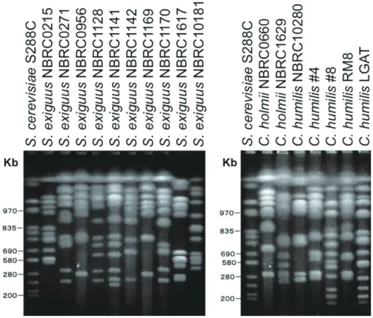
The CHEF electrophoretic pattern of all the strains was shown in Figure 1. Different karyotypes were observed among tested strains of S. exiguus, indicating that phenotypic classification may not be correct. The same
result was shown in two other strains, C. holmii and C. humilis. Since the wild yeast is usually polyploid, the exact chromosome number is not clear. But the chromosome number can be estimated roughly by means of a combination of the band numbers and the intensity of the band. When looking at the smaller sized chromosome, the second and the third chromosomes of S. exiguus NBRC1142 exist closer for S. exiguus NBRC0956. And those two chromosomes seem to overlap for S. exiguus NBRC1169. Therefore these three strains are very close evolutionally. S. exiguus NBRC0271 showed similar karyotype to S. exiguus NBRC1128, S. exiguus NBRC1141, and S. exiguus NBRC1170 (Group A). Group A is very similar to S. cerevisiae. S. exiguus NBRC0956 showed a similar karyotype to S. exiguus NBRC1142 and S. exiguus NBRC1169 (Group B).
S. exiguus NBRC0215, S. exiguus NBRC1617 and S. exiguus NBRC10181 were different to each other from Group A and B. Therefore five different patterns of karyotypes were found among S. exiguus strains.
The karyotype of C. holmii NBRC0660 is different from C. holmii NBRC1629. The karyotype of C. holmii NBRC0660 is very similar to S. exiguus NBRC0956 (Group B) though the chromosome number is one or two excess in C. holmii NBRC0660 compared with S. exiguus NBRC0956 (Group B). On the other hand, C. holmii NBRC1629 showed a similar karyotype to S. exiguus NBRC1170 (Group A).
There were two groups for C. humilis. C. humilis NBRC10280 showed a very similar karyotype to C. humilis #4 and C. humilis RM8 (Group “a”). Group “a” showed a similar karyotype to S. exiguus NBRC0956 (Group B). C. humilis #8 showed a very similar karyotype to C. humilis LGAT (Group “b”). Since C. holmii is the asexual form of S. exiguus and S. cerevisiae9), the karyotype should resemble each other. Actually, the karyotype of Group “a” is similar to the karyotype of Group B. And the karyotype of Group “b” is similar to the karyotype of Group A. As the result mentioned above, it is
suggested that the karyotype was classifi ed roughly into two groups and one other group, totally three groups.
Relationships of yeast strains
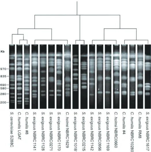
Generally, the phylogenic tree of yeast is drawn by means of rDNA sequences11, 12). When creatures are classifi ed, the chromosome number is often used as an eff ective means. In the case of yeast, the number of chromosomes can be ascertained by pulsed-fi eld gel electrophoresis (PFGE), but there are few examples which made a phylogenic tree among yeast strains by their karyotypes. Electrophoretic patterns of microbes DNA cleaved with a restriction enzyme can be analyzed by Fingerprinting II Software ver. 3 (Bio-Rad Laboratories)13). The software is designed to analyze banding patterns and fi ngerprints from 1-D gels, chromatograms, and density curves. This software is the only a tool at present in the
world to analyze the electrophoretic pattern of PFGE. Because this software canʼt consider the chromosome number for cluster analysis, a phylogenic tree for yeast strains is hard to draw. Thus, a manual grouping was performed on the result of the software by the chromosome number, as the result of which the phylogenic tree was estimated. The phylogenic tree drawn according to the electrophoretic patterns of Figure 1 is shown in Figure 2.
As previously mentioned, Figure 2 demonstrates that strains were classifi ed into two big groups and one other. The strains of S. exiguus were divided into fi ve categories, and, as for this classifi cation, it is strongly suggested that reconsideration is necessary. C. humilis was divided into two groups, and one group was considerably similar to S. cerevisiae, and the karyotype resembles it, with only one or two chromosome numbers being diff erent.
Foschino et al.2) performed a comparison of
Figure 1. The result of PFGE analysis on yeast strains found in panettone mother dough.
classifying the yeast strains found in Panettone mother dough between the recent genotypic method11) (rDNA analysis) and the traditional phenotypic methods14, 15). They showed that a big discrepancy occurred in the classifi cation by the phenotype according to the diff erence in the analytical technique. Also, big diff erence was observed in the classifi cation between the recent taxonomic method whose analysis is based on rDNA sequence and the past taxonomic method whose analysis is based on phenotype, that is, physiological characteristics and assimilation capabilities based on diff erent carbon sources. In their study, among the three phenotypic methods, surprisingly there was not even one case that matched a result accorded. As a matter of course, the phenotypic methods did not match with the recent genotypic method
either. According to the recent rDNA analysis, they reclassifi ed Panettone yeasts into three species, that is, S. cerevisiae, C. humilis, S. pastorianus.
By our result using the karyotype analysis, the Panettone yeasts were classifi ed into at least fi ve categories, and if S. cerevisiae is included into the Panettone yeats as shown by Foschino et al.2), the yeasts were classifi ed into six categories. The recent taxonomic method for yeast is not taking into account the chromosome karyotype for yeast classifi cation. But our result lead us to strongly urge that the classifi cation method of the genus class of yeast should be reviewed on the standing point of considering the balance among the chromosome karyotype, rDNA sequence, and phenotypic feature.
ACKNOWLEDGMENTS
This study was made possible partially by a grant from Seinan Jo Gakuin University. We wish to acknowledge the kind assistance of research fellows in Kai Lab. of the Department of Nutritional Sciences, Seinan Jo Gakuin University.
REFERENCES
1) Kai, T. and Furukawa, K. : The recent baking method of Italian panettone. The bulletin of Seinan Jo Gakuin University 16, 103-112, 2012. 2) Foschino R., Gallina S., Andrighetto C., Rosetti
L. and Galli A. : Comparison of cultural methods for the identification and molecular investigation of yeasts from sourdoughs for Italian sweet baked products. FEMS Yeast Res. 4, 609-618, 2004.
3) Okada S., Ishikawa M., Yoshida I., Uchimura T., Ohara N. and Kozaki M. : Identification and charactertistics of lactic acid bacteria isolated from sour dough sponges. Biosci. Biotech. Biochem. 56(4), 572-575, 1992.
4) Gobbetti M., Corsetti A., Rossi J., La Rossa F. and De vincenzi S. : Identification and clustering of lactic acid bacteria and yeasts from wheat sourdoughs of central Italy. Ital. J. Food Sci. 6, 85-94, 1994.
5) Gobbetti M. and Corsetti A. : Lactobacillus sanfrancisco: a key sourdough lactic acid bacterium-a review. Food Microbiol. 14, 175-187, 1997.
6) Sugihara T.F., Kline L. and Miller M.W. :
Microorganisms of the Sanfrancisco sourdough bread process. I. Yeasts responsible for the leavening action. Appl. Microbiol. 22, 87-96, 1971. 7) Galli A. and Ottogalli G. : Aspetti della microflora
degli impasti per panettone. Ann Microbiol. Enzimol. 23, 39-49, 1973.
8) Galli A., Franzetti L. and Fortina M.G. : Isolation and identication of sourdough microflora. Micr. Alim. Nutr. 6, 345-351, 1988.
9) Foschino R., Terraneo R., Mora D. and Galli A. : Microbial characterization of sourdoughs for sweet baked products. Ital. J. Food Sci. 11, 19-28, 1999.
10) Chu, G., Voiirath, D. and Davis, R.W. : Separation of large DNA molecules by contour-clamped homogenous electric field. Science 234:1582-1585, 1986.
11) Kurtzman C.P., Fell J.W. and Boekhout T. : The yeasts, A taxonomic study 5th ed. Elsevier
Science Publishers B.V., Amsterdam, 2011. 12) Blackwell M., Kurtzman C.P. and Lachance
M-A. : Phylogenetics of Saccharomycetales, the ascomycete yeasts. Mycologia 98(6), 1006-1017, 2006.
13) Sonobe K., Norose Y., Miura Y., Shinoyama A., Okawa S., Washio W., Maehara S., Nakagawa H., Fujita M. and Maeda M. : Different pulsed-field gel electrophoresis patterns in the same VRE carrier. Japanese J. Infection Prevention and Control 28(1), 13-17, 2013.
14) Deák T. and Beuchat L.R. : Handbook of food spoilage yeasts. CRC Press, Boca Raton, USA, 1996.
15) Kurtzman C.P. and Fell J.W. : The yeasts, A taxonomic study 4th ed. Elsevier Science
パネットーネ酵母染色体の核型分析
甲斐 達男 高本 なつみ 石本 祐子
︿要 旨﹀ パネットーネとはイタリア北部でクリスマスシーズンに食される伝統的なスイートブレッドのひとつである。パ ンの分類上はサワーブレッドのひとつであり、自然種製法によって作られる。酵母と乳酸菌の共生によってパン生 地の発酵がなされることが広く知られている。これまで多くの研究者が母種から酵母と乳酸菌の分離・同定を試 みてきている。乳酸菌については、主に、Lactobacillus sanfranciscencisが分離されてきている。酵母については、主なものとしてSaccharomyces exiguus、Saccharomyces cerevisiae、Candida holmii、Candida humilisが分離さ れてきている。これら4種の酵母は表現型による属種の分類が難しく、各種表現型やリボソームDNAのスペーサー 配列による遺伝子型による同定方法などによって、研究者によってそれぞれ異なる学名が付されているのが現状で ある。そこで、パネットーネ酵母の分類上、有力な情報源としてこれら4種の酵母の核型解析をパルスフィールド ゲル電気泳動(PFGE)によって行った。その結果、核型から観ると、これまでの研究による属種の分類方法は全 体に見直しが必要であり、大きく3群に分けられることが示唆された。 キーワード:パネットーネ、酵母、染色体、核型分析、分類