Title
Successful vitrification of pronuclear-stage pig embryos with a novel cryoprotective agent, carboxylated ε-poly-L-lysine
Author(s)
Kamoshita, Maki; Kato, Tsubasa; Fujiwara,
Katsuyoshi; Namiki, Takafumi; Matsumura, Kazuaki; Hyon, Suong-Hyu; Ito, Junya; Kashiwazaki, Naomi
Citation PLOS ONE, 12(4): e0176711
Issue Date 2017-04-27
Type Journal Article
Text version publisher
URL http://hdl.handle.net/10119/14093
Rights
Maki Kamoshita, Tsubasa Kato, Katsuyoshi Fujiwara, Takafumi Namiki, Kazuaki Matsumura, Suong-Hyu Hyon, Junya Ito, Naomi Kashiwazaki (2017) Successful vitrification of pronuclear-stage pig embryos with a novel cryoprotective agent, carboxylated ε-poly-L-lysine. PLoS ONE 12(4): e0176711. doi:10.1371/journal.pone.0176711 ©2017 Kamoshita et al. This is an open access article distributed under the terms of the Creative Commons Attribution License, which permits unrestricted use, distribution, and reproduction in any medium, provided the original author and source are credited.
Successful vitrification of pronuclear-stage pig
embryos with a novel cryoprotective agent,
carboxylated
ε-poly-L-lysine
Maki Kamoshita1, Tsubasa Kato1, Katsuyoshi Fujiwara1, Takafumi Namiki1, Kazuaki Matsumura2, Suong-Hyu Hyon3, Junya Ito1,4*, Naomi Kashiwazaki1,4
1 Laboratory of Animal Reproduction, Graduate School of Veterinary Science, Azabu University,
Sagamihara, Japan, 2 Japan Advanced Institute of Science and Technology, Nomi, Japan, 3 Center for Fiber and Textile Science, Kyoto Institute of Technology, Matsugasaki, Kyoto, Japan, 4 School of Veterinary Medicine, Azabu University, Sagamihara, Japan
Abstract
Vitrification is a powerful tool for the efficient production of offspring derived from cryopre-served oocytes or embryos in mammalian species including domestic animals. Genome editing technologies such as transcription activator-like effector nucleases (TALENs) and clustered regularly interspaced short palindromic repeats (CRISPR)/ CRISPR-associated (Cas)9 are now available even for domestic species, suggesting that the vitrification of embryos at the pronuclear stage (PN) will be more important because they could provide genomic host cells to be targeted by TALENs or CRISPR/Cas9. Although we reported the successful production of piglets derived from vitrified PN embryos by a solid-surface vitrifica-tion method with glutathione supplementavitrifica-tion, further improvements are required. The cryo-protective agent (CPA) carboxylatedε-poly-L-lysine (COOH-PLL) was introduced in 2009. COOH-PLL reduces the physical and physiological damage caused by cryopreservation in mammalian stem cells and the vitrification of mouse oocytes and embryos. Those results suggested that vitrification of COOH-PLL may help improve the developmental ability of pig embryos vitrified at the PN stage. However, it remains unclear whether COOH-PLL is avail-able as a CPA for the vitrification of embryos in domestic species. In this study, we evalu-ated COOH-PLL as a CPA with ethylene glycol (EG) and Cryotop as a device for the vitrification of PN pig embryos. Exposure to vitrification solution supplemented with COOH-PLL up to 30% did not decrease developmental ability to the 2-cell stage and the blastocyst stage. After warming, most of the vitrified embryos survived regardless of the concentra-tion of COOH-PLL (76.0±11.8% to 91.8±4.6%). However, the vitrified embryos without COOH-PLL showed a lower development rate up to the blastocyst stage (1.3±1.0%) com-pared to the fresh embryos (28.4±5.0%) (p<0.05). In contrast, supplementation of 20% (w/v) COOH-PLL in the vitrification solution dramatically improved the developmental ability to blastocysts of the vitrified embryos (19.4±4.6%) compared to those without COOH-PLL (p<0.05). After the transfer of embryos vitrified with 30% (v/v) EG and 20% (w/v) COOH-PLL, we successfully obtained 15 piglets from 8 recipients. Taken together, our present findings demonstrate for the first time that COOH-PLL is an effective CPA for embryo
a1111111111 a1111111111 a1111111111 a1111111111 a1111111111 OPEN ACCESS
Citation: Kamoshita M, Kato T, Fujiwara K, Namiki
T, Matsumura K, Hyon S-H, et al. (2017) Successful vitrification of pronuclear-stage pig embryos with a novel cryoprotective agent, carboxylatedε-poly-L-lysine. PLoS ONE 12(4): e0176711.https://doi.org/10.1371/journal. pone.0176711
Editor: Yang Yu, Peking University Third Hospital,
CHINA
Received: November 10, 2016 Accepted: April 15, 2017 Published: April 27, 2017
Copyright:© 2017 Kamoshita et al. This is an open access article distributed under the terms of the
Creative Commons Attribution License, which permits unrestricted use, distribution, and reproduction in any medium, provided the original author and source are credited.
Data Availability Statement: All relevant data are
within the paper.
Funding: This work was supported in part by
Grants-in-Aid for Scientific Research from JSPS KAKENHI Grant Number JP25242050 (SH.H. and K.M.), JP25660215 (N.K.) and JP15H04584 (J.I.). This research was partially supported by a research project grant awarded by the Azabu University Research Services Division to J.I. and N.K.. This work was also supported by Collaborative
vitrification in the pig. COOH-PLL is a promising CPA for further improvements in the vitrifi-cation of oocytes and embryos in mammalian species.
Introduction
For the production of transgenic animals, pre-implantation embryos at the pronuclear (PN) stage are usually used, as the injection of foreign DNA into pronuclei contributes to the recom-bination of genomic DNA [1]. It was demonstrated that PN embryos can be used not only for transgenic animals but also for knockout animals by using a genome-editing system such as zinc-finger nucleases (ZFNs) [2], transcription activator-like effector nucleases (TALENs) [3], or clustered regularly interspaced short palindromic repeats (CRISPR)/ CRISPR-associated (Cas)9 [4] through the induction of these nucleotides. Even in the pig, gene-modified pigs have been successfully produced using a genome-editing system via microinjection or somatic cell nuclear transfer [5–8] because gene-modified pigs can contribute to the expansion of lots of bio-medical researches, for example generating human organs and tissues, designing new dung screening methodologies and developing new human disease models [9]. In addition, it has been recently reported successful interspecies human-pig blastocyst complementation [10]. These results lead us the idea that the cryopreservation of embryos will become more important, because such embryos are ready-to-use after warming. However, it is well known that pig embryos are very sensitive to damage caused by low temperature and osmotic stress [11].
The vitrification method is now common for the cryopreservation of oocytes and embryos instead of conventional freezing methods. The vitrification method was first reported by Rall and Fahy [12]. The major advantages of the vitrification method are (1) the elimination of the physiological damage caused by intracellular or extracellular ice crystal formation, and (2) the reduction of chilling damage by shortening the exposure to suboptimal temperature [13]. One of the major factors that can affect the efficiency of vitrification is the cryoprotective agent (CPA). Various CPAs such as dimethyl sulfoxide (DMSO), ethylene glycol (EG), glycerol, and propylene glycol have been widely used for the vitrification of oocytes and embryos in various mammalian species [14–16].
Our previous study demonstrated that compared to DMSO, EG had a lesser toxic effect on the vitrification of unfertilized oocytes in mice [17]. It was also suggested that the current CPAs, even EG, have toxic effects on cell viability in a dose-dependent manner [18], indicating that the development of a new CPA showing high efficiency and low toxicity is necessary for further improvements in vitrification. Carboxylatedε-poly-L-lysine (COOH-PLL) was intro-duced as a CPA in 2009 by Matsumura and Hyon [18]. They demonstrated that COOH-PLL reduced the risks of damage by ice recrystallization during freezing and thawing, with anti-freezing protein-like activities [19]. We succeeded in producing mouse offspring derived from unfertilized oocytes and PN embryos vitrified with COOH-PLL and EG [20,21]. Although the production of piglets derived from vitrified PN embryos was reported using another vitrifica-tion method and CPA, i.e., a SSV method and EG [22], our results from the mouse indicated that COOH-PLL is a more suitable CPA for the vitrification of oocytes and embryos, even in the pig, compared to the other CPAs that have been used to date. We speculated that vitrifica-tion using COOH-PLL as a CPA could thus provide a high success rate for embryo vitrificavitrifica-tion even in pigs.
The objective of the present study was to clarify whether COOH-PLL is effective as a CPA for the vitrification of PN porcine embryos. We also evaluated thein vitro and in vivo develop-ment of PN porcine embryos that were vitrified using COOH-PLL.
Research Project organized by the Interuniversity Bio-Backup Project (IBBP) to K.M..
Competing interests: The authors declare that
there is no conflict of interest that could be perceived as prejudicing the impartiality of the research reported.
Materials and methods
All chemicals and reagents were purchased from Sigma-Aldrich (St. Louis, MO, USA) unless otherwise stated. The study was approved by the Ethical Committee for Vertebrate Experi-ments at Azabu University (ID#140219–4) [23].
Oocyte collection and in vitro maturation (IVM)
The collection of porcine follicular oocytes andin vitro maturation (IVM) were performed as described by Kikuchiet al [24]. In brief, porcine ovaries were collected at a local slaughterhouse and transported to the laboratory at 37.5˚C. Cumulus oocyte complexes (COCs) were collected from 2–6 mm in diameter follicles. Fifty COCs were cultured for 22 h in four-well dishes (Nunc™ Cell-Culture Treated Multidishes; Thermo Fisher Scientific Inc., Waltham, MA, USA), each con-taining 500μL of a modified North Carolina State University-37 (NCSU-37) solution [25] which contained 10% (v/v) porcine follicular fluid, 0.6 mM cysteine, 20μM beta-mercaptoethanol, 1 mM dibutyryl cAMP (dbcAMP), 10 IU/mL eCG (1000 units; PMS; Nippon Zenyaku Kogyo, Fukushima, Japan), and 10 IU/mL hCG (3000 units; Puberogen; Novartis Animal Health, Tokyo). The COCs were subsequently cultured for 22 h in NCSU-37 solution without dbcAMP, eCG or hCG. The maturation culture was performed under 5% CO2in air at 38.5˚C.
In vitro fertilization and in vitro culture
Thein vitro fertilization (IVF) and in vitro culture (IVC) were performed as described by Kiku-chiet al [24]. After 44 h IVM, COCs were washed three times in modified pig fertilization medium (Pig-FM) [26] and 20–25 COCs were transferred into each 90μL droplet of Pig-FM covered with paraffin oil (Kanto Chemicals, Tokyo). Epididymal spermatozoa were collected and frozen as described by Kikuchiet al [27]. Frozen-thawed epididymal spermatozoa were washed in Medium 199 with Earle salts (Gibco) adjusted to pH 7.8 [22] and preincubated for 15 min at 38.5˚C in Pig-FM.
After preincubation, 10μL of sperm was added to the droplets of Pig-FM containing COCs. The final concentration of sperm was 1.0 x 10⁶sperm/mL. The COCs and sperm were co-cul-tured for 3 h at 38.5˚C under 5% CO2in air. At 3 h after the IVF, the cumulus cells and sperm
were removed from the oocytes with the use of a fine glass pipette. Denuded oocytes were cul-tured in NCSU-37 without glucose supplemented with 50μM beta-mercaptoethanol, 0.17 mM sodium pyruvate, 2.73 mM sodium lactate, and 4 mg/mL albumin from bovine serum (BSA) (IVC-PyrLac) [24] for 48 h.
The embryos were subsequently cultured in NCSU-37 supplemented with 5.55 mM glu-cose, 50μM beta-mercaptoethanol and 4 mg/mL BSA (IVC-Glu) [24] for 120 h. IVC was per-formed at 38.5˚C under 5% CO2in air. At 10 h after the IVF, the oocytes were centrifuged at
17,860g at 38˚C for 10 min in a 1.5-mL tube for the visualization of the pronuclei. Oocytes with two or three pronuclei (PN) were defined as porcine embryos at the PN stage and further used for vitrification [22].
Vitrification and warming
Vitrification and warming by the Cryotop method was performed as describe [28] with some modification. PN embryos were selected and exposed for 10 min to PB1, i.e., phosphate-buff-ered saline (PBS) supplemented with 20% (v/v) fetal calf serum (FCS), 15% (v/v) ethylene gly-col (EG) and COOH-PLL. Embryos were exposed to PB1 supplemented with 20% (v/v) FCS, 0.5 M sucrose, 30% (v/v) EG and COOH-PLL for 1 min before being plunged into liquid nitro-gen on Cryotop. In each Cryotop, 10–20 embryos were loaded.
The concentrations of COOH-PLL in the vitrification solution (w/v, P0, P1, P10, P20 and P30, respectively), and the concentrations in the equilibration solution were one-half of those in the vitrification solution (Table 1). Vitrified embryos were preserved in liquid nitrogen for at least 1 week. Embryos that had been vitrified on the Cryotop were immersed in PB1 supple-mented with 20% (v/v) FCS and 1.0 M sucrose for 1 min. The embryos were then transferred to PB1 supplemented with 20% (v/v) FCS and 0.5 M sucrose for 3 min, and transferred to sup-plemented PB1 with 20% (v/v) FCS for 5 min. After warming, the embryos were cultured at 38.5˚C under 5% CO2in air. To investigate the effect of COOH-PLL on the developmental
ability of the embryos, some embryos were only exposed to equilibration solution, and not vitrified.
Evaluation of survival and developmental ability in vitro
We morphologically evaluated the survival of the vitrified embryos at 1 h after warming. The membrane integrity of vitrified-warmed embryos under a microscope according to the method by Zeronet al [29]. Embryos with normal and spherical shape, without lysis, and not shrunken, swollen, or blackened were regarded as surviving. To evaluate the developmental ability of vitrified or exposed embryosin vitro, we determined the cleavage rates (at 38 h after warming) and the rates of blastocyst formation (at 158 h after warming). Blastocysts were har-vested and stained with orcein for the determination of the number of cells in each blastocyst [22].
Evaluation of the permeability of COOH-PLL to embryos
COOH-PLL was prepared as reported previously [20]. To clarify the permeability of PN embryos to COOH-PLL as a CPA, we used fluorescein isothiocyanate (FITC)-labeled COOH-PLL. After centrifugation, PN embryos were exposed to PB1 supplemented with FITC-labeled COOH-PLL (5% (w/v)) for 5 min. After exposure, some of the embryos were washed 3 times with PBS. Both washed and unwashed embryos were used for the experiments to evaluate the interaction of COOH-PLL with the embryos under confocal laser microscopy (TCS-SP5; Leica Co. Ltd, Wetzlar, Germany). As control, mouse PN embryos were also col-lected by in vitro fertilization as previously reported [20] and then used for the experiments as described above.
Embryo transfer
The embryo transfer was performed as described [30]. Estrus synchronization of the recipient nonpregnant gilts (>140 days old) was achieved by an intramuscular injection of 1,500 IU eCG followed by an injection of 500 IU hCG 72 h later. Embryos that were vitrified with P20
Table 1. The concentrations of the cryoprotectants in the equilibration solution (ES) or vitrification (VS).
Treatments ES1 VS2
P0 15%EG 30%EG
P1 15%EG + 0.5%COOH-PLL 30%EG + 1%COOH-PLL
P10 15%EG + 5%COOH-PLL 30%EG + 10%COOH-PLL
P20 15%EG + 10%COOH-PLL 30%EG + 20%COOH-PLL
P30 15%EG + 15%COOH-PLL 30%EG + 30%COOH-PLL
1These cryoprotectants were added into PB1 supplemented with 20% FCS. 2
These cryoprotectants were added into PB1 supplemented with 20% FCS and 0.5 M sucrose.
and warmed were surgically transferred into the oviducts of estrus-synchronized recipient gilts. After warming, 97–143 embryos (5–10 Cryotops) were transferred within 1h for each experiment. On Day 28 (Day 0 was the day of transfer), pregnancy was confirmed in the recip-ients with an ultrasound pregnancy detector (Doppler Pregtector; Rotech Livestock Equip-ment, Chichester, England). The offspring was confirmed on Days 115–116.
Statistical analysis
Each experiment had at least five replicates. All percentage data were subjected to arcsine transformation before the statistical analysis. Kruskal-Wallis test or Scheffe’s method was used for the analysis. Data are shown as mean± standard error of the mean (SEM).
Results
The developmental ability of PN embryos exposed to various
concentrations of COOH-PLL
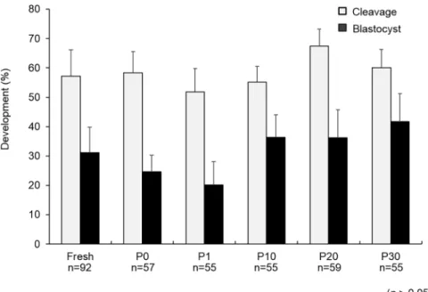
There were no significant differences (p>0.05) in the cleavage rates of the PN embryos ex-posed to different concentrations of COOH-PLL (P0: 59.6± 7.1%, P1: 52.7 ± 5.3%, P10: 56.4± 5.8%, P20: 67.8 ± 5.8%, and P30: 60.0 ± 6.2%, respectively) (Fig 1). There were also no significant differences (p>0.05) in the blastocyst rates of the PN embryos exposed to different concentrations of COOH-PLL (P0: 24.6± 5.6%, P1: 20.0 ± 8.0%, P10: 36.4 ± 7.7%, P20: 37.3 ± 9.6%, and P30: 41.8± 9.5%, respectively) (Fig 1). To confirm whether COOH-PLL enters to the cytoplasm of embryos, we exposed mouse and pig PN embryos to FITC-tagged COOH-PLL. The results are illustrated inFig 2. FITC-tagged COOH-PLL was partially observed in cytoplasm of both unwashed and washed mouse embryos (Fig 2A and 2B). On the other hand, FITC was not observed in cytoplasm of both unwashed and washed pig embryos.
Fig 1. The developmental ability of PN porcine embryos exposed to various concentrations of COOH-PLL. Data are mean±SEM. Data was analyzed by Kruskal-Wallis test. There were no significant differences in the cleavage rate or blastocyst rate among the treatments (p>0.05). Number of oocytes used in each group were described under each treatment group.
The survival and developmental ability in vitro of the PN embryos vitrified
with various concentrations of COOH-PLL
Typical morphology of embryos after vitrification was shown inFig 3A.
Embryos with normal and spherical shape, without lysis, and not shrunken, swollen, or blackened were regarded as surviving (Fig 3A) and others were defined as non-survived (Fig 3A).
Fig 2. The permeability of PN mouse and pig embryos to COOH-PLL. The embryos were exposed to
FITC-labeled COOH-PLL (5% (w/v)) for 5 min. After exposure, embryos with or without washing were examined under a laser scanning microscope. Scale bars denote 50 um.
There were no significant differences (p>0.05) in the survival rates of the PN embryos vitri-fied with different concentrations of COOH-PLL (P0: 91.8± 4.6%, P1: 91.7 ± 5.7%, P10: 83.2± 3.6%, P20: 72.7 ± 7.3%, and P30: 76.0 ± 11.8%, respectively) (Fig 3B). There were also no significant differences (p>0.05) in the cleavage rates of the PN embryos vitrified with differ-ent concdiffer-entrations of COOH-PLL (P0: 23.1± 5.8%, P1: 27.5 ± 3.8%, P10: 37.8 ± 7.5%, P20: 41.8± 4.6% and P30: 39.7 ± 11.8%) (p>0.05) (Fig 3C).
The blastocyst rate of the PN embryos vitrified with P20 (19.4± 4.6%) was significantly higher than that of the P0 (1.3± 1.0%) and P1 (3.8 ± 2.1%) groups (p<0.05). There were no sig-nificant differences (p>0.05) in blastocyst rates between the P20 group and the fresh group (28.4± 5.0%) (Fig 2B). The total numbers of cells in the blastocysts developed from the em-bryos vitrified with P10 (22.7± 2.3 cells) were significantly lower than those of the fresh embryos (36.1± 2.9 cells) (p<0.05), but there was no difference in the total numbers of cells between the P20 group (30.2± 2.1 cells) and the fresh group (p>0.05) (Table 2).
Fig 3. The effects of COOH-PLL on the morphology (A), survival (B) and developmental abilities (C) of vitrified porcine embryos at the PN stage. Data are mean±SEM. Scheffe’s method was used for the analysis. Different superscripts denote significant differences (p<0.05). Number of oocytes used in each group were described under the treatment group. Scale bars denote 50 um.
https://doi.org/10.1371/journal.pone.0176711.g003
Table 2. The effect of COOH-PLL concentration on the number of cells in a blastocyst. Blastocysts No. of total cells
Fresh 31 36.1±2.9a
P10 14 22.7±2.3b
P20 24 30.2±2.1ab
P30 13 21.8±2.2b
Data are shown as mea ns±S.E.M. Different superscripts denote a significant difference (p<0.01) by Scheffe’s method.
The in vivo developmental ability of PN embryos vitrified with COOH-PLL
In our preliminary study, 79 fresh embryos were transferred to a recipient and 13 piglets were obtained (data not shown). The transfer of PN embryos vitrified with P20 into eight recipients resulted in two pregnancies, which were maintained until term (Table 3). Fifteen piglets were obtained from the two pregnant recipients; two of them were stillborn (Fig 4).
Discussion
In mammalian species, the vitrification of germ cells is an essential tool in various basic biology and clinical areas. Since genome-editing technology has become available even for domestic species, the vitrification of embryos at the PN stage will be more important for the efficient production of genome-edited domestic animals. In this study, we succeeded in the production of pig offspring derived from vitrified PN embryos.
Table 3. In vivo development of porcine embryos vitrified with P20. Recipients No. of embryos
transferred Pregnant (Yes/No) No. of piglets (No. of stillborn) #1 143 No 0 (0) #2 103 No 0 (0) #3 111 No 0 (0) #4 105 Yes 5 (0) #5 121 No 0 (0) #6 121 No 0 (0) #7 117 Yes 10 (0) #8 97 No 0 (0) https://doi.org/10.1371/journal.pone.0176711.t003
Fig 4. Piglets derived from PN porcine embryos vitrified with COOH-PLL. https://doi.org/10.1371/journal.pone.0176711.g004
Vitrification is simpler and quicker than the cryopreservation technique called the conven-tional freezing method [31]. Successful vitrification depends on several factors. One of the fac-tors affecting the survival and developmental ability of vitrified oocytes/embryos is how the device or protocol is used for vitrification. An increased volume of vitrification solution inter-feres with the survival of vitrified-warmed embryos, because a large volume decreases the cool-ing rate [31]. The use of only a small volume of vitrification solution in the container is thus a key to achieving vitrification at a high success rate. Many devices and methods have been developed to decrease the total volume of vitrification solution [14]. We previously succeeded in a more efficient production of offspring derived from vitrified-warmed mouse oocytes [17,
32], PN mouse embryos [21], rat oocytes [33], and PN rat embryos [34] using the Cryotop method. In matured pig oocytes, the Cryotop method yielded the highest survivability com-pared to other vitrification methods [11,35]. These results of our present study strongly sug-gested that the Cryotop is one of the most powerful and superior devices for vitrification, even in PN pig embryos.
Although we previously succeeded in the generation of offspring derived from unfertilized mouse oocytes vitrified with EG alone because EG showed lower toxicity than DMSO [17,32], the developmental ability of PN pig embryos vitrified with EG alone was low. Many reports support the idea that pig oocytes are more sensitive than mouse oocytes to physiological stress such as cooling and warming [11,36,37]. It was suggested that many lipids exist in the cyto-plasm of pig oocytes, which negatively affect developmental ability after thawing [38].
Nagashimaet al. [39] demonstrated that the physical removal of lipids (delipation) dramati-cally improved the developmental ability of cryopreserved pig embryos. A latter study con-firmed that the uneven distribution of lipids by centrifugation can improve the developmental ability of cryopreserved embryos [22]. In the present study, we also used centrifugation for the uneven distribution of PN pig embryos, but the developmental ability was still low. These results suggest that additional improvements such as the use of a CPA are required for success-ful vitrification.
In this study, we used COOH-PLL as a CPA for the first time for the vitrification of embryos in a domestic species, and our findings demonstrated that the vitrification with EG and COOH-PLL dramatically improved the embryonic developmental abilityin vitro and suc-ceeded in production of offspring. Our previous study showed that the vitrification of mouse oocytes with EG and COOH-PLL was successful in mouse oocytes [20]. It has been reported that higher concentration of CPA such as EG increases toxicity of cells after cryopreservation but up to 20% (w/v) of COOH-PLL did not decease the survivability of frozen-thawed cells [19]. In addition, recent studies demonstrated that COOH-PLL inhibited of ice crystallization and recrystallization during freezing and thawing with anti-freezing protein-like activitiesin vitro [40,41]. Although detailed mechanism of how COOH-PLL works in the vitrification of mammalian oocytes and embryos is still unclear, inhibition of growth of crystallization and recrystallization during vitrification seems to be a benefit using COOH-PLL.
On the other hand, developmental ability of mouse oocytes vitrified with COOH-PLL alone was very low [20] because permeability of COOH-PLL was low in mouse oocytes [20]. Higher concentration of EG had detrimental effect on survivability of the cells [19]. Therefore, the concentration of EG can be decreased by using COOH-PLL as a CPA, results in the improvement of developmental ability of vitrified embryos. We also demonstrated improve-ment of embryo vitrification at PN [21], 2-cell, morulae and blastocysts in the mouse (Kawa-sakiet al., unpublished data). We reported that FITC-tagged COOH-PLL entered to mouse oocyte cytoplasm to some extent [20]. In the present study however, FITC-tagged was ob-served in PN embryos of mouse embryos to some extent but not pig embryos (Fig 2). It is very difficult to explain the reason why there is a species-dependent differences in the permeability
of COOH-PLL. However, in our previous study, vitrification solution composed of 15% EG and 15% COOH-PLL was effective for vitrification of mouse PN embryos [21]. In case of pig PN embryos, 30% EG and 20% COOH-PLL was required for successful vitrification. These differences seem to be from species-dependent permeability. In addition, one of the positive effects of COOH-PLL for vitrifcaiton of pig embryos may be to protect the membrane from cryoinjury by binding to the membrane as other non-permeable CPAs do [42,43] and/or by inhibition of crystallization and recrystallization as described above.
Taken together, our present findings show for the first time that COOH-PLL is effective as a CPA for embryo vitrification, even in the pig. Our findings will also contribute to the improvement of oocyte/embryo vitrification in other domestic species.
Acknowledgments
We would like to express our gratitude to Mrs. Mayumi Mizuno and also thank the members of the Laboratory of Animal Reproduction, School of Veterinary Medicine, Azabu University.
Author Contributions
Conceptualization: MK JI NK. Data curation: MK JI.
Formal analysis: MK JI.
Funding acquisition: SHH KM JI NK. Investigation: MK JI NK. Methodology: MK TK KF TN KM SHH JI NK. Project administration: JI NK. Resources: MK KM SHH JI KN. Supervision: JI NK. Validation: MK KM SHH JI NK. Visualization: MK JI.
Writing – original draft: MK JI.
Writing – review & editing: MK KM SHH JI NK.
References
1. Brinster RL, Chen HY, Trumbauer ME, Avarbock MR. Translation of globin messenger RNA by the mouse ovum. Nature. 1980;31; 283(5746):499–501. PMID:7352032
2. Geurts AM, Cost GJ, Freyvert Y, Zeitler B, Miller JC, Choi VM, et al. Knockout rats via embryo microin-jection of zinc-finger nucleases. Science. 2009;24; 325(5939):433.https://doi.org/10.1126/science. 1172447PMID:19628861
3. Sung YH, Baek IJ, Kim DH, Jeon J, Lee J, Lee K, et al. Knockout mice created by TALEN-mediated gene targeting. Nat Biotechnol. 2013; 31(1):23–4.https://doi.org/10.1038/nbt.2477PMID:23302927 4. Ran FA, Hsu PD, Lin CY, Gootenberg JS, Konermann S, Trevino AE, et al. Double nicking by
RNA-guided CRISPR Cas9 for enhanced genome editing specificity. Cell. 2013;12; 154(6):1380–9.https:// doi.org/10.1016/j.cell.2013.08.021PMID:23992846
5. Lillico SG, Proudfoot C, Carlson DF, Stverakova D, Neil C, Blain C, et al. Live pigs produced from genome edited zygotes. Sci Rep. 2013;10; 3:2847.https://doi.org/10.1038/srep02847PMID:24108318
6. Whitworth KM, Lee K, Benne JA, Beaton BP, Spate LD, Murphy SL, et al. Use of the CRISPR/Cas9 sys-tem to produce genetically engineered pigs from in vitro-derived oocytes and embryos. Biol Reprod. 2014; 91(3):78.https://doi.org/10.1095/biolreprod.114.121723PMID:25100712
7. Carlson DF, Tan W, Lillico SG, Stverakova D, Proudfoot C, Christian M, et al. Efficient TALEN-mediated gene knockout in livestock. Proc Natl Acad Sci USA. 2012;23; 109(43):17382–7.https://doi.org/10. 1073/pnas.1211446109PMID:23027955
8. Hauschild J, Petersen B, Santiago Y, Queisser AL, Carnwath JW, Lucas-Hahn A, et al. Efficient genera-tion of a biallelic knockout in pigs using zinc-finger nucleases. Proc Natl Acad Sci USA. 2011;19; 108 (29):12013–7.https://doi.org/10.1073/pnas.1106422108PMID:21730124
9. Wu J, Izpisua Belmonte JC. Dynamic Pluripotent Stem Cell States and Their Applications. Cell Stem Cell. 2015;5; 17(5):509–25.https://doi.org/10.1016/j.stem.2015.10.009PMID:26544113
10. Wu J, Platero-Luengo A, Sakurai M, Sugawara A, Gil MA, Yamauchi T, et al. Interspecies Chimerism with Mammalian Pluripotent Stem Cells. Cell. 2017;26; 168(3):473–86.e15.https://doi.org/10.1016/j. cell.2016.12.036PMID:28129541
11. Somfai T, Kikuchi K, Nagai T. Factors affecting cryopreservation of porcine oocytes. J Reprod Dev. 2012; 58(1):17–24. PMID:22450280
12. Rall WF, Fahy GM. Ice-free cryopreservation of mouse embryos at -196 degrees C by vitrification. Nature. 1985;14–20; 313(6003):573–5. PMID:3969158
13. Kuwayama M, Leibo S. Efficiency of the cryotop method to cryopreserve human oocytes; analysis of in vitro and in vivo results at eleven IVF clinics. Fertil Steril. 2008; 90:281–2.
14. Saragusty J, Arav A. Current progress in oocyte and embryo cryopreservation by slow freezing and vitri-fication. Reproduction. 2011; 141(1):1–19.https://doi.org/10.1530/REP-10-0236PMID:20974741 15. Kono T, Suzuki O, Tsunoda Y. Cryopreservation of rat blastocysts by vitrification. Cryobiology. 1988; 25
(2):170–3. PMID:3371062
16. Smorag Z, Gajda B, Wieczorek B, Jura J. Stage-dependent viability of vitrified rabbit embryos. Therio-genology. 1989; 31(6):1227–31. PMID:16726641
17. Kohaya N, Fujiwara K, Ito J, Kashiwazaki N. High developmental rates of mouse oocytes cryopreserved by an optimized vitrification protocol: the effects of cryoprotectants, calcium and cumulus cells. J Reprod Dev. 2011; 57(6):675–80. PMID:21778666
18. Matsumura K, Bae JY, Hyon SH. Polyampholytes as cryoprotective agents for mammalian cell cryo-preservation. Cell Transplant. 2010; 19(6):691–9.https://doi.org/10.3727/096368910X508780PMID:
20525437
19. Matsumura K, Hyon SH. Polyampholytes as low toxic efficient cryoprotective agents with antifreeze pro-tein properties. Biomaterials. 2009; 30(27):4842–9.https://doi.org/10.1016/j.biomaterials.2009.05.025
PMID:19515417
20. Watanabe H, Kohaya N, Kamoshita M, Fujiwara K, Matsumura K, Hyon SH, et al. Efficient production of live offspring from mouse oocytes vitrified with a novel cryoprotective agent, carboxylated epsilon-poly-L-lysine. PLoS One. 2013;23; 8(12):e83613.https://doi.org/10.1371/journal.pone.0083613PMID:
24376724
21. Shibao Y, Fujiwara K, Kawasaki Y, Matsumura K, Hyon SH, Ito J, et al. The effect of a novel cryoprotec-tive agent, carboxylated epsilon-poly-L-lysine, on the developmental ability of re-vitrified mouse embryos at the pronuclear stage. Cryobiology. 2014; 68(2):200–4.https://doi.org/10.1016/j.cryobiol. 2014.01.008PMID:24491796
22. Somfai T, Ozawa M, Noguchi J, Kaneko H, Nakai M, Maedomari N, et al. Live piglets derived from in vitro-produced zygotes vitrified at the pronuclear stage. Biol Reprod. 2009; 80(1):42–9.https://doi.org/ 10.1095/biolreprod.108.070235PMID:18768913
23. Ito J, Yoshida T, Kasai Y, Wakai T, Parys JB, Fissore RA, et al. Phosphorylation of inositol 1,4,5-triphos-phate receptor 1 during in vitro maturation of porcine oocytes. Anim Sci J. 2010; 81(1):34–41.https:// doi.org/10.1111/j.1740-0929.2009.00699.xPMID:20163670
24. Kikuchi K, Onishi A, Kashiwazaki N, Iwamoto M, Noguchi J, Kaneko H, et al. Successful piglet produc-tion after transfer of blastocysts produced by a modified in vitro system. Biol Reprod. 2002; 66(4):1033– 41. PMID:11906923
25. Petters RM, Wells KD. Culture of pig embryos. J Reprod Fertil Suppl. 1993; 48:61–73. PMID:8145215 26. Suzuki K, Asano A, Eriksson B, Niwa K, Nagai T, Rodriguez-Martinez H. Capacitation status and in vitro
fertility of boar spermatozoa: effects of seminal plasma, cumulus-oocyte-complexes-conditioned medium and hyaluronan. Int J Androl. 2002; 25(2):84–93. PMID:11903657
27. Kikuchi K, Nagai T, Kashiwazaki N, Ikeda H, Noguchi J, Shimada A, et al. Cryopreservation and ensu-ing in vitro fertilization ability of boar spermatozoa from epididymides stored at 4 degrees C. Theriogen-ology. 1998; 50(4):615–23. PMID:10732152
28. Hirose M, Kamoshita M, Fujiwara K, Kato T, Nakamura A, Wojcikiewicz RJ, et al. Vitrification procedure decreases inositol 1,4,5-trisphophate receptor expression, resulting in low fertility of pig oocytes. Anim Sci J. 2013; 84(10):693–701.https://doi.org/10.1111/asj.12061PMID:23607492
29. Zeron Y, Pearl M, Borochov A, Arav A. Kinetic and temporal factors influence chilling injury to germinal vesicle and mature bovine oocytes. Cryobiology. 1999; 38(1):35–42.https://doi.org/10.1006/cryo.1998. 2139PMID:10079127
30. Hisamatsu S, Sakaue M, Takizawa A, Kato T, Kamoshita M, Ito J, et al. Knockout of targeted gene in porcine somatic cells using zinc-finger nuclease. Anim Sci J. 2015; 86(2):132–7.https://doi.org/10. 1111/asj.12259PMID:25187232
31. Kuwayama M. Highly efficient vitrification for cryopreservation of human oocytes and embryos: the Cryotop method. Theriogenology. 2007;1; 67(1):73–80.https://doi.org/10.1016/j.theriogenology.2006. 09.014PMID:17055564
32. Kohaya N, Fujiwara K, Ito J, Kashiwazaki N. Generation of live offspring from vitrified mouse oocytes of C57BL/6J strain. PLoS One. 2013; 8(3):e58063.https://doi.org/10.1371/journal.pone.0058063PMID:
23516430
33. Fujiwara K, Kamoshita M, Kato T, Ito J, Kashiwazaki N. Generation of rats from vitrified oocytes with surrounding cumulus cells via in vitro fertilization with cryopreserved sperm. Anim Sci J. 2017; 88 (1):180–4.https://doi.org/10.1111/asj.12666PMID:27481698
34. Seita Y, Okuda Y, Kato M, Kawakami Y, Inomata T, Ito J, et al. Successful cryopreservation of rat pro-nuclear-stage embryos by rapid cooling. Cryobiology. 2009; 59(2):226–8.https://doi.org/10.1016/j. cryobiol.2009.07.007PMID:19631202
35. Ogawa B, Ueno S, Nakayama N, Matsunari H, Nakano K, Fujiwara T, et al. Developmental ability of por-cine in vitro matured oocytes at the meiosis II stage after vitrification. J Reprod Dev. 2010; 56(3):356– 61. PMID:20332589
36. Vajta G. Vitrification of the oocytes and embryos of domestic animals. Anim Reprod Sci. 2000;2; 60– 61:357–64. PMID:10844207
37. Mandawala AA, Harvey SC, Roy TK, Fowler KE. Cryopreservation of animal oocytes and embryos: Current progress and future prospects. Theriogenology. 2016;15; 86(7):1637–44.https://doi.org/10. 1016/j.theriogenology.2016.07.018PMID:27555525
38. Nagashima H, Kashiwazaki N, Ashman RJ, Grupen CG, Seamark RF, Nottle MB. Removal of cyto-plasmic lipid enhances the tolerance of porcine embryos to chilling. Biol Reprod. 1994; 51(4):618–22. PMID:7819441
39. Nagashima H, Kashiwazaki N, Ashman RJ, Grupen CG, Nottle MB. Cryopreservation of porcine embryos. Nature. 1995;30; 374(6521):416.https://doi.org/10.1038/374416a0PMID:7700349 40. Vorontsov DA, Sazaki G, Hyon SH, Matsumura K, Furukawa Y. Antifreeze effect of carboxylated
epsi-lon-poly-L-lysine on the growth kinetics of ice crystals. J Phys Chem B. 2014;28; 118(34):10240–9.
https://doi.org/10.1021/jp507697qPMID:25113284
41. Matsumura K, Kawamoto K, Takeuchi M, Yoshimura S, Tanaka D, Hyon S. Cryopreservation of a Two-Dimensional Monolayer Using a Slow Vitrification Method with Polyampholyte to Inhibit Ice Crystal For-mation. ACS Biomater Sci Eng. 2016; 2(6):1023–29.
42. Eroglu A, Bailey SE, Toner M, Toth TL. Successful cryopreservation of mouse oocytes by using low concentrations of trehalose and dimethylsulfoxide. Biol Reprod. 2009; 80(1):70–8.https://doi.org/10. 1095/biolreprod.108.070383PMID:18815355
43. Lagares MA, Castanheira PN, Amaral DC, Vasconcelos AB, Veado JC, Arantes RM, et al. Addition of ficoll and disaccharides to vitrification solutions improve in vitro viability of vitrified equine embryo. Cryo Letters. 2009; 30(6):408–13. PMID:20309496