ORIGINAL
Electrons released from both flavins of NADPH-P450 reductase
contribute to the reductive mobilization of iron from ferritin
Kazumi Takaishia and Hiroshi Kitahatab
aDepartment of Dental Anesthesiology, Tokushima University Hospital, Tokushima, Japan
bDepartment of Dental Anesthesiology, Tokushima University Graduate School of Biomedical Sciences, Tokushima, Japan
Abstract : Ferritin, an iron storage protein, plays an important role in iron homeostasis. The mechanism of reductive mobilization of iron from ferritin has not been clarified yet despite many studies. The aim of this study was to assess the mechanisms of the mobilization of iron from ferritin by NADPH P-450 reductase. Nucleotide-dependent flavoenzymes generated significant mobilization of iron from ferritin. The possibility of reductive mobilization of iron from ferritin by electrons released from flavin sites or heme site of two flavoenzymes was investigated to elucidate the mediator-independent mechanisms of such reductive mobilization. The mobilization by NADPH-P450 reductase in the presence of ferricyanide increased threefold, while in the presence of cyto-chrome C increased thirteen-fold. These results indicate that electrons released from both flavins of NADPH-P450 reductase contribute to the reductive mobilization of iron from ferritin. The mechanism of the mobilization of iron from ferritin is discussed. J. Med. Invest. 66 : 230-232, August, 2019
Keywords : Ferritin, Iron, Electron, Flavoenzyme, Reductive mobilization
INTRODUCTION
Ferritin is an iron storage protein in many mammalian cells (1). Cellular iron homeostasis is maintained by regulation of the expression of ferritin and the transferrin receptor.
The ferritin molecule consists of a mineral core of hydrated ferric oxide and 24 protein shells, and the iron content in the core consists of a maximum of 4500 atoms. Two types of channels, six four-fold hydrophobic channels and eight three-fold hydrophilic channels, are generated by assembly of the protein shells. The hydrophobic channel is 0.3-0.4 nm wide and 1.2 nm long, and the hydrophilic channel is 0.3-0.4 nm wide and 0.5 nm long, but is blocked by metal irons (2, 3). The route of iron deposition and mobilization via the hydrophobic channel has been the subject of investigations. The iron in the core can be mobilized from ferritin by reduction of ferric iron, and iron mobilization has been observed on the addition of biological reductants such as dihydroflavin (4-7), dihydrolipoate (7, 8), superoxide anion radical (9, 10), and nitric oxide (11). In recent studies, it was shown that the iron re-lease from ferritin occurred through lysosomal proteolysis or Nu-clear Receptor Coactivator-4 – mediated ferritinophagy (12, 13). However, this process is relatively slow from the perspective of rapid reactivity of iron ion and is related to degradation of ferritin cage (12, 14). It has been suggested that the reductant must pen-etrate into the interior of the channel in order to reduce the iron. Whether ferritin iron reduction involves direct access by the reductant into the central cavity of the molecule or electron tunneling via the hydrophobic channel has remained unclear. The purpose of our study was to investigate the molecular mech-anisms of the mobilization of iron from ferritin by the nucleo-tide-dependent flavoenzymes.
MATERIALS AND METHODS
Ferritin (horse spleen) and NADPH-P 450 reductase (rabbit liver) were obtained from Sigma-Aldrich Co. (St. Louis, MO).
The standard reaction mixture for iron mobilization from ferritin contains 1.9 μM ferritin, 1U NADPH-P 450 reductase, 20 μM bathophenanthroline disulfonic acid 2Na salt in 20 mM phosphate buffer, pH 7.4. Bathophenanthroline is used as a quantity regent of iron by binding to ferrous iron and forming orange-red chelate. We measured the quantity of released fer-rous iron in real time using the spectrophotometer (U-3900, Hitachi High-Tsch Science Corporation, Japan) after adding 100 μM NADPH to the standard reaction mixture. The quantity of released ferrous iron was calculated from absorbance at 530 nm by using a molar absorbance coefficient of 22.1 × 10³ M-1 cm-1 for bathophenanthroline.
To investigate the effect of superoxide dismutase on the reduc-tive mobilization of iron, 10 μM superoxide dismutase added to the standard reaction mixture. In another experiment to eluci-date the mechanism of ferritin iron reduction, 2 mM ferricyanide or 6.7 μM cytochrome C was added each to the standard reaction mixture.
No less than 4 experiments were performed separately. The results are expressed as the representative data for all the experiments.
RESULTS
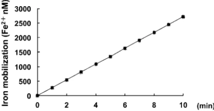
Mobilization of iron from ferritin by NADPH-P 450 reductase NADPH-P 450 reductase mobilized iron from ferritin as shown in Fig.1. This reductive mobilization was reduced by 6% in the presence of superoxide dismutase (Table 1). It was not significantly reduced in the presence of superoxide dismutase, indicating that the superoxide anion radical did not mediate the reduction of ferritin iron.
The Journal of Medical Investigation Vol. 66 2019
Received for publication February 8, 2018 ; accepted February 2, 2019. Address correspondence and reprint requests to Kazumi Takaishi, Department of Dental Anesthesiology, Tokushima University Hospital, 3-18-15 Kuramoto, Tokushima 770-8504, Japan and Fax : +81-88-633-7368.
231
The Journal of Medical Investigation Vol. 66 August 2019
The effects of ferricyanide and cytochrome C on the mobilization of iron from ferritin by NADPH-P 450 reductase
We performed another study to elucidate the mechanism of ferritin iron reduction. It is recognized that the external electron acceptors of FAD and FMN of diflavin reductase family are ferricyanide and cytochrome C, respectively. Here, the reductive mobilization of iron from ferritin is investigated in the presence of external electron acceptors. The mobilization in the presence of ferricyanide increased threefold, while in the presence of cyto-chrome C increased thirteen-fold as shown in Table 2.
DISCUSSION
In this study, NADPH-P450 reductase was found to be able to catalyze the mobilization of iron from ferritin. It was reported that the reductive mobilization of iron from ferritin is dependent on the relative rate of NADH oxidations, dissolved O2 consump-tion and mineral core reducconsump-tion in the study using recombinant frog M ferritin (15). It was also reported that the iron mobiliza-tion from ferritin is dependent on the concentramobiliza-tion of dissolved oxygen in solution (16). However, Johnson LE et al. reported that
the reductive mobilization of iron by reduced flavin mononu-cleotide (FMNH2) was limited by the concentration of FMNH2 under their experimental conditions excluded the possibility of oxygen diffusion into the reaction mixture, and that iron reduc-tive mobilization was independent on the presence of chaotropes (17). In our experiment, the influence of oxygen diffusion on iron release was extremely limited because the iron mobilization was reduced by only 6% in the presence of superoxide dismutase. Moreover, Johnson LE et al. showed that the diffusion of FMNH2 through the ferritin pores was unlikely mechanism by the ex-periments about encapsulation of FMN inside the ferritin cavity (17).
A further investigation is needed to clarify the mechanism of ferritin iron reduction by the nucleotide-dependent flavoen-zymes. One possibility is thought to be that the electron from either enzyme could gain direct access into the core through the ferritin channel and thus activate the reduction of ferritin iron.
NADPH-P 450 reductase exerts their ferricyanide reductive activity by means of electrons from the FAD site and the cyto-chrome C reductive activity by means of electrons from the FMN site (18, 19). In our experimental conditions, the reductive mobi-lization of iron from ferritin is about three folds by the addition of ferricyanide. On the other hand, the reductive mobilization of iron from ferritin is about thirteen folds by the addition of cyto-chrome C, indicating that electrons released from both flavins of NADPH-P450 reductase contribute to the reductive mobilization of iron from ferritin.
The hop distance of electron transfer from site to site of the metal complex model is 1-2 nm. The rate constant is 8 × 10 sec at a distance of 1.13 nm and drops during metal ion separation by 10 seconds for each 0.3 nm increment (20). While it can be assumed that a hop distance of 2 nm is close to the limit of use-fulness, it can occur in proteins over a distance of more than 1 nm (21).
Access to the interior of the shell of the ferritin molecule is possible only via a channel. There are two types of channels through which small molecules can gain access to the central core of the molecule. The hydrophobic channel is 0.3-0.4 nm in diameter and 1.2 nm long, while the hydrophilic channel is 0.3-0.4 nm in diameter and 0.5 nm long (2, 3). In theory, therefore, the electrons could gain direct access via one of these channels to the central core of the ferritin molecule.
Ferritin iron cores can be reduced by one electron per iron accompanied by an uptake of two protons per electron from the surrounding medium (22). The midpoint potential of ferritin iron is E = − 190mV (23). The midpoint potential of NADPH-P 450 reductase is E = − 274 mV (ox/sq), E = − 371 mV (sq/hq) for FAD and E = − 109 mV (ox/sq), E = − 279 mV (sq/hq) for FMN (24). Since the midpoint potential for FAD of NADPH-P 450 reductase is much lower than the midpoint potential of ferritin iron, flavin semiquinone of the enzyme is provided thermody-namically for the mechanism of electron transfer to ferritin iron. For these reasons, the reductive mobilization of iron from ferritin may be induced by direct electron transfer from the flavin site of NADPH-P 450 reductase to the ferritin core.
Iron homeostasis is related to a variety of important physiolog-ical functions (25). A large quantity of free iron released from fer-ritin generates highly reactive hydroxyl radical from superoxide and hydrogen peroxide. It can lead the control of the pathophys-iology related to free radicals to explore the mechanism of iron release from ferritin. It is mandatory to investigate more about the mobilization of iron from ferritin.
Table 1 Effects of Superoxide dismutase on iron mobilization from ferritin by NADPH P-450 reductase
Rate of iron mobilization (Fe2+ μM / 10min)
Control 2.71
Superoxide dismutase (10 μM) 2.55
Fig 1. Time course of iron mobilization from ferritin by means of NADPH P-450 reductase.
The reaction mixture contains 1.9μM ferritin, 1U NADPH-P 450 re-ductase, 100μM NADPH, 20μM bathophenanthroline disulfonic acid 2Na salt in 20mM phosphate buffer, pH 7.4. The quantity of released ferrous iron was calculated from absorbance at 530 nm by using a molar absorbance coefficient of 22.1 × 10⁻³ M-1 cm-1 for
bathophenanth-roline.
Table 2 Effects of ferricyanide and Cytochrome C on iron mobiliza-tion from ferritin by NADPH-P450 reductase
Rate of iron mobilization (Fe2+ μM / 10min)
Control 2.71
Ferricyanide (2 mM) 8.58 Cytochrome C (6.7 μM) 34.29
232
K. Takaishi and H Kitahata Reductive mobilization of iron by electronsCONCLUSION
The iron mobilization by NADPH-P450 reductase from fer-ritin in the presence of ferricyanide or cytochrome C increased markedly. It is suggested that electrons released from both flavins of NADPH-P450 reductase contribute to the reductive mobilization of iron from ferritin.
CONFLICT OF INTEREST
None of the authors have any conflicts of interest to declare.
ACKNOWLEDGEMENT
We are greatly indebted and wish to express our profound gratitude to Prof. Emeritus Aki K for his advice and direct guid-ance throughout the entire course of this study.
This study was supported in part by a Gran-in-Aid for Scien-tific Research from Japan Society for the Promotion of Science (#23792350).
REFERENCES
1. Arosio P, Ingrassia R, Cavadini P: Ferritins : a familiy of molecules for iron storage, antioxidation and more. Biochim Biophys Acta 1790 : 589-599, 2009
2. Crichton RR, Charloteaux-Wauters M : Iron transport and storage. Eur J Biochem 164 : 485-506, 1987
3. Crichton RR, Ward RJ : Iron metabolism--new perspectives in view. Biochemistry 31 : 11255-11264, 1992
4. Sirivech S, Frieden E, Osaki S : The release of iron from horse spleen ferritin by reduced flavins. Biochem J 143 : 311-315, 1974
5. Jones T, Spencer R, Walsh C : Mechanism and kinetics of iron release from ferritin by dihydroflavins and dihydroflavin analogues. Biochemistry 17 : 4011-4017, 1978
6. Funk F, Lenders JP, Crichton RR, Schneider W : Reductive mobilisation of ferritin iron. Eur J Biochem 152 : 167-172, 1985
7. Bonomi F, Pagani S : Removal of ferritin-bound iron by DL-dihydrolipoate and DL-dihydrolipoamide. Eur J Biochem 155 : 295-300, 1986
8. Bonomi F, Cerioli A, Pagani S : Molecular aspects of the re-moval of ferritin-bound iron by DL-dihydrolipoate. Biochim Biophys Acta 994 : 180-186, 1989
9. Biemond P, Swaak AJ, Beindorff CM, Koster JF : Superox-ide-dependent and -independent mechanisms of iron mobi-lization from ferritin by xanthine oxidase. Implications for oxygen-free-radical-induced tissue destruction during isch-aemia and inflammation. Biochem J 239 : 169-173, 1986
10. Bolann BJ, Ulvik RJ : Release of iron from ferritin by xan-thine oxidase. Role of the superoxide radical. Biochem J 243 : 55-59, 1987
11. Reif DW, Simmons RD : Nitric oxide mediates iron release from ferritin. Arch Biochem Biophys 283 : 537-541, 1990 12. Kidane TZ, Sauble E, Linder MC : Release of iron from
fer-ritin requires lysosomal activity. Am J Physiol Cell Physiol 291 : C445-C455, 2006
13. Gryzik M, Srivastava A, Longhi G, Bertuzzi M, Gianoncelli A, Carmona F, Poli M, Arosio P : Expression and characteri-zation of the ferritin binding domain of Nuclear Receptor Co-activator-4 (NCOA4). Biochim Biophys Acta Gen Subj 1861 : 2710-2716, 2017.
14. De Domenico I, Ward DM, Kaplan J : Specific iron chelators determine the route of ferritin degradation. Blood 114 : 4546-4551, 2009.
15. Koochana PK, Mohantly A, Das S, Subhadarshanee B, Satpati S, Dixit A, Sabat SC, Behera RK: Releasing iron from ferritin protein nanocage by reductive method : The role of electron transfer mediator. Biochim Biophys Acta Gen Subj 1862 : 1190-1198, 2018.
16. Melman G, Bou-Abdallah F, Vane E, Maura P, Arosio P, Melman A : Iron release from ferritin by flavin nucleotides. Biochim Biophys Acta 1830 : 4669-4674, 2013.
17. Johnson LE, Wilkinson T, Arosio P, Melman A, Bou-Abdallah F : Effect of chaotropes on the kinetics of iron release from ferritin by flavin nucleotides. Biochim Biophys Acta Gen Subj 1861 : 3257-3262, 2017.
18. Vermilion JL, Coon MJ : Identification of the high and low potential flavins of liver microsomal NADPH-cytochrome P-450 reductase. J Biol Chem 253 : 8812-8819, 1978
19. Vermilion JL, Ballou DP, Massey V, Coon MJ : Separate roles for FMN and FAD in catalysis by liver microsomal NADPH-cytochrome P-450 reductase. J Biol Chem 256 : 266-277, 1981
20. Marcus RA, Sutin N : Electron transfers in chemistry and biology. Biochim Biophys Acta 811 : 265-322, 1985
21. Williams RJ : Electron transfer in biology. Biochem Int 18 : 475-499, 1989
22. Jacobs DL, Watt GD, Frankel RB, Papaefthymiou GC : Redox reactions associated with iron release from mammalian ferri-tin. Biochemistry 28 : 1650-1655, 1989
23. Watt GD, Frankel RB, Papaefthymiou GC : Reduction of mammalian ferritin. Proc Natl Acad Sci U S A 82 : 3640-3643, 1985
24. Iyanagi T, Makino N, Mason HS : Redox properties of the reduced nicotinamide adenine dinucleotide phosphate-cyto-chrome P-450 and reduced nicotinamide adenine dinucleo-tide-cytochrome b5 reductases. Biochemistry 13 : 1701-1710, 1974
25. Bogdan AR, Miyazawa M, Hashimoto K, Tsuji Y : Regulators of iron homeostasis : New players in metabolism, cell death, and disease. Trends Biochem Sci 41: 274-286, 2016