INTRODUCTION
Acanthamoeba keratitis (AK) is a rare corneal
in-fectious disease that can cause severe complications in contact lens wearers. Previously, AK was fre-quently misdiagnosed as herpetic keratitis or kerato-mycosis and was thought to be an intractable type of corneal ulcer. Therefore, many AK patients would visit one eye clinic after another but would not be cured despite long term administration of several medications (1). In Japan, however, recent develop-ments in diagnostic techniques and the increased knowledge about AK have facilitated early diagnosis and adequate treatment. Herein, we present 3 cases of AK that were diagnosed and treated in the early stages of the disease. Further, we review the differ-ences in the disease course in these cases from an etiological point of view.
CASES
Case 1A 23-year-old male who used two-weekly dispos-able soft contact lenses visited a medical practitio-ner on September 21, 2007, presenting with con-junctival hyperemia and foreign body sensation in his right eye. He was administered various antimi-crobial medications, including an ophthalmic solu-tion of tobramycin 0.3% and moxifloxacin 0.3%, topi-cal and systemic fluconazole, acyclovir 3% ointment and systemic valacyclovir, and fluorometholone 0.1% ophthalmic solution administered. Despite this treatment, his symptoms worsened. He was subse-quently referred to Tokushima University Hospital on October 9, 2007. On his first visit to the hospital, the patient presented with a relatively strong pain in his right eye, and his visual acuity was (0.02) in right eye and (1.5) in left. The ocular tension was normal in both eyes. Slit-lamp examination of the right eye revealed radial keratoneuritis, multifocal stromal infiltrates and diffuse corneal edema, and marked ciliary injection (Fig. 1). Fungi flora Y!!
staining and immunostaining with a fluorescence
CASE REPORT
Three cases of
Acanthamoeba
keratitis diagnosed and
treated in the early stage
Nami Ueki
1), Hiroshi Eguchi
1), Yaeko Oogi
1), Hiroshi Shiota
1), Shinta Yamane
2)Hiromichi Umazume
3), and Kenji Mizui
4) 1)Department of Ophthalmology, Institute of Health Biosciences, the University of Tokushima Graduate School ;2)
Yamane Eye Clinic ; 3)
Umazume Eye Clinic ; and4)
Ito Eye Clinic, Tokushima, Japan
Abstract : Acanthamoeba keratitis (AK) is a severe infectious corneal ulcer that usually oc-curs in contact lens wearers. Although the number of AK cases in Japan has been increas-ing, many of these cases are diagnosed in the early stage and are treated adequately. This is probably because of the increased availability of various diagnostic techniques and the ever-increasing knowledge about AK among ophthalmologists. In this article, we described 3 cases of AK that were diagnosed and treated in the early stages of the disease, and we dis-cuss why 1 of the cases had a less favorable prognosis than the other 2 cases, which had ex-cellent prognoses, from an etiological point of view. J. Med. Invest. 56 : 166-169, August, 2009 Keywords : acanthamoeba keratitis, early diagnosis, steroid, compliance
Received for publication March 23, 2009 ; accepted June 25, 2009.
Address correspondence and reprint requests to Hiroshi Shiota, M.D., Ph. D., Department of Ophthalmology, Institute of Health Biosciences, the University of Tokushima Graduate School, Kuramoto cho, Tokushima 770 8503, Japan and Fax : +81 88 -631 - 4848.
The Journal of Medical Investigation Vol. 56 2009
antibody to detect for herpes simplex virus sug-gested the presence of an Acanthamoeba cyst in the corneal scraping. An isolation culture was performed on the multi purpose solution that the patient used to clean and store his contact lenses. The culture, performed on a BactoTMAgar plate containing Difco
Bacto"!yeast extract (Becton, Dickinson and
Com-pany Sparks, USA) revealed Acanthamoeba cysts and trophozoites (Fig. 2). Treatment for stage 2 AK (2)
was initiated ; the treatment involved corneal scrap-ing and frequent use of eye drops of chlorhexidine 0.02%, moxifloxacin 0.3% (5 times daily) and topical application of ofloxacin ointment 0.3% at bedtime. During a follow-up visit on October 22, the corneal
findings gradually improved, however marked cili-ary injection persisted. The next examination on No-vember 14 indicated recovery, after which the pa-tient was lost to follow-up. Five months later, on April 14, 2008, the patient visited our hospital once again, presenting with marked ciliary injection and superficial punctate keratitis in the central area of his right cornea. Repeat treatment with the abovemen-tioned medications reduced ciliary injection decreas-ing and increased visual acuity in the right eye to (0.1). A topical steroid, fluorometholone 0.1% oph-thalmic solution, which is not commonly used to treat AK, was administered to treat persistent inflam-mation of the anterior chamber of the eye on Octo-ber 22, 2008. Thereafter, the patient did not visit our hospital regularly.
Case 2
A 29-year-old female who used two-weekly dis-posable soft contact lenses developed pain and blurred vision in her right eye on March 25, 2007. She visited a medical practitioner on the next day and was administered levofloxacin 0.5% and bromfe-nac sodium 0.1% ophthalmic solutions to treat cor-neal edema and opacity. On March 26, she was re-ferred to Tokushima University Hospital because of faint ring infiltration suggestive AK. On the first visit, she presented with a relatively strong pain in her right eye and visual acuity was (0.4) in the right eye and (1.0) in the left eye. Ocular tension was nor-mal in both eyes. Slit-lamp examination of the right eye revealed faint ring infiltrates and marked ciliary injection (Fig. 3). Staining of the corneal epithelial specimen with Diff Quick"!revealed the presence of
an Acanthamoeba cyst. Treatment for stage 1 or early stage 2 AK was initiated ; this involved corneal
Fig. 1 Marked ciliary injection, multifocal stromal infiltrations, diffuse corneal edema, and radial keratoneuritis ($) in case 1
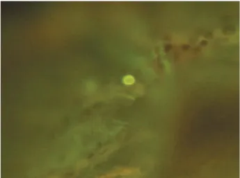
Fig. 2 Colonies of Acanthamoeba and moving tracks of tropho-zoites (original magnification 100
!
)Fig. 3 Ciliary injection and faint ring infiltration (#) in case 2
scraping and the frequent use of eye drops of chlor-hexidine 0.02% and levofloxacin 0.5% ophthalmic solution (5 times a day), and ofloxacin 0.3% ointment (twice a day). The corneal infiltration decreased gradually, and it disappeared completely on May 21, 2007.
Case 3
A 43-year-old female who used two-weekly dis-posable soft contact lenses visited a medical practi-tioner on October 2, 2007, presenting with a foreign body sensation and visual disturbance in her right eye. On the same day, a gatifloxacin 0.3% ophthal-mic solution was administered to treat superficial punctate keratitis. An acyclovir 3% ointment was ad-ministered the following day. Three days later, linear and ring opacities were observed, and the patient was then referred to Tokushima University Hospital. On the first visit, she presented with an intense pain in the right eye, and her visual acuity was (0.4) in right and (1.0) in the left eye. Ocular tension was normal in both eyes. Slit-lamp examination of the right eye revealed ring infiltration, and marked cili-ary injection (Fig. 4). We suspected AK because of
the ring infiltration and the intense pain in her eye. Specimen of a corneal epithelial scraping stained with fungi flora Y"!revealed an Acanthamoeba cyst
(Fig. 5). Treatment for stage 2 AK was initiated ; this involved a corneal scraping and the use of hourly eye drops of chlorhexidine 0.02% during day-time and gatifloxacin 0.3% ophthalmic solution (5 times a day). Ten days later, ring infiltrates disap-peared and the patient’s visual acuity increased ; but
a radial keratoneuritis persisted. The visual acuity in her right eye increased to (1.5) seven days after the second visit, and all treatment was ceased on December 12, 2007.
DISCUSSION
Advanced stage AK is relatively easy to diagnose because of the presence of ring infiltrates or ring ulcers, which are most specific to AK (3) and the presence of Acanthamoeba cysts, which can often be detected in corneal specimens by light microscopy. On the other hand, the clinical features of early-stage AK are nonspecific and the condition may pre-sent with various morphological manifestations such as epithelial microerosions, irregularities, opacities, microcystic edema and stromal infiltrate. Although a radial keratoneuritis is thought to be pathogno-monic for early-stage AK, a similar manifestation has also been reported in Pseudomonas keratitis previously (4). Therefore, it is possible to confuse the symptoms of AK with those of other corneal dis-eases, especially with herpetic keratitis as described in case 1. In recent years, however, the availability of various diagnostic techniques and the increased knowledge about AK presumably has facilitated early diagnosis and adequate treatments of the con-dition. This is reflected in cases 2 and 3, wherein the patients were referred to our hospital within 5 days after treatments were initiated by local medical practitioners. Furthermore, it is necessary to create awareness about the importance of the early diag-nosis of AK because of potential increase in the number of cases of AK among young contact lens
Fig. 4 Ring infiltration (#), superficial punctate keratopathy and ciliary injection in case 3
Fig. 5 Fungi flora Y"!staining of Acanthamoeba cyst in corneal scraping in case 3 (original magnification 200
!
)N. Ueki, et al. Acanthamoeba keratitis treated in early stage
wearers in Japan.
Since very specific and potent anti-Acanthamoeba drugs have not been available for years, various drugs such as neomycin, miconazole, ketoconazole, itraconazole, fluconazole, dibromopropamidine, propamidine isethionate, polyhexamethylene biguanide or chlorhexidine were used for the treat-ment of AK. Among them polyhexamethylene biguanide and chlorhexidine are thought to be best, because these two drugs are effective against not only trophozoites, but also cysts. However, medical treatment with these drugs are not always satisfac-tory. Therefore, recent recommended treatment for AK is a combination of corneal scraping, antifungal drugs and antibiotics. Meanwhile, the use of steroid for AK is pros and contras. Topical steroids are oc-casionally contraindicated because they can sup-press the host immune response. If steroids can in-hibit endocytosis as has been reported previously (5), they may prove beneficial for the treatment of AK in the very early stage. However, McClellan, et
al (6). reported that exposure of Acanthamoeba
tro-phozoite and cysts to dexamethasone increases the pathogenicity of these organisms. No standardized guideline describing the optimal protocol for AK treatment with topical steroid, i.e., the type of ster-oid to be used, appropriate time of treatment, and concentration has been established so far. There-fore, topical steroids ought to be used with caution only when the symptoms of infection are undoubt-edly exclusive of the presence of Acanthamoeba tro-phozoite and cysts. In case 1, we used a fluorome-tholone 0.1% ophthalmic solution as an anti-inflam-matory agent because no symptoms of infection remained after long-term treatment with non - ster-oidal drugs. However, in case 1, the use of topical steroids at the onset of antiamoebic therapy prob-ably contributed to the unsatisfactory prognosis.
Another possible reason for the poor therapeutic outcomes noted in case 1 is the poor patient com-pliance. The patient did not revisit the hospital for scheduled follow-up session but only revisited when the clinical symptoms recurred. Moreover, we as-sume that the patient may have discontinued the treatment and resumed wearing contact lenses as
per his own judgment. It is possible that we did not adequately emphasize to the patient the need for hospitalization, but we think that the patient’s nature may have contributed to the poor clinical outcome. Currently, we lose a return appointment for his next examination.
In conclusion, excellent results can be obtained when the AK is appropriately treated in the early stage, as observed in cases 2 and 3. The best thera-peutic outcome can be achieved with early diagnosis of AK, adequate treatment, and high level of patient compliance. If necessary, topical steroids can be used with caution when the patient has received suf-ficient antiamoebic therapy, after the symptoms of the infections have disappeared. Hospitalization should be considered in cases of poor patient com-pliance.
REFERENCES
1. Ishibashi Y, Miyanaga Y : Acanthamoeba kerati-tis. Nihon No Gannka 79 : 721-726, 2008 (in Japanese)
2. Shiota H, Yano M, Kamada Y, Katayama T, Mimura Y : Classification of clinical stages of
Acanthamoeba keratitis. Japanese Journal of Clinical Ophthalmology 48 : 1149-1154, 1994 (in
Japanese)
3. Theodore FH, Jakobiec FA et al : The diagnos-tic value of a ring infiltrate in Acanthamoebic keratitis. Ophthalmology 92 : 1471-1479, 1985 4. Feist RM, Sugar J, Tessler H : Radial
kerato-neuritis in Pseudomonas keratitis. Archives of
Ophthalmology 109 : 774-775, 1991
5. Osato M, Robinson N, Wilhelmus K, Jones D : Morphogenesis of Acanthamoeba castellanii : ti-tration of the steroid effect (abstract).
Investi-gation of Ophthalmology & Visual Science 27
(Suppl) : 37, 1986
6. McClellan K, Howard K, Niederkorn JY, Alizadeb H : Effect of steroids on Acanthamoeba cysts and trophozoites. Investigation of
Ophthal-mology & Visual Science 42 : 2885-2893, 2001