Abstract Pulmonary manifestation of Mycobacterium avium complex (MAC) disease is an unusual event in patients with advanced HIV infection. Here, we present a case of disseminated MAC disease in a 29-year-old acquired immune defi ciency syndrome (AIDS) patient that had both pulmonary and enteral involvement. The mycobacteria isolated from the pulmonary and enteral lesions were genetically identical. Based on a dendro-gram analysis with variable-number tandem-repeat typing, the isolate appears to cluster with M.avium subsp. avium and M.avium subsp. paratuberculosis. Biopsies of both the pulmonary lesion and the enteral lesion were conducted; the resulting histology showed infections of the epithelia, implicating both sites as being the source of the transepithelial infection. The patient was then treated with mycobacterial therapy, and anti-retroviral therapy for the treatment of AIDS was introduced on the 13th day. The patient’s general condition improved, so he was discharged on the 69th day. To our knowledge, this is the fi rst case report of a MAC transepithelial infection through the airways in the lung of a patient with advanced human immunodefi ciency virus infection. However, the information collected in the present study is insuffi cient to determine the mech-anism by which the enteral lesion developed. Further study will be required to determine whether or not the specifi city of the isolates is related with the mechanism of lesion development.
Key words: Mycobacterium avium complex, AIDS, Pulmonary involvement
1Department of Infectious, Respiratory, and Digestive Medicine,
University of the Ryukyus, 2Department of Oral and Maxillofacial
Surgery, Matsue City Hospital, 3Northern Okinawa Medical Center, 4National Hospital Organization Okinawa National Hospital
Correspondence to : Kenji Hibiya, Department of Infectious, Respi-ratory, and Digestive Medicine, University of the Ryukyus, 207, Uehara, Nishihara-cho, Okinawa 903_0215 Japan.
(E-mail: kenjihibiya@gmail.com)
(Received 8 Aug. 2017/Accepted 15 Jan. 2018) −−−−−−−−Case Report−−−−−−−−
RESPIRATORY AND ENTERAL TRANSEPITHELIAL INFECTIONS
OF MYCOBACTERIUM AVIUM COMPLEX IN A PATIENT
WITH ADVANCED HIV INFECTION
1, 2
Kenji HIBIYA,
1Masao TATEYAMA,
3Morifumi INAMINE,
1Kazuya MIYAGI,
1
Shusaku HARANAGA,
1Satomi YARA,
4Futoshi HIGA, and
1Jiro FUJITA
INTRODUCTION
Mycobacterium avium complex (MAC) contains two genet-ically distinct species: Mycobacterium intracellulare and M. avium1). M.intracellulare is more common among
immuno-competent individuals. Although M.avium also occasionally infects immunocompetent individuals, it typically invades pa-tients with human immunodefi ciency virus (HIV) infection1)2).
In immunocompetent subjects, pulmonary MAC disease is caused by airborne infection. In contrast, MAC disseminated disease is generally caused through enteral invasion in patients with HIV infection2). Additionally, pulmonary involvement
during MAC disease is an unusual event in patients with HIV infection; the percentage of pulmonary involvement is reported to be 0%_22% in these patients3)∼5). It is generally
thought that these pulmonary lesions are formed following bacteremia2). However, it was previously unknown if airborne
infection could occur in patients with advanced HIV infec-tion. In this report, we present a case of disseminated MAC disease in a patient with HIV infection. This case had dual
pulmonary involvement and enteral involvement, and both sites were histologically suggested as being the transepithelial infection. This is the fi rst reported case demonstrating diverse infectious routes (airborne and enteral) in the same individual.
CASE REPORT (1) Clinical presentation
A 29-year-old homosexual man with herpes zoster con-sulted the ophthalmologic department at the University of the Ryukyus Hospital with the complaint of an unpleasant sensation in his right eye (Fig. 1). Based on the results of an ophthalmologic screening test, cytomegalovirus retinitis was diagnosed, and a subsequent hematological examination detected the presence of anti-HIV antibodies. Based on these fi ndings, he was admitted to our department, the Department of Infectious, Respiratory, and Digestive Medicine. Upon admission, the patient’s level of HIV RNA was 1.57×10⁵
copies/mL, and his CD4+ T cell count was 7 cells/µL, so he
was diagnosed as having AIDS. The patient had a history of sex with men over the last nine years but no history of drug
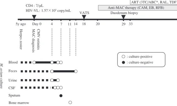
Fig. 1 Clinical course of the patient. Schematic illustrating the clinical course of the patient. ART: anti-retroviral therapy, ABC: abacavir, CAM: clarithromycin, CMV: cytomegalovirus, CSF: cerebral spinal fl uid, EB: ethambutol, MAC: Mycobacterium avium complex, RAL: raltegravir, RFB: rifabutin, TDF: tenofovir disoproxil fumarate, VATS: video-assisted thoracic surgery, VL: viral load, 3TC: lamivudine.
*TDF treatment was replaced with ABC treatment due to kidney dysfunction on the 56th day. Feces Blood Urine Sputum CSF Bone marrow M. avium culture
Anti-MAC therapy (CAM, EB, RFB) Duodenum biopsy
ART (3TC/ABC*, RAL, TDF)
33
5y ago Day 0 4 7 11 14 18 20 29
VATS CD4 : 7/ µL
HIV-VL: 1.57×10⁵ copy/mL
Herpes zoster MAC diagnosis CMV retinitis
: culture-positive : culture-negative
mycobacterial therapy, anti-retroviral therapy with tenofovir, lamivudine, and raltegravir was introduced for the treatment of AIDS. On the 23rd day after the introduction of anti-retroviral therapy, the administration of tenofovir was discon-tinued due to kidney dysfunction. Abacavir with 3TC was then administered as a replacement for tenofovir, while the lamivudine and raltegravir were administered continuously. The patient’s general condition improved, so he was discharged on the 69th day.
(2) Pathological observation
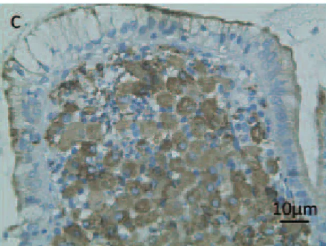
Histological examinations of the specimens obtained by the VATS and duodenum biopsy were conducted. The results from the VATS samples showed an organized proliferative granuloma that was extended along the submucosa of the peripheral bronchiole to the alveoli (Fig. 4A). Additionally, foamy cells with necrosis were observed extensively in the alveolar areas (Fig. 4B), and phagocytized bacilli inside alveolar macrophages were positively stained by anti-myco-bacterial antibody (Fig. 5A). The results for a specimen from the duodenum biopsy showed that acid-fast bacilli had invaded into the mucosal epithelial cells and that the lamina propria was fi lled with histiocytes packed with acid-fast bacilli (Fig. 5B, C and Fig. 6A, B). The histiocytes containing the acid-fast bacilli were positively stained with anti-CD68 antibody (Fig. 6C).
(3) Genetic tests of the isolates
PCR assays detected M.avium DNA from fecal, bone marrow, and lung tissue specimens but did not detect M. avium DNA in the sputum. Cultures of peripheral blood, bone marrow, feces, urine, and lung tissue each resulted in abuse. At the initial visit, no clinical fi ndings were observed
that suggested liver dysfunction, nor did the patient’s medical history include the occurrence of gastroesophageal refl ux disease (GERD).
During the examination for HIV infection, a chest x-ray showed a nodular shadow in the lower lung fi eld (Fig. 2A). The subsequent chest computed tomography (CT) scan re-vealed nodules in the middle lobe, as well as in the lingular lobe (Fig. 2B, C), in addition to a right mediastinal lymphade-nopathy (Fig. 2D). The results of positron-emission tomog-raphy/thoracic CT showed an accumulation of fl uorine-18 deoxyglucose in the mediastinal lymph nodes (Fig. 2E). To further investigate these fi ndings, video-assisted thoracoscopic surgery (VATS) was conducted on the left upper lobe. An abdominal CT scan revealed a tumor mass adjacent to the abdominal descending aorta and mesenteric artery (Fig. 3A), but no abnormal liver fi ndings were observed on the image. The results of positron-emission tomography/abdominal CT showed an accumulation of fl uorine-18 deoxyglucose in the abdominal lymph nodes (Fig. 3B). Esophagogastroduo-denoscopy was conducted for further evaluation; the endo-scopic examination showed that the color of the duodenum mucosa was faded and the microvilli were destroyed. To investigate the cause of these changes, the lesion was biopsied. Mycobacteria were isolated from the lung specimen, duo-denum specimen, feces, and blood. These bacteria were all genetically identifi ed as M.avium, so we diagnosed the patient with disseminated MAC disease (Fig. 1). Clarithromycin, ethambutol, and rifabutin were then administered as mycobacterial therapy. On the 13th day after introducing
anti-Fig. 2 Radiologic fi ndings of the patient’s pulmonary lesions.
A) Image from the patient’s chest x-ray. Encircled area indicates a nodular shadow in the lower lung fi eld. B_D) Images from the patient’s chest computed tomography scan. Arrows indicate small nodules in the middle lobe (B), a nodule in the lingular lobe (C), or a right mediastinal lymphadenopathy (D). E) Image from the patient’s positron-emission tomography/thoracic computed tomography scan. Arrow head indicates accumulations of fl uorine-18 deoxyglucose in the right mediastinal lymph nodes.
the growth of M.avium. Genotyping of these isolates was performed using the pulsed-fi eld gel electrophoresis method6).
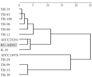
Interestingly, the genotyping patterns of all the isolates from this patient were identical (Fig. 7A, B). Dendrogram analyses were conducted on the results of a variable-number tandem-repeat typing using the M.avium tandem tandem-repeat loci for the isolates from this case along with other reference strains7). We
found that the isolates from our patient formed a cluster with M. avium subsp. avium (MAA; K-10) and M.avium subsp. paratuberculosis (MAP; ATCC25291) (Fig. 8).
DISCUSSION (1) Primary infection of M. avium
Primary complex is the commonly accepted morphological evidence of primary Mycobacterium tuberculosis infection. It consists of a small peripheral focus of infection with regional lymph node involvement8)9). MAC also causes respiratory
tract infections and contributes to the development of granu-lomatous lesions in alveolar areas and bronchioles in immuno-competent subjects6). In our previous work, we demonstrated
that the mechanism involved in the progression of pulmonary M. avium infection from a pulmonary focus to the regional lymph nodes occurred via the lymphatic vessels in a case with HIV-infection6). In the present case, the pulmonary lesions
were localized to the middle lobe and the lingular lobe. The granulomatous lesion was distributed in the submucosa of the
Fig. 3 Radiologic fi ndings of the patient’s abdominal lesions. A) Images from the patient’s abdominal computed tomogra-phy scan. Arrows indicate lymph node swellings around the aorta and mesenteric artery. B) Images from the patient’s pos-itron-emission tomography/abdominal computed tomography scan. Asterisks indicate accumulations of fl uorine-18 deoxy-glucose in the abdominal lymph nodes.
Fig. 5 Transepithelial infection in the lung and the intestine. A) Image of immunohistochemical staining for mycobacterial antigen (color), performed as previously described6), in a lung
sample. B, C) Images of the duodenum lumen (B) and the intestinal epithelium (C) stained with Fite-Faraco staining (color) to mark the acid-fast bacilli (arrows). Scale bars indi-cate 10 µm.
Fig. 4 Distribution of granulomatous lesions in the lung. Two representative histology images from the video-assisted thoracic surgery sample stained with Victoria Blue van Gieson staining (A, B) and hematoxylin eosin staining (the insets). A) The asterisk in the top image shows an organized prolif-erative granuloma in the submucosa of the peripheral bron-chiole. Green indicates elastic fi bers, red indicates collagen fi bers, and yellow indicates the cell cytoplasm, muscle fi bers, and caseous necrotic matter. B) Abundant foamy cells occu-py alveoli around necrotic matter. Scale bars indicate 100 µm.
Fig. 6 Histological characteristics of enteric M.avium infec-tion in patients with AIDS.
A) Image of the granulomatous lesion in the lamina propria of the intestine stained with hematoxylin and eosin. B) Image of the granulomatous lesion in the lamina propria stained with Fite-Faraco staining. C) Image of immunohistochemical staining with anti-CD68 monoclonal antibody (brown) of the foamy histiocytes in the granulomatous lesion in the lamina propria. Scale bars indicate 10 µm.
1∼3, 5: PB (Day 0, 4, 6, 7) 4, 7: feces (Day 4, 14) 6: bone marrow (Day 11) 8: lung (Day 18) M: DNA ladder marker
1, 2, 3, 4 5, 6, 7, 8 80 90 100 % A B 436.5 533.5 485.0 388.0 339.5 291.0 242.5 194.0 145.5 97.0 48.5 kb
Fig. 7 Pulsed-fi eld gel electrophoresis patterns of isolates from various samples.
The pulsed-fi eld gel electrophoresis pattern of isolates from the peripheral blood, the feces, the bone marrow, and the pulmonary lesions (A) and a schematic diagram showing the differences (B).
Fig. 9 Proposed mechanism for the dissemination of Mycobacterium avium infection in the present case. Airborne infection might fi rst occur (①), then granulomatous lesion formed in the lower lung fi eld (②). Subsequently, mediastinal lymphadenopathy occurred. Then, the organism may spread through the blood circulation to reticuloendothelial organs such as the bone marrow (③). Meanwhile, the organism reaches the intestinal mucosa after ingestion of the organism (④). The organism may then infect epithelial cells and may be transported to mesenteric lymph nodes by phagocytic cells (⑤). Further spread through the lymphatic system to the superior deep lymph nodes may occur (⑥).
25 20 15 10 5 0 TH-83 TH-39 TH-33 TH-29 TH-99 ATCC19978 K-10 RU-AIDS3 ATCC25291 TH-12 TH-80 TH-98 TH-100 TH-35
Fig. 8 Dendrogram of isolates from the present case. A dendrogram caused by the Ward s method and based on the results of M.avium tandem repeat -variable number tandem repeat (MATR-VNTR) typing. TH: strainers isolated from patients with pulmonary disease. The MATR-VNTR profi le data of TH strains carry a quotation from the study by Inagaki et al.24). All of the TH-strains are M.avium subsp.
hominissuis24). ATCC25291: Mycobacterium avium subsp.
avium, K-10: Mycobacterium avium subsp. paratuberculosis.
peripheral bronchiole, and an exudative lesion was observed in the alveolar areas. Additionally, the patient showed medi-astinal lymphadenopathy. These fi ndings indicate that this pathological condition is similar to the primary complex of tuberculosis. However, the hematogenous dissemination that typically occurs as multiple well-defi ned small nodular le-sions spread over the entire surface of the affected organ10)11)
was not observed. Notably, the present case also showed a localized duodenum lesion in the submucosal layer with regional lymphadenopathy. Aggregated histiocytes with acid-fast bacilli were focally distributed in the lamina propria of the duodenum, indicating that the pathogenic organisms had penetrated into the mucosal epithelial cells. These fi ndings may suggest a primary enteral infection of MAC12).
(2) Potential mechanisms for the development of the M.avium enteral infection.
In the present case, the pulmonary lesions formed a proliferative granuloma, whereas the enteral lesion was an immature granuloma. This suggests that either the pulmonary lesions began developing fi rst or the pulmonary granuloma grew at a faster rate than the enteral granuloma (Fig. 9). Notably, both the pulmonary lesion and the intestinal lesion were still in an active infectious state and were also relatively localized when we observed them. Even in AIDS patients
receiving highly active antiretroviral therapy (HAART), the gastrointestinal tract, which is a major site for early viral replication and CD4+ T-cell destruction, may be a major viral
reservoir of human immunodefi ciency virus (HIV)13). This
suggests that there is a difference in the immune respon-siveness into mucosal membranes between the intestine and lung. In light of this difference, despite the lesions in the lung and the intestine being different sizes and maturities when detected, these lesions may have begun forming at similar times. Based on previous work, there is a small chance that the enteral lesion in this case was formed via the swallowing of infected sputum from the pulmonary lesion14). Alternately,
previous studies have found that GERD can contribute to the development of pulmonary lesions15)16); however, the
present case did not have history of GERD so this is unlikely to have been the cause of the pulmonary lesions in this case. At present, it is not possible to prove that there was a time lag between the airborne infection and enteral infection of M. avium in this case.
(3) Potential portals of entry for the bacteria
Genetically identical strains were isolated from the duo-denum, lung, bone marrow, and peripheral blood in the present case. Therefore, hematogenous dissemination must have taken place from either the pulmonary lesions or duo-denum lesion (Fig. 9). It is well known that M.avium pre-dominantly disseminates via the bowel mucosa to the mes-enteric lymph nodes and the superior lymphatic system in patients with advanced AIDS17)18). The distribution of lesions
in the present case appears to be compatible with this patho-genesis; however, 80% of cases in which the bacteremia originated from the gastrointestinal tract have liver involve-ment4), and liver involvement was not clinically observed
in the present case.
Mazurek et al. (1997) reported that M.avium can be isolated from respiratory secretions before the detection of bacteremia and that the same strain can be recovered latterly from the stool in some cases with advanced HIV-infection19). This
evidence indicates that pulmonary lesions are developed initially and the respiratory tracts are entry portals from which M. avium can disseminate. Interestingly, non-HIV cases with primary or secondary immunodefi ciency states often show MAC dissemination with lung involvement20)∼22). O’Connell
et al. (2012) reported that the characteristic radiographic fi ndings in patients with non-HIV disseminated non-tuber-culous mycobacterial disease with lung involvement were miliary nodules, which were diffusely scattered throughout the lung parenchyma without aggregation around the air-ways20). Song et al. (2006) described a healthy pregnant
wom-an with MAC dissemination who presented placenta wom-and pulmonary involvement21). Myojo et al. (2003) reported a
case who presented multiple osteomyelitis and pulmonary M. avium disease with infi ltration in the right lower lobe of the lung, with pleural effusion23). Notably, none of the patients in
these cases had a known underlying immunological disease.
The mechanism of MAC development in an immuno-competent host may be different from that in a patient with an AIDS-related MAC infection22). Immunocompromised hosts
with comparatively high cellular immunity show relatively restricted MAC infection with bone marrow involvement, but AIDS patients show systemic infection over a relatively wide range in the abdomen22). Thus, the present case may have
acquired M.avium through the airway passage in a stage of AIDS with a relatively higher cellular immunity, and the lung may have been the portal of entry for the bacteria. (4) Characteristics of the isolates
Based on the results of variable-number tandem-repeat typing using the M.avium tandem repeat loci, the isolates from the present case formed a cluster with MAA and MAP. Until now, isolates from AIDS patients have always formed a cluster distinct from the isolates from patients with pulmonary MAC disease or MAA/MAP24)∼28). To our knowledge, no
M. avium subsp. hominissuis (MAH) isolated from an AIDS patient have previously formed a cluster with MAA/MAP25) 26).
If there is such a cluster, it is unclear what conclusions can be drawn from its existence.
The infectiveness and pathogenicity of MAP in humans are uncertain29). Most mycobacteria isolated from patients with
HIV-infection are MAH, but MAA is occasionally observed in HIV-infected patients30∼34). It has been reported that Th-1
cytokine responses are higher in human blood mononuclear cells after in vitro stimulation with MAA than they are after stimulation with MAH34). MAH-infected pigs had an
unvary-ing level of specifi c antibodies and showed low cell-mediated immunity, whereas MAA infection induced a signifi cant in-crease in both types of immune responses in pigs35). In addition,
MAA also replicates intracellularly to a greater extent than does MAH36). Furthermore, the results of restriction fragment
length polymorphism for the insertion sequences IS1245 and IS901 indicate that MAA and MAH have different genetic patterns37).
It is not obvious whether or not the fi ndings from these previous studies apply to the isolates from the case presented here because the currently available data do not show if MAP or MAA share genetically homology with the present isolates. Further investigation is needed to determine how the uniqueness in the present case relates to the immunological diversity and/ or genetic diversity between the newly identifi ed cluster formed among MAA isolates and the isolates from this case and the conventional clusters that include MAH isolated from AIDS patients.
ACKNOWLEDGMENTS
We thank Katie Oakley, PhD, from Edanz Group (www. edanzediting.com/ac) for editing a draft of this manuscript. Confl icts of interest: None to declare.
REFERENCES
1 ) Inderlied CB, Kemper CA, Bermudez LE: The Mycobac-terium avium complex. Clin Microbiol. 1993; Rev. 6 : 266 310.
2 ) Horsburgh CR Jr: The pathophysiology of disseminated Mycobacterium avium complex disease in AIDS. J Infect Dis. 1999 ; 179 Suppl 3 : S461 465.
3 ) Klatt EC, Jensen DF, Meyer PR: Pathology of Mycobacte-rium avium-intracellulare infection in acquired immuno-defi ciency syndrome. Hum Pathol. 1987 ; 18 : 709 714. 4 ) Torriani FJ, McCutchan JA, Bozzette SA, et al.: Autopsy
fi ndings in AIDS patients with Mycobacterium avium com-plex bacteremia. J Infect Dis. 1994 ; 170 : 1601 1605. 5 ) Abdel-Dayem HM, Omar WS, Aziz M, et al.: Disseminated
Mycobacterium avium complex. Review of Ga-67 and TI-201 scans and autopsy fi ndings. Clin Nucl Med. 1996 ; 21 : 547 556.
6 ) Hibiya K, Tateyama M, Tasato D, et al.: Mechanisms involved in the extension of pulmonary Mycobacterium avium infection from the pulmonary focus to the regional lymph nodes. Kekkaku. 2011 ; 86 : 1 8.
7 ) Inagaki T, Nishimori K, Yagi T, et al.: Comparison of a variable-number tandem-repeat (VNTR) method for typ-ing Mycobacterium avium with mycobacterial interspersed repetitive-unit-VNTR and IS1245 restriction fragment length polymorphism typing. J Clin Microbiol. 2009 ; 47 : 2156 2164.
8 ) Ranke KE: Primaeraffekt, sekudaere und teriaere Stadien der Lungentbk auf Grund von histlogiscen Untersuchungen der Lymphdruesen der Lungenpforte. Dtsch Arch, Klin Med. 1916 ; 119 : 201 375.
9 ) Shimao T: Tuberculosis primary infection. Kekkaku. 2009 ; 84 : 499 502. (In Japanese with an English abstract) 10) Oka H, Kumabe H: Hematogenous dissemination of
tuber-culosis. Kansenshogakuzasshi. 1926 ; 14 : 819. (in Japanese). 11) Esteve E, Supervía A, Pallàs O, et al.: Miliary tuberculosis
coinfection with human immunodefi ciency virus. West J Emerg Med. 2010 ; 11 : 405 407.
12) Hibiya K, Teruya K, Tateyama M, et al.: Enteral entrance of Mycobacterium avium in patients with disseminated mycobacterial disease. Int J Mycobacteriol. 2013a ; 2 : 121 122.
13) Dandekar S: Pathogenesis of HIV in the gastrointestinal tract. Curr HIV/AIDS Rep. 2007 ; 4 : 10 15.
14) Sheer TA, Coyle WJ: Gastrointestinal tuberculosis. Curr Gastroenterol Rep. 2003 ; 5 : 273 278.
15) Thomson RM, Armstrong JG, Looke DF: Gastroesophageal refl ux disease, acid suppression, and Mycobacterium avium complex pulmonary disease. Chest. 2007 ; 131 : 1166 1172. 16) Koh WJ, Lee JH, Kwon YS, et al.: Prevalence of gastro-esophageal refl ux disease in patients with nontuberculous mycobacterial lung disease. Chest. 2007 ; 131 : 1825 1830. 17) Torriani FJ, Behling CA, McCutchan JA, et al.: Disseminated
Mycobacterium avium complex: correlation between blood and tissue burden. J Infect Dis. 1996 ; 173 : 942 949. 18) Hibiya K, Tateyama M, Teruya K, et al.: Depression of
local cell-mediated immunity and histological characteristics of disseminated AIDS-related Mycobacterium avium infec-tion after the initiainfec-tion of antiretroviral therapy. Intern Med. 2013b ; 52 : 1793 803.
19) Mazurek GH, Chin DP, Hartman S, et al.: Genetic similarity among Mycobacterium avium isolates from blood, stool, and sputum of persons with AIDS. J Infect Dis. 1997 ; 176 : 976 983.
20) O’Connell ML, Birkenkamp KE, Kleiner DE, et al.: Lung manifestations in an autopsy-based series of pulmonary or disseminated nontuberculous mycobacterial disease. Chest. 2012 ; 141 : 1203 1209.
21) Song JY, Park CW, Kee SY, et al.: Disseminated Myco-bacterium avium complex infection in an immunocompe-tent pregnant woman. BMC Infect Dis. 2006 ; 22 : 154. 22) Hibiya K, Higa F, Tateyama M, et al.: The pathogenesis
and the development mechanism of Mycobacterium avium complex infection. Kekkaku. 2007 ; 82 : 903 918. Review. (in Japanese).
23) Myojo M, Fujiuchi S, Matsumoto H, et al.: Disseminated Mycobacterium avium complex (DMAC) in an immuno-competent adult. Int J Tuberc Lung Dis. 2003 ; 7 : 498 501. 24) Inagaki T, Nishimori K, Yagi T, et al.: Comparison of a
variable-number tandem-repeat (VNTR) method for typing Mycobacterium avium with mycobacterial interspersed repetitive-unit-VNTR and IS1245 restriction fragment length polymorphism typing. J Clin Microbiol. 2009 ; 47 : 2156 2164.
25) Hibiya K, Tateyama M, Niimi M, et al.: Acquired immune-defi ciency syndrome with focal onset of Mycobacterium avium infection displaying a histological/genetic pattern of disseminated mycobacteria. Intern Med. 2012 ; 51 : 3089 3094.
26) Agdestein A, Olsen I, Jørgensen A, et al.: Novel insights into transmission routes of Mycobacterium avium in pigs and possible implications for human health. Vet Res. 2014 ; 45 : 46.
27) Adachi T, Ichikawa K, Inagaki T, et al.: Molecular typing and genetic characterization of Mycobacterium avium subsp. hominissuis isolates from humans and swine in Japan. J Med Microbiol. 2016 ; 65 : 1289 1295.
28) Imperiale BR, Moyano RD, DI Giulio AB, et al.: Genetic diversity of Mycobacterium avium complex strains isolated in Argentina by MIRU-VNTR. Epidemiol Infect. 2017 ; 1 10.
29) Timms VJ, Hassan KA, Mitchell HM, et al.: Comparative genomics between human and animal associated subspecies of the Mycobacterium avium complex: a basis for patho-genicity. BMC Genomics. 2015 ; 16 : 695.
30) Garriga X, Cortés P, Rodríguez P, et al.: Comparison of IS1245 restriction fragment length polymorphism and
RESPIRATORY AND ENTERAL TRANSEPITHELIAL INFECTIONS
OF MYCOBACTERIUM AVIUM COMPLEX IN A PATIENT
WITH ADVANCED HIV INFECTION
(進行したHIV感染者におけるMycobacterium avium complexの経気道および経腸感染) 日比谷建司 健山 正男 稲嶺 盛史 宮城 一也
原永 修作 屋良さとみ 比嘉 太 藤田 次郎
要旨:進行した HIV 感染者の播種性 MAC 症において,経気道感染は稀な現象である。われわれは 29 歳の HIV 感染者で肺感染と消化器感染を同時に合併した播種性 MAC 症の 1 例を経験したので報告す る。患者の腸感染病巣と肺感染病巣から分離された抗酸菌は遺伝子学的に同一のものであり,VNTR 解析によれば M.avium subsp. avium および M.avium subsp. paratuberculosis と同一のクラスターを形成 した。病理組織学的所見から両病巣は共に上皮感染を示唆するものであった。すなわち経気道感染お よび経腸感染が生じたことを意味している。しかし,経腸感染のメカニズムは今回の結果だけからは 明らかでない。本報告は,進行した HIV 感染者における肺と腸の経上皮感染を病理学的に示した最初 の報告である。
キーワーズ:Mycobacterium avium complex,エイズ,肺感染 pulsed-fi eld gel electrophoresis for typing clinical isolates
of Mycobacterium avium subsp. avium. Int J Tuberc Lung Dis. 2000 ; 4 : 463 472.
31) Johansen TB, Olsen I, Jensen MR, et al.: New probes used for IS1245 and IS1311 restriction fragment length poly-morphism of Mycobacterium avium subsp. avium and Myco-bacterium avium subsp. hominissuis isolates of human and animal origin in Norway. BMC Microbiol. 2007 ; 7 : 14. 32) Ichikawa K, Yagi T, Moriyama M, et al.: Characterization
of Mycobacterium avium clinical isolates in Japan using subspecies-specifi c insertion sequences, and identifi cation of a new insertion sequence, ISMav6. J Med Microbiol. 2009 ; 58 : 945 950.
33) Radomski N, Thibault VC, Karoui C, et al.: Determination of genotypic diversity of Mycobacterium avium subspecies from human and animal origins by mycobacterial interspersed repetitive-unit-variable-number tandem-repeat and IS1311 restriction fragment length polymorphism typing methods.
J Clin Microbiol. 2010 ; 48 : 1026 1034.
34) Thegerström J, Jönsson B, Brudin L, et al.: Mycobacterium avium subsp. avium and subsp. hominissuis give different cytokine responses after in vitro stimulation of human blood mononuclear cells. PLoS One. 2012 ; 7 : e34391.
35) Stepanova H, Pavlova B, Stromerova N, et al.: Different immune response of pigs to Mycobacterium avium subsp. avium and Mycobacterium avium subsp. hominissuis infec-tion. Vet Microbiol. 2012 ; 159 : 343 350.
36) Agdestein A, Johansen TB, Kolbjørnsen Ø, et al.: A comparative study of Mycobacterium avium subsp. avium and Mycobacterium avium subsp. hominissuis in experi-mentally infected pigs. BMC Vet Res. 2012 ; 8 : 11. 37) Mijs W, de Haas P, Rossau R, et al.: Molecular evidence to
support a proposal to reserve the designation Mycobac-terium avium subsp. avium for bird-type isolates and M. avium subsp. hominissuis for the human/porcine type of M. avium. Int J Syst Evol Microbiol. 2002 ; 52 : 1505 1518.