characteristics
著者 ウェン チョン ロン
著者別表示 Nguyen Trong Luan journal or
publication title
博士論文本文Full 学位授与番号 13301甲第3956号
学位名 博士(学術)
学位授与年月日 2013‑09‑26
URL http://hdl.handle.net/2297/39351
Creative Commons : 表示 ‑ 非営利 ‑ 改変禁止 http://creativecommons.org/licenses/by‑nc‑nd/3.0/deed.ja
Trace elements in Japanese precious corals as indicators for habitat and growth characteristics
Graduate School of Natural Science & Technology Kanazawa University
Major Subject: Division of Material Science Course: Material Information Analysis
School Registration No: 1023132312 Name : Nguyen Trong Luan
Chief Advisor: Prof. Hiroshi Hasegawa
i
japanicum), Japanese pink coral (Corallium elatius) and Japanese white coral (Corallium konojoi) are considered as ecologically as well as economically important natural resources of Japan, and are characterised by slow growth rates compared to other precious corals of other geographical locations. The study reveals that the trace elements in skeletons of precious corals are habitat-specific rather than species-specific.
The Mg/Ca and Ba/Ca ratios in skeletons of precious corals, particularly, are the indicators of their habitats and environments. The study also show the spatial distribution of trace elements (S, P, Mg, and Sr) in the skeleton of P. japonicum. The distribution pattern of the trace elements, particularly Mg, S and P, illustrates linkage between the trace element distribution and the formation of growth bands in the coral skeleton.
The petrographic method is a popular technique for estimating the age and growth rates of corals based on growth ring density in their axial skeleton. The organic matrix staining (OMS) method, a modified method of petrographic method, has been used for measuring age and growth rate of the Mediterranean red coral (Corallium rubrum) by staining the organic matrix in the calcite skeleton. Since the OMS method is based on the concentration of organic matrix in the coral skeleton, this method may not be suitable for coral species with low organic matrix. In the presnt study, growth characteristics and growth rates of three Japanese precious corals (Paracorallium japonicum, Corallium elatius and Corallium konojoi) were determined based on the principles of the petrographic method using a VHX-1000 digital microscope, termed as
ii
slabs of the Japanese red coral were more clearly visible through the VHX-1000. This may be due to the low concentration of organic matrix in the Japanese precious corals compared to the Mediterranean red coral. Growth rates of Japanese precious corals differ significantly depending on coral species, habitat and environmental conditions.
Diametric and linear growth rates of the Japanese red coral (P. japonicum) were slower (0.20±0.08–0.27±0.01 and 2.22±0.82–6.66±5.52 mm yr-1, respectively) than the Japanese pink (C. elatius; 0.30±0.04 and 2.76±2.09 mm yr-1, respectively) and white (C.
konojoi; 0.44±0.04 and 7.60±0.75 mm yr-1, respectively) corals. In addition, the diametric growth rate of the Japanese precious corals (P. japonicum) is slower (0.24±0.05-0.44±0.04 mm yr-1) than the Mediterranean red coral (C. rubrum;
0.20-0.62±0.19 mm yr-1).
Precious corals have been commercially exploited for many centuries all over the world. The skeletons of these corals consist of calcium carbonate, and have been used as amulets or gemstones since ancient times. Different Corallium species of Coralidae family (e.g., C. rubrum, C. elatus, C. konojoi, and P. japonicum) were collected from different locations of the Mediterranean Sea (off Italy) and Pacific Ocean (off Japan and off Midway Island), and trace elements in their skeletons were analyzed.
Results show that trace element concentrations in the skeletons of Corallium spp. were attributable to their habitat and origin. In particular, Mg/Ca and Ba/Ca ratios in the skeletons of Corallium spp. from the Mediterranean Sea and Japanese and the Midway Islands’ waters were found to be habitat-specific. This study also reveals that trace
iii
of precious corals. Findings of this study will also be of great relevance to the coral industry to authenticate and identify the habitat and origin of the corals.
This study also investigated the distribution of magnesium (Mg), phosphorus (P), sulfur (S) and strontium (Sr) using micro X-ray fluorescence (µ-XRF), and the speciation of sulfur using X-ray absorption near edge spectroscopy (XANES) along the annual growth rings (AGRs) in the skeleton of Japanese red coral (Paracorallium japonicum). The Mg, P and S distribution in µ-XRF mapping images correspond to the dark and light bands along the AGRs in microscopic images of the coral skeleton.
µ-XRF mapping data showed a strong positive correlation (r = 0.6) between P and S distribution in the coral skeleton. A contrasting distribution pattern of S and Mg along the axial skeleton of the coral indicates a weak negative correlation (r = -0.2) between these two elements. The distribution pattern of S, P and Mg in the axial skeleton of P.
japonicum reveals linkage between the trace element distribution and the formation of dark/light bands along the AGRs. S and P were distributed in the organic matrix (OM) rich dark bands, while Mg was distributed in the light bands of the AGRs. XANES analysis showed that inorganic sulphate is the major species of S in the skeleton of P.
japonicum with a ratio of 1:20 for organic and inorganic sulphate.
iv
I am grateful to the many people who have made completing this research I am very grateful to my advisor, Dr Hiroshi Hasegawa (Professor, Institute of Science and Engineering, Kanazawa University, Japan) for giving me the opportunity to work with him and for his cheerful attitude to work, his solemn instruction, valuable suggestions and constant encouragement during the entire period of the research work and in the preparation of this dissertation. Grateful acknowledgment is made to Dr.
Nozomu Iwasaki (Professor, Faculty of Geo-environmental Science, Rissho University, Japan) for their support and many helpful discussion throughout this research.
I would like to thank Dr. M.Azizur Rahman, School of Environmental Sciences, University of Technology Sydney (UTS), Australia, for reviewing the manuscript and making a number of helpful suggestions. I am also grateful to Dr.
Yusuke Tamenori and Dr. Toshio Ninomiya for their guidance and expertise with XRF and XANES measurement in his beam line during my visit and would like to thank Dr.
Toshihiro Yoshimura (Atmospheric and Ocean Research Institute, University of Tokyo, Japan) and Ms. Satomi Takahashi (Rissho University) for his cooperation and comments.
I would like to thank to Dr. Maki Teruya (Associate Professor, Institute of Science and Engineering, Kanazawa University, Japan) for his helps and supports throughout the course of my study. I would like to thank Mr. M.D Mamunur Rahman for his cooperation during the whole research period.
Thanks to the crews of the ROV ‘Hakuyo 2000’, ‘Hakuyo 3000’ and its mother vessel ‘Shinsei Maru’ and the SNK Ocean Co. Ltd for sample collection. The authors also thank the Kochi prefectural government for providing coral samples for
v
Research and Development Projects for Application in Promoting New Policy of Agriculture, Forestry and Fisheries (22032) in Japan. I really appreciate Soc Trang 150 Project, Mekong 1000 Project’s financial support for my expenses within three years in Japan.
I am grateful to my friends and lab-mates for being encouraging and helpful.
Special thanks to my dear parents, sisters, brother and relatives for their encouragements, wishes and all kind of helps during my study.
Nguyen Trong Luan June, 2013
vi
Dedicated to My Family
vii
ABSTRACT i
ACKNOWLEDGEMENTS iv
TABLE OF CONTENTS vii
LIST OF TABLES xi
LIST OF FIGURES xii
CHAPTER 1: INTRODUCTION 1
References 6
CHAPTER 2: LITERATURE REVIEW 9
2.1 Japanese precious coral species and distribution 9
2.2 Reproduction 12
2.3 Skeletal structure and feeding 13
2.4 Growth characteristics of DPCs 14
2.4.1 Growth banding 14
2.4.2 Age and growth rate 16
2.5 Trace element distribution in the skeletons of precious coral and its roles in paleoenvironment reconstruct
19
References 26
CHAPTER 3: GROWTH CHARACTERISTICS AND GROWTH RATE ESTIMATION OF JAPANESE PRECIOUS CORALS
3.1 Introduction
31
31
viii
3.3.1 Sample collection 34
3.3.2 Cross-section preparation of the coral skeleton 34 3.3.3 Staining of organic matrix in the coral skeleton 35 3.3.4 Determination of the age and growth rates of Japanese
precious corals
35
3.4 Results 37
3.4.1 Growth characteristics of Japanese precious corals 37 3.4.2 Age and diametric growth rates of Japanese precious corals 40 3.4.3 Linear growth rates of Japanese precious corals 41
3.5 Discussion 42
3.5.1 Growth characteristics of Japanese precious corals 42 3.5.2 Growth rates of Japanese precious corals 46 3.5.3 Correlation between number of growth rings and diameter 48 3.5.4 Determination of the ages and growth rates of Japanese
precious corals
50
References 60
CHAPTER 4: RESULTS AND DISCUSSION:
TRACE ELEMENTS IN Corallium spp. as INDICATORS FOR ORIGIN AND HABITAT
65
4.1 Introduction 65
ix
4.3.1 Sampling sites 69
4.3.2 Chemical analysis of skeleton composition 69 4.3.3 Analysis of inorganic elements with electron probe
micro-analyzer (EPMA)
71
4.4 Results and Discussion 71
4.4.1 Trace element distribution in the skeletons of precious coral
72
4.4.2 Trace element compositions reflecting the characteristics of coral habitats
73
References 80
CHAPTER 5: RESULTS AND DISCUSSION:
DISTRIBUTION OF TRACE ELEMENT IN JAPANESE RED CORAL (Paracorallium japonicum) BY µ-XRF AND SULFUR SPECIATION BY XANES: LINKAGE BETWEEN TRACE ELEMENT DISTRIBUTION AND GROWTH RING FORMATION
84
5.1 Introduction 84
5.2 Focus 88
5.3 Materials and Methods 88
5.3.1 Sample collection and preparation 88
5.3.2 Extraction of organic matrix from fraction 89
5.3.3 µ-XRF and XANES analysis 89
x
skeleton
5.4.2 Distribution of trace elements and the formation of annual growth rings
92
5.4.3 Speciation of S in Japanese red coral using XANES 95
References 102
CHAPTER 6: CONCLUSION 107
APPENDIX 110
xi
Table 1.1 Precious corals refers to about 31 species that belong to the Corallium and Paracorallium genera (Bayer & Cairns, 2003)
3
Table 2.1 Actual age and estimate obtain by staining the organic matrix and by the petrographic for five colonies collected from the experiment panels (Marschal et al, 2004)
18
Table 2.2 Biology of Japanese precious corals species 23 Table 3.1. Diametric growth rates and ages of the Japanese red
(Paracorallium japonicum), pink (Corallium elatius), and white (Corallium konojoi) corals collected from Japanese waters.
54
Table 3.2. Linear growth rates of the Japanese red (Paracorallium japonicum), pink (Corallium elatius), and white (Corallium konojoi) corals collected from Japanese
55
Table 3.3. Diametric and linear growth rate of Japanese precious corals (Luan et al., unpublished)
57
Table 3.4 Diametric growth rates of precious corals from different geographical locations estimated by different methods
58
Table 4.1. Different types of precious corals collected for ICP-AES analysis. Samples were collected from Japanese waters, Midway Islands’ waters and Mediterranean Sea from fishermen, coral traders, and research institutes.
76
xii
Figure 2.1 Japanese precious corals. Above left : Shiro (white) (Corallium konojoi). Above right: Momo (Pink) (Corallium elatius).
Below: Aka (red) (Paracorallium japonicum)
11
Figure 2.2 A cross-section of the coral skeleton of DPC-19 (Paracorallium japonicum) (left). Two types of growth bands, light and dark, as seen under the VHX-1000 digital microscope. The dark bands indicate the zones of high organic matrix (OM) content,
15
Figure 2.3 Microscopic images of a corallium rubrum colony. A gerneral view of cross section obtained suing the petrographic and B the cross section after staining the oragnic matrix (Marschal et al., 2004)
15
Figure 2.4 Living colony of Mediterranean red coral (Corallium rubrum) (A). Axial skeleton of C. rubrum (B) and Sclerites of this coral (C)
25
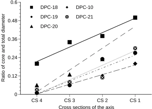
Figure 3.1 Axial skeleton of Japanese red coral Paracorallium japonicum (DPC-18). Numbers on the skeleton indicate the sample sites, and the images are the cross-sections of the corresponsing samples. The cross-sections were polished to make the growth rings clear to count them and estimate growth rate and age of the coral. The diameter of the core become wider at the younger part (tip;1) than at the older part (base; 6) of the colony.
38
Figure 3.2 The ratio of the core diameter and total (including the core) 39
xiii
colony. The cross-section numbers (CS4-CS1) in x-axis of the figure represent the consecutive slabs from the base to the tip of coral axis.
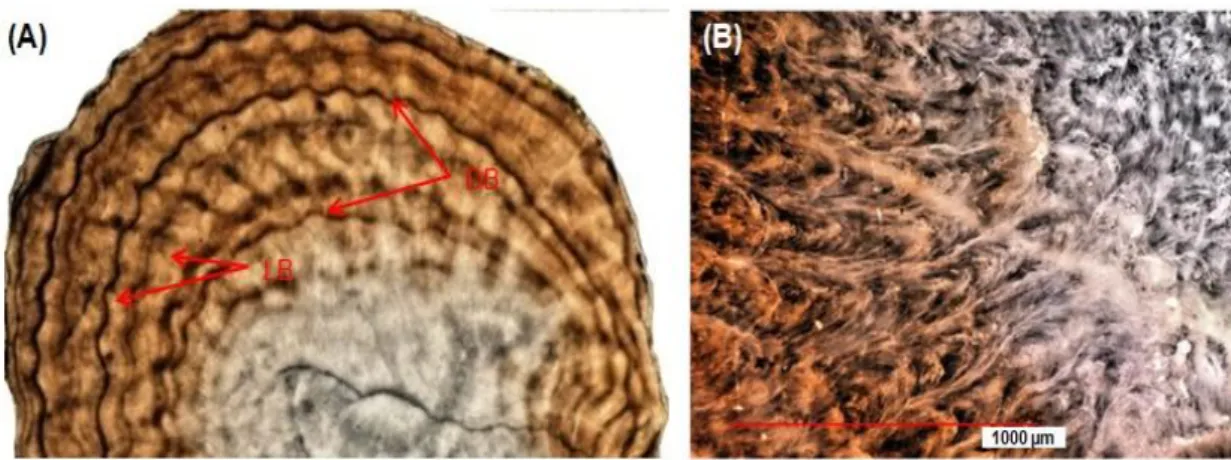
Figure 3.3 (A) Two types of growth bands, light and dark, as seen under the VHX-1000 digital microscope. The dark bands indicate the zones of high organic matrix (OM) content, while the light bands represent the zones of low OM content in the coral skeleton. (B) A cross-section at the tip on the younger part of the coral skeleton (Paracorallium japonicum). The calcium carbonate deposited in the core on the younger part of the axis does not generate/form growth rings.
44
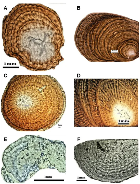
Figure 3.4 Growth rings in thin cross-sections of the axial skeleton of different Japanese precious coral species under the VHX-1000 digital microscope. (A-B) red coral (Paracorallium japonicum), (C-D) pink coral (Corallium elatius), (E-F) white coral (Corallium konojoi).
45
Figure 3.5 Correlation between the number of growth rings and the diameter of the base of Japanese precious corals (Paracorallium japonicum, Corallium elatius, and Corallium konojoi).
49
Figure 3.6 Cross-section of the axial skeleton of the Japanese red coral (Paracorallium japonicum) observed under the VXH-1000
51
xiv
organic matrix in the skeleton with 0.05% Toluidine blue for 10-30 s (C). Magnified images of the internal region near center before and after staining the organic matrix with 0.05%
Toluidine blue (D).
Figure 4.1 Skeleton of Japanese white coral. Photograph of white coral (A), mapping analysis of Mg (B), Ca (C), Ba (D), and Sr (E) in the skeleton of precious white coral by EPMA.
78
Figure 4.2 Distribution of Mg and Ba in skeleton of precious corals collected from different depth and geographical locations indicating the origin of the corals. Values are mean ± SD.
79
Figure 5.1 X-ray fluorescence spectrum of trace elements (Na, S, P, Mg and Sr) in powdered sample of Japanese red coral (DPC-19) skeleton measured at 2500 eV of excitation photon energy
97
Figure 5.2 X-ray fluorescence spectrum of trace elements (Na, S, P, Mg and Sr) in Japanese red coral skeleton (DPC-19) using 2500 eV energy, 5s/point measuring time, and 10 µm step size. White arrow (7 mm) is the position from where the section was taken for µ-XRF analysis (A). Trace element spectrum in the sample (B). Relationship between the distributions of S and Mg (C) and S and P (D) in the skeleton of DPC-19
98
Figure 5.3 Micro-XRF mapping analysis of Mg and S in axial skeleton of Japanese red coral (DPC-18). Cross section of DPC-18 skeleton
99
xv
Distribution maps of Mg and S in the skeleton of the coral (c).
The colour bar represents the signal intensity with the blue and red color correspond to lower and higher distributions, and the black and red color regions in the maps indicate the lowest and highest distribution of the elements, respectively. Micro-XRF signal was collected at 10 µm step size
Figure 5.4 Micro-XRF mapping analysis of trace elements in polished cross section of Japanese red coral skeleton (DPC-19). Two copper rings of one mm diameter were used to select two positions on the cross section of the coral skeleton where the distribution of trace elements were detected. The colour bars represent the signal intensity with the blue and red color correspond to lower and higher distributions, and the black and red color regions in the maps indicate the lowest and highest distribution of the elements, respectively. Micro-XRF signal was collected at 10 µm step size
100
Figure 5.5 XANES spectra of sulfur (S) in axial skeleton and organic matter extract of DPC-19. XANES spectra of S reference compounds (A). Each of the cysteine and methionine spectra display a single peak at 2473 eV photon energy, while condroitine, protamine, and gypsum show peaks at 2482 eV photon energy. Saccharine exhibits a single pick at 2477.5 eV
101
xvi
sulfate is the main species of S and organic matter content in the DPC-19 skeleton is very low
xvii
1. The Twelfth Asian Conference on Analytical Sciences, August 22-24, 2013, Maidashi campus of Kyushu University , Fukuoka-city, Japan. (Presentation: Luan Trong Nguyen, M. Azizur Rahman, Toshihiro Yoshimura, Yusuke Tamenori, Teruya Maki, Nozomu Iwasaki, Hiroshi Hasegawa. ‘‘Determination of Growth Rings and Microstructure in Skeleton of Japanese Precious Corals’’
2. Forum of Division of Material Science. November, 7.2011, Kanazawa University, Japan. (Presentation: Luan Trong Nguyen. ‘‘Distribution and Chemical Forms in Skeleton of Japanese precious corals by Micro-XRF and XANES analysis’’
3. Forum of Division of Material Science. November, 5.2012, Kanazawa University, Japan. (Presentation: Luan Trong Nguyen. ‘‘Estimation of Growth Rate of Japanese Precious Red Coral, Paracorallium japonicum)’’
1
CHAPTER 1
INTRODUCTION
Precious corals (PCs) belong to family Corallidae, class Anthozoa, subclass Octocorallia, order Gorgonacea. They refer to roughly 31 species that belong to the Corallium and Paracorallium genera (Table 1.1) (Bayer and Cairns, 2003). They are found mainly in the Mediterranean Sea and Pacific Ocean (Japanese waters and off Taiwan, off the Midway Islands and off the Hawaiian Islands (Iwasaki and Suzuki, 2010). Many precious coral species are long-lived, grow slowly (diametric growth rate usually less than 1mm per year) and reaching more than 100 years of age (Andrews et al., 2005; Luan et al., 2013). Precious corals have been highly valued because their skeletons have been used as jewellery, souvenir industries, medicine and are one of the most valuable living marine resource (Nonaka and Muzik, 2009; Iwasaki and Suzuki, 2010; Tsounis et al., 2010). Precious corals have attracted worldwide attention as sparse biological resources, and Corallidae have been recently proposed for inclusion in Appendix II of the Convention on International Trade in Endangered Species of Wild
2
Fauna and Flora (CITES) that regulates the international trade in endangered species by listing them in its appendices (Hasegawa and Yamada, 2010; Tsounis et al., 2010;
Hasegawa et al., 2012)
Knowledge of the biology, growth rates, and durability of deep-sea corals (DSC) is important since they provide habitat to commercially important fishes and are under great threat from benthic fishing techniques (Witherell et al., 2000; Roark et al., 2006). Geochemical and isotopic data derived from DSC provide an understanding of past climate and environmental change (Roark et al., 2006). Understanding of growth rates and ages of these long-lived DSC also contributes to our knowledge of their biology, ecology, and physico-chemical characteristics. In addition, conservation and management of marine ecosystems and DSC, and reconstruction of the paleo-environment using the paleoceanographic archive of DSC require knowledge of the growth rates and longevity of these organisms (Goldberg, 1991; Roark et al., 2006).
Chemical analyses of carbonate skeletons in precious corals found that the mineralised hard tissues of precious corals are composed of a skeletal axis and spicules.
Both spicules and skeletons of corals are mainly made of calcium carbonate (CaCO3) crystallized in the form of calcite and trace elements (Maté et al., 1986; Hasegawa and Iwasaki, 2010). Precious corals are different from reef-building corals in that their skeletons are closely-packed with magnesium calcite, while the reef-building corals consist mostly of aragonite, and are porous because of its loosely-packed crystals (Hasegawa et al., 2012).
Reef-building coral is better understood, and concentrations of trace elements in its carbonate skeletons have been determined. The validity of their use as indicators of past environmental conditions, such as water temperatures, nutrients and pollution
3
levels has been confirmed in earth and environmental science studies (Weber and Woodhead, 1970; Weber, 1973; Mitsuguchi et al., 1996; Mitsuguchi et al., 2001;
Mitsuguchi et al., 2003). In contrast, studies on trace elements in precious coral have been focused mostly on Mediterranean red coral (C. rubrum) (Weinbauer and Vellmirov, 1995; Weinbauer et al., 2000). Studies on trace elements in other precious corals, especially the Japanese precious coral is limited.
Table 1.1 Precious corals refer to about 31 species that belong to the Corallium and Paracorallium genera (Bayer & Cairns, 2003)
Corallium abyssale Corallium niobe Corallium variabile Corallium borneense Corallium niveum Corallium sp. nov Corallium boshuense Corallium porcellanum Paracorallium inutile Corallium ducale Corallium pusillum Paracorallium japonicum Corallium elatius Corallium regale Paracorallium nix
Corallium halmaheirense Corallium reginae Paracorallium salomonense Corallium imperiale Corallium rubrum Paracorallium stylasteroides Corallium johnsoni Corallium secundum Paracorallium thrinax Corallium konojoi Corallium sulcatum Paracorallium tortuosum Corallium maderense Corallium tricolor
Corallium medea Corallium vanderbilti
4
Objectives of the Study
The aim objective of this thesis was to describe the growth rate and growth characteristics of Japanese precious corals. Concentration and distribution of trace elements in precious corals related to their origin and habitat were investigated as well.
In addition, we also have examined the linkage between the distribution pattern of trace elements and the formation of annual growth rings along the coral skeleton.
The detailed thesis aims can be summarised as follows:
1. To investigate the skeletal structure, estimating growth rates and ages of three Japanese precious corals Paracorallium japonicum, Corallium elatius, Corallium konojoi using a high resolution VHX-1000 digital microscope.
2. To compare growth rings in the stained (the staining the organic matrix in the skeleton with toluidine blue) with unstained cross section.
3. To investigate if concentration and distribution of trace elements in precious corals related to their origin and habitat.
4. To investigated the distribution of magnesium (Mg), phosphorus (P), sulfur (S) and strontium (Sr) using micro X-ray fluorescence (µ-XRF), and the speciation of sulfur using X-ray absorption near edge spectroscopy (XANES) along the annual growth rings in the skeleton of Japanese red coral (Paracorallium japonicum).
5
Organization of the Study
Chapter 2 provides an overall view of the findings of biological and biochemical research on precious corals, introduces classification, ecology of corals, growth characteristics and trace elements distribution in the skeleton of corals.
Chapter 3 describes the growth characteristics and growth rates of Japanese precious corals by examining their skeletal structure and estimating growth rates using digital microscope. Comparison between the growth rings in both stained (toluidine blue, organic matrix staining methods) and unstained slabs of the corals
Chapter 4 discusses about the trace elements in Corallium Spp as indicator for origin and Habitat.
Chapter 5 is distribution of trace elements in Japanese red coral by µ-XRF and sulfur speciation by XANES focus on the linkage between trace element distribution and growth ring formation.
Chapter 6 provides a summary of overall experiments and scope of the future research.
6
References
Andrews, A., Cailliet, G., Kerr, L., Coale, K., Lundstrom, C., DeVogelaere, A., 2005.
Investigations of age and growth for three deep-sea corals from the Davidson Seamount off central California. In: Freiwald, A., Roberts, J.M. (Eds.), Cold-Water Corals and Ecosystems. Spinger, Berlin, pp. 1021-1038.
Bayer, F.M., Cairns, S.D., 2003. A new genus of the scleraxonian family Coralliidae (Octocorallia: Gorgonacea). Proc. Biol. Soc. Wash. 116, 222-228.
Goldberg, W.M., 1991. Chemistry and structure of skeletal growth rings in the black coral Antipathes fiordensis (Cnidaria, Antipatharia). Hydrobiologia 216, 403-409.
Harvell, C.D., Kim, K., Burkholder, J.M., Colwell, R.R., Epstein, P.R., Grimes, D.J., Hofmann, E.E., Lipp, E.K., Osterhaus, A., Overstreet, R.M., 1999. Emerging marine Hasegawa, H., Iwasaki, N., 2010. Chemical analyses of carbonate skeletons in precious
corals. Biohistory of Precious corals.
Hasegawa, H., Rahman, M.A., Luan, N.T., Maki, T., Iwasaki, N., 2012. Trace elements in Corallium spp. as indicators for origin and habitat. J. Exp. Mar. Biol. Ecol.
414-415, 1-5.
Iwasaki, N., H. Hasegawa, T. Suzuki, Yamasa, a.M., 2009. Biology of Japanese Corallium and Paracorallium, paper presented at Proceedings of the First International Workshop on Corallium Science, Management, and Trade. NOAA Technical Memorandum NMFS-OPR-43 and CRCP-8, Silver Spring, MD, Hong Kong, China., 149.
Iwasaki, N., Suzuki, T., 2010. Biology of precious corals. In: Iwasaki, N. (Ed.), Biohistory of precious corals- scientific,cultural and historical perspectives.
7
Tokai Univeristy Press, Tokai, Japan.
Jackson, J.B.C., Kirby, M.X., Berger, W.H., Bjorndal, K.A., Botsford, L.W., Bourque, B.J., Bradbury, R.H., Cooke, R., Erlandson, J., Estes, J.A., 2001. Historical overfishing and the recent collapse of coastal ecosystems. Science 293, 629-637.
Luan, N.T., Rahman, M.A., Maki, T., Iwasaki, N., Hasegawa, H., 2013. Growth characteristics and growth rate estimation of Japanese precious corals. J. Exp.
Mar. Biol. Ecol. 441, 117-125.
Maté, P., Revenge, S., Masso, C., 1986. Estudio preliminar de la composicion quimica del coral rojo (Corallium rubrum L.) de distintas zonas del Mediterraneo Espanol.
Bol. Inst. Esp. Oceanogr. 3, 53-60.
Mitsuguchi, T., Matsumoto, E., Abe, O., Uchida, T., Isdale, P.J., 1996. Mg/Ca thermometry in coral skeletons. Science 274, 961.
Mitsuguchi, T., Uchida, T., Matsumoto, E., Isdale, P.J., Kawana, T., 2001. Variations in Mg/Ca, Na/Ca, and Sr/Ca ratios of coral skeletons with chemical treatments:
implications for carbonate geochemistry. Geochim. Cosmochim. Acta 65, 2865-2874.
Musick, J.A., 1999. Ecology and conservation of long-lived marine animals. In: Musick, J.A. (Ed.), Life in the Slow Lane: Ecology and Conservation of Long-Lived Marine Animals. American Fisheries Society, Maryland, USA, pp. 1-10.
Nonaka, M., Muzik, K., 2009. Recent harvest records of commercially valuable precious corals in the Ryukyu Archipelago. Mar. Ecol. Prog. Ser. 397, 269-278.
Roark, E.B., Guilderson, T.P., Dunbar, R.B., Ingram, B.L., 2006a. Radiocarbon-based ages and growth rates of Hawaiian deep-sea corals. Mar. Ecol. Prog. Ser.
Seki, K., 1991. Study of precious coral diver survey with open circuit air SCUBA
8
diving at 108 m. Ann. Physiol. Anthropol. 10(3), 189-192.
Tsounis, G., Rossi, S., Aranguren, M., Gili, J.M., Arntz, W., 2006. Effects of spatial variability and colony size on the reproductive output and gonadal development cycle of the Mediterranean red coral (Corallium rubrum L.). Mar. Biol. 148, 513-527.
Weber, J.N., 1973. Incorporation of strontium into reef coral skeletal carbonate.
Geochim. Cosmochim. Acta 37, 2173-2190.
Weinbauer, M., Brandstätter, F., Velimirov, B., 2000. On the potential use of magnesium and strontium concentrations as ecological indicators in the calcite skeleton of the red coral (Corallium rubrum). Mar. Biol. 137, 801-809.
Weinbauer, M.G., Vellmirov, B., 1995. Calcium, magnesium and strontium concentrations in the calcite sclerites of Mediterranean gorgonians (Coelenterata:
Octocorallia). Estuar. Coast. Shelf Sci. 40, 87-104.
Witherell, D., Pautzke, C., Fluharty, D., 2000. An ecosystem-based approach for Alaska groundfish fisheries. ICES J. Mar. Sci. 57, 771-777.
9
CHAPTER 2
LITERATURE REVIEW
Precious corals are a group of about 31 species that belong to the Corallium and Paracorallium genera. Trade has mainly focused on species that are in highest demand by the jewellery, art objects, medicine such as Corallium rubrum from the Mediterranean and North East Atlantic, and C. Secundum, C. konojoi, C. elatius, C.
regale and Paracorallium japonicum from the Pacific (2003). Three Japanese precious corals are Paracorallium japonicum (aka sango), C. elatius (momo sango), C. konojoi (shiro sango) belong to this precious group. In this thesis we focus on these species.
2.1 JAPANESE PRECOUS CORAL SPECIES AND DISTRIBUTION
Japanese precious corals (JPCs) refer to Japanese red coral (Paracorallium japanicum), Japanese pink coral (Corallium elatius) and Japanese white coral (Corallium konojoi), which are distributed and harvested in waters near Japan (Iwasaki
10
et al., 2009). The first studies in Japan of JPCs (Kishinouye, 1903, 1904; Seki, 1991) found that P. japonicum habitat at depths of 76 to 280 m on the rocky bottom in Sagami Bay, Pacific coast of Japan, in the waters from the Ogasawara Islands to Taiwan and off the coast near the Goto Islands, Nagasaki. It was measured up to 30 cm in height. In the waters near Wakayama, Pacific coast of Japan, from the Ogasawara Islands to the northern South China Sea and off the Goto Islands, Nagasaki, C. elatius distributed on the rocky bottom at a depth of 100 – 276 m , with a height of up to 1.1m and 1.7 m in width and weighed 67 kg (Iwasaki and Suzuki, 2010). C. konojoi distributed on the rocky bottom at a depth of 76 to 276 m in the waters of Wakayama, pacific coast of Japan, in the waters from the Ogasawara Islands to the northern South China Sea and off the Goto Islands, Nagasaki, with a height of up to 30 cm (Seki, 1991; Nonaka et al., 2006). .
In Japan, JPC fishing for P. japonicum, C. elatius, C. konojoi began in Kochi during the 19th century and maintain the present day in Kochi, Kagoshima and the Ryukyu Archipelago (Nonaka and Muzik, 2009). Different areas have different fishing methods. For example, in order to collect the coral, the fishermen in Kochi drag a kind of tangle net with a stone weight the sea, while manned or unmanned underwater vehicles are used to exploit the coral in Kagoshima and off Okinawa. Coral fisheries in Japan authorized by prefecture governors and fishing methods, areas and periods are different in prefectures. In a case of manned or manned underwater vehicles, fishermen follow self imposed minimum size limits that allow them to harvest corals only above a certain size (Iwasaki et al., 2009).
11
Fig. 2.1 Japanese precious corals. Above left : Shiro (white) (Corallium konojoi). Above right: Momo (Pink) (Corallium elatius). Below: Aka (red) (Paracorallium japonicum)
12
2.2. REPRODUCTION
Precious coral species have both sexual and asexual production. Concerning gorgonian sexual reproduction, Ribes et al (2007) reported that there are 3 types. Firstly, fertilization occurs when eggs and sperms are released into the water. Secondly, eggs are fertilized and develop into larvae within female polyp. Thirdly, eggs are fertilised and develop into larvae on the surface of the mother colonies. Red coral (Corallium rubrum) can attain sexual maturity from 2 – 10 years (Santangelo et al., 2003; Torrents et al., 2005) while Grigg (1993) reported that C. secundum needed 12- 13 years to attain maturity. Gallmetzer et al., (2010) reported that sexual maturation between male and female of precious corals is different. Male can achieve sexual maturation only approximately 6 years, while those of female is 10 years. Early maturation in males could point to a strategy against possible sperm limitation in environments where the presence of constant currents or turbulences enhances the dilution of gametes released into the water and reduces the fertilization success, if not compensated by early and high sperm production (Gallmetzer et al., 2010). Therefore, these information have been helpful for management of the species. For example, colonies achieve sexual maturity only after approximately 10 years, but need at least 20 years or more to reach a colony size able to ensure a higher reproductive potential (Garrabou and Harmelin, 2002;
Marschal et al., 2004; Torrents et al., 2005)
This thesis focuses on Japanese precious corals. However, information on reproduction of this species is very limited. (Kishinouye, 1904) reported that the reproductive season of P. japonicum was spring. Eggs and spermatids of this species
13
collected in March and September and it is noted that eggs and spermatids in March were larger in number and bigger than in size than in September.
2.3. SKELETAL STRUCTURE AND FEEDING
Iwasaki and Suzuki (2010) reported that JPC live attached directly to the bottom of 10-20 m high rock, between rocks, on slopes of 20-30 m high sea cliffs and rocks scattered on the sandy bottom. Colonies of C. elatius and C. Konojoi usually growth in the shape of fan, oriented at right angles to the water current, and their outer branches, were polyps grow, curve inwards slightly toward the current (Nishijima and Kamura, 1969). Precious coral skeletons are a complex of protein or calcium carbonate, its task is considered as a support system (Kishinouye, 1903, 1904; Muzik and Wainwright, 1977). In fact, the axis must be hard to support colonies away from seabed as well as limiting impact of water velocities (Muzik and Wainwright, 1977).
Iwasaki,(2010) also showed that the axial skeleton of precious coral is hard with a Mohs hardness of 3.5 and specific gravity of 2.6 to 2.7.
Jones et al.(1994) suggested that if precious coral colonises form a three-dimension structure and other marine organisms make it their habitat for creating communities. In the fact that numerous organisms , such as galatheid crabs, brittle stars, mollusks , polychaetes and zoanthids, live in the colonies (Kishinouye, 1904; Reimer et al., 2008). Precious corals not only provide shelter but also feed on suspended matter coming down form ocean surface turns it into organic matter, and pass it down to benthic communities (Gili and Coma, 1998). Stable carbon and nitrogen isotope
14
composition of a JPC, its associated organisms, and plankton collected from the sea off Kagoshima, Japan were shown by Iwasaki (2010)
2.4 GROWTH CHARACTERISTICS OF JAPANSE PRECIOUS CORALS
2.4.1 Growth banding
In the fact the information on growth characteristics of precious corals and reef building corals were described by (Marschal et al., 2004; Vielzeuf et al., 2008).
However, research on determining the growth characteristic of JPCs has been limited.
Some growth characteristics of JPCs were described by (Luan et al., 2013). For instance, The skeletons of the JPC were narrow at the tip and wide at the base of colony. Distinct growth rings did not form in the core of skeleton (detailed discussion in Chapter 3)
Using digital microscope (Luan et al., 2013) reported that axis cross section of JPC showed light and dark bands. The light bands were wider than the dark bands.
Growth rings of axis cross section (a few hundred micrometers) were clearly visible and identifiable under the VHX-1000 and It was difficult to identify the growth rings in thicker sections. These characteristics were confirmed by (Marschal et al., 2004). They reported that a growth band consist a thick light colour and a thin dark colour that forms during the late autumn and winter months, corresponding to slow growth seasons. The dark bands indicate the zones of high organic matrix (OM) concentration, while the wider light bands represent the zones of low OM concentration in the coral skeleton (Marschal et al., 2004; Vielzeuf et al., 2008).
15
Fig. 2.2 A cross-section of the coral skeleton of DPC-19 (Paracorallium japonicum) (left). Two types of growth bands, light and dark, as seen under the VHX-1000 digital microscope. The dark bands indicate the zones of high organic matrix (OM) content
Fig 2.3. Microscopic images of a corallium rubrum colony. A gerneral view of cross section obtained suing the petrographic and B the cross section after staining the oragnic matrix (Marschal et al., 2004)
16
2.4.2 Age and Growth rate
For some species of coral, ages can be estimated by counting skeletal growth zones in cross sections of the axial skeleton (Grigg, 1974; Andrews et al., 2002; Risk et al., 2002b; Marschal et al., 2004; Roark et al., 2006).
The growth ring is annual and estimating age of corals based on counting the rings is more accurate than based on growth rate (Grigg, 1974). This author also suggested the number rings and estimates of age based on the growth age was suitable for young colonies.
Studies of age and growth characteristics of coral as shown by (Andrews et al., 2002), those who counted growth rings in cross section to estimate age and growth and using a radiometric method to validate the estimated age and growth from ring counts.
Risk et al.(2002a) and Casu et al (2008) also studied age of deep sea corals based on counting skeletal growth zones in cross sections of the axial skeleton. The results showed that the growth band of skeletons was approximately 10μm wide,which may be annual, and these authors suggested that the growth of these corals was very slow and long-lived.
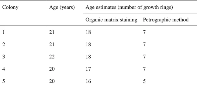
Marschal et al.(2004) also reported that conventional petrographic method of direct microscope observation detected about 7 rings on the cross section of a 21 – 22 year old colony, while organic matrix staining method showed 17 – 18 rings. The organic matrix staining method showed underestimation of 3 – 4 rings in comparison with the actual age (Table 2.1)
However most these study focusing on reef-building corals or red coral (C.
rubrum) and research on determining growth bands of JPCs is limited.
17
Growth rates of JPCs differ significantly depending on coral species, habitat and environmental conditions. Diametric growth rates of the P. japonicum were slow.
Different species showed different growh rate eventhough the same species and the same habitat. For example the same species but one colony showed 0.20±0.08, while another one was (0.27±0.01). While linear growth rate of this species range from 2,22±0.82-6.66±5.52 mm per year (Luan et al., 2013). C. elatius showed a diametric growth rate of 0.30±0.04 and linear growth rate of 2.76±2.09 mm yr-1. The diametric and linear growth rate of C. konojoi showed a 0.44±0.04 and 7.60±0.75 mm yr-1, respectively (Luan et al., 2013).
Other PC species have been found growth faster: for Mediterranean red coral (C. rubrum), 0.35±0.15 (Marchal et al., 2004); 0,24 ± 0.05 (Gallmetzer et al., 2010);
0.62±0.19 (Bramanti et al., 2005). Information on growth rate of other species are listed in Table 2.2
18
Table 2.1: Actual age and estimate obtain by staining the organic matrix and by the petrographic for five colonies collected from the experiment panels (Marschal et al, 2004)
Colony Age (years) Age estimates (number of growth rings)
Organic matrix staining Petrographic method
1 21 18 7
2 21 18 7
3 22 18 7
4 20 17 7
5 20 16 5
19
Marschal et al (2004) used staining organic matrix matrix method for aging C.
rubrum (Fig. 2.3). . This method provided high resolution and clearer growth rings of C.
rubrum. However, our recent study on P. japonicum showed that the annual growth rings were not more clearly visible in the stained slabs (using organic matrix staining method) than un-stained slab (using a high resolution VHX-1000 digital microscope) of skeleton of JPC because it is likely that the low concentration of organic matrix in the JPC (Luan et al., 2013).
2.5 TRACE ELEMENT DISTRIBUTION IN THE SKELETONS OF PRECIOUS
CORALS OF PRECIOUS CORAL AND ITS ROLES IN
PALEOENVIRONMENT RECONSTRUCT.
Chemical analyses of carbonate skeletons in precious corals found that the mineralised hard tissues of precious corals are composed of axial skeleton and sclerites (Fig. 2.4). Both sclerites and skeletons of corals are mainly made of calcium carbonate (CaCO3) crystallized in the form of calcite and small amounts of trace elements also found (Maté et al., 1986; Hasegawa and Iwasaki, 2010; Debreuil et al., 2011)
Trace elements are incorporated in corals in the formation of skeleton and this can results in the production of growth rings composed of inorganic elements. In order to get this information, several studies have determined surface composition of the skeleton with other methods.
Using X-ray Fluorescence (XRF) method, Mg, Sr, Ba, I, and Mo concentrations have also been confirmed in P. japonicum by two-dimensional images (Hasegawa et al., 2010). The results showed that strong correlation between Mg and
20
growth rings on the skeleton of P. japonicum obtained by XRF mapping analysis has been reported by Hasegawa et al. (2010), while a weak correlation between Sr concentration and growth rings on the skeleton of JPC obtained by XRF and Japanese white coral obtained by EPMA analysis (Hasegawa et al., 2012). Hasegawa et al (2012) also reported that Ca is distributed homogeneously while Mg concentration is distributed concentrically forming growth rings.
The Mg/Ca and Ba/Ca ratios in the skeletons of JPC can reflect the characteristics of corals habitats. Hasegawa et al (2012) reported that P. japonicum, C.
elatius, C. konojoi co-habit in sea-floors around Japan and the Mg/Ca and Ba/Ca ratios in these three corals collected from the same area were within similar ranges without species-specific differences. Comparison between Mg/Ca and Ba/Ca ratios in corals from the Mediterranean Sea and Japanese waters and the sea around the Midway Islands, Hasegawa et al (2012) suggested that these trace element composition differed depending on their habitats (detailed discussion in the chapter 4)
Mg/Ca and Sr/Ca ratios in the skeletons of JPC of present study determined by EPMA analysis are well agreed with those in the skeletons of C. rubrum measured by XRF (Weinbauer and Vellmirov, 1995).
Luan et al.,(unpublished) investigated the distribution of magnesium (Mg), phosphorus (P), sulfur (S) and strontium (Sr) along annual growth rings of JPC skeleton using micro X-ray fluorescence (µ-XRF). The results showed that The Mg, P and S distribution in µ-XRF mapping images correspond to the dark and light bands along the AGRs in microscopic images of the coral skeleton. The report also showed the distribution pattern of S, P and Mg in the axial skeleton of JPC (P. japonicum) reveals linkage between the trace element distribution and the formation of dark/light bands
21
along the AGRs. S and P were distributed in the organic matrix (OM) rich dark bands, while Mg was distributed in the light bands of the AGRs (detailed discussion in the chapter 5).
Besides, the speciation of sulfur using X-ray absorption near edge spectroscopy (XANES) along the annual growth rings (AGRs) in the skeleton of Paracorallium japonicum were also investigated. The results revealed XANES analysis showed that inorganic sulphate is the major species of S in the skeleton of P. japonicum with a ratio of 1:20 for organic and inorganic sulphate (Luan et al., unpublished) (in the chapter 5)
Previous reports also suggested that trace elements roles in the skeleton of coral to reconstruct paleoenvironment. For instance, Weinbauer et al (2000b) showed that the Mediterranean red coal was sensitive to increase in temperature and light. The incorporation of Mg and Sr in the skeleton may be affected under changes in climate and increase in temperature and ultraviolet radiation. As a result, the Mg/Ca and Sr/Ca ratios of red coral skeletons has been considered as ecological indicator.
The variability of Mg, Sr and Ca concentrations in the skeleton types of the red coral (C. rubrum) based on X-ray fluorescence spectroscopy and microprobe analysis as a prerequisite for their use as ecological indicators (Weinbauer et al., 2000a).
By using secondary ion mass spectrometry (SIMS), Heikoop et al.(2002) also reported a temperature dependency of skeletal Mg/Ca ratios in the precious coral, while Bond et al (2005) also using SIMS to analyze Mg/Ca ratios of high-Mg calcite loculi with skeleton of a shallow water gorgonian Plexaurella dichotoma from Bermusa and found a positive correction between Mg/Ca ratios and annual sea surface temperature.
Skeletal Mg/Ca ratio in deep-sea PCs have strong potential as a geochemical indicator for temperature (Suzuki et al., 2010). These authors also analyzed chemical
22
compositions of C. konojoi which is collected off Kochi, Japan including skeletal Mg/Ca and Sr/Ca ratios. This result showed skeletal Mg/Ca ratios from core to the margin of the section declined by about 10 mmol mol-1 and this decrease in the Mg/Ca ratio corresponded to a cooling of approximately 1.7-2.5oC because Mg/Ca ratios depended on temperature with ratio of 4-6 mmol mol-1oC-1, while the increase in oxygen isotope ratios from the centre to the margin along the transect was about 2‰, suggesting approximately 8oC of warming if typical temperature dependency of oxygen isotope ratios (- 0.22‰. C-1) (Weinbauer et al., 2000a)
The studies on biochemistry of PC in general and JPCs in particular is very limited so these studies provide vital important to better understanding about these species and marine organism as well.
23
Species Location Depth Basal
diameter (mm)
Linear growth rate
Diametric growth rate
Age Method Reference
Paracorallium japonicum
(Japanese red coral)
Off Amami Island, Kagoshima, Japan
207 4.20±1.48 5.68 ±2.6 0.24±0.02 17.60±6.3 5
Petrographic method (VHX-1000 digital microscope)
(Luan et al., 2013)
Off Amami Island, Kagoshima, Japan
212 13.05±6.58 5.82 ±
4.86
0.27±0.01 50.00±24.
79
Petrographic method (VHX-1000 digital microscope)
(Luan et al., 2013)
Off Muroto, Kochi, Japan
94 8.23±5.60 1.78±0.67 0.20±0.08 44.50±24.
15
Petrographic method (VHX-1000 digital microscope)
(Luan et al., 2013)
Off Kochi, Japan 100 0.3±0.08 OMS method (Iwasaki and
Suzuki, 2010)
Off Goyo, Japan 140 0.34-0.50 Infrared
radiation
(Iwasaki and Suzuki, 2010)
24
Corallium elatius (pink coral)
Okinawa, Japan 200-300 0.26-0.28 Infrared radiation method
(Bruckner and Roberts, 2009)
Okinawa, Japan 100-300 0.15 210Pb dating
method
(Hasegawa and Yamada, 2010) Off Ryukyu Islands,
Okinawa, Japan
200-300 17.00±13.20 2.76 ± 1.70
0.30±0.04 53.50±33.
19
(Luan et al., 2013)
Corallium konojoi (Japanese white coral)
Japan to northern Philippines Islands
50-382 0.58 (Iwasaki and
Suzuki, 2010)
Off Ashizuri, Kochi, Japan
108 7.18±4.21 7.15 ±
0.53
0.44±0.04 16.25±9.4 3
(Luan et al., 2013)
25
Fig. 2.4 Living colony of Mediterranean red coral (Corallium rubrum) (A). Axial skeleton of C. rubrum (B) and Sclerites of this coral (C) (Debreuil et al., 2011)
26
REFERENCES
Andrews, A.H., Cordes, E.E., Mahoney, M.M., Munk, K., Coale, K.H., Cailliet, G.M., Heifetz, J., 2002. Age, growth and radiometric age validation of a deep-sea, habitat-forming gorgonian (Primnoa resedaeformis) from the Gulf of Alaska.
Hydrobiologia 471, 101-110.
Bayer, F.M., Cairns, S.D., 2003. A new genus of the scleraxonian family Coralliidae (Octocorallia: Gorgonacea). Proc. Biol. Soc. Wash. 116, 222-228.
Bond, Z.A., Cohen, A.L., Smith, S.R., Jenkins, W.J., 2005. Growth and composition of high-Mg calcite in the skeleton of a Bermudian gorgonian (Plexaurella dichotoma):
Potential for paleothermometry. Geochemistry, Geophysics, Geosystems 6, 1-10.
Bramanti, L., Magagnini, G., De Maio, L., Santangelo, G., 2005. Recruitment, early survival and growth of the Mediterranean red coral Corallium rubrum (L 1758), a 4-year study. J. Exp. Mar. Biol. Ecol. 314, 69-78.
Bruckner, A.W., Roberts, G.G. (Eds.), 2009. Proceedings of the First International Workshop on Corallium Science, Management, and Trade. Silver Spring, Hong Kong.
Casu, M., Casu, D., Lai, T., Cossu, P., Curini-Galletti, M., 2008. A molecular tool for genetic surveys in the red coral (Corallium rubrum): An Inter-Simple Sequence Repeats (ISSRs) perspective. Biochem. Syst. Ecol. 36, 77-83.
Debreuil, J., Tambutté, S., Zoccola, D., Segonds, N., Techer, N., Allemand, D., Tambutté, É., 2011. Comparative analysis of the soluble organic matrix of axial skeleton and sclerites of Corallium rubrum: Insights for biomineralization.
Comparative Biochemistry and Physiology Part B: Biochemistry and Molecular Biology 159, 40-48.