Usefulness of human papillomavirus detection in oral rinse as a biomarker of oropharyngeal cancer.
著者 吉田 博
著者別表示 Yoshida Hiroshi journal or
publication title
博士論文本文Full 学位授与番号 13301甲第4581号
学位名 博士(医学)
学位授与年月日 2017‑06‑30
URL http://hdl.handle.net/2297/00049029
doi: 10.1080/00016489.2016.1274426.
Creative Commons : 表示 ‑ 非営利 ‑ 改変禁止 http://creativecommons.org/licenses/by‑nc‑nd/3.0/deed.ja
Full Terms & Conditions of access and use can be found at
http://www.tandfonline.com/action/journalInformation?journalCode=ioto20
Download by: [Fukushima Medical College] Date: 25 June 2017, At: 22:13
Acta Oto-Laryngologica
ISSN: 0001-6489 (Print) 1651-2251 (Online) Journal homepage: http://www.tandfonline.com/loi/ioto20
Usefulness of human papillomavirus detection in oral rinse as a biomarker of oropharyngeal cancer
Hiroshi Yoshida, Shigeyuki Murono, Takayoshi Ueno, Yosuke Nakanishi, Akira Tsuji, Miyako Hatano, Kazuhira Endo, Satoru Kondo, Hisashi Sugimoto, Naohiro Wakisaka & Tomokazu Yoshizaki
To cite this article: Hiroshi Yoshida, Shigeyuki Murono, Takayoshi Ueno, Yosuke Nakanishi, Akira Tsuji, Miyako Hatano, Kazuhira Endo, Satoru Kondo, Hisashi Sugimoto, Naohiro Wakisaka
& Tomokazu Yoshizaki (2017) Usefulness of human papillomavirus detection in oral rinse as a biomarker of oropharyngeal cancer, Acta Oto-Laryngologica, 137:7, 773-777, DOI:
10.1080/00016489.2016.1274426
To link to this article: http://dx.doi.org/10.1080/00016489.2016.1274426
Published online: 17 Jan 2017.
Submit your article to this journal
Article views: 94
View related articles
View Crossmark data
RESEARCH ARTICLE
Usefulness of human papillomavirus detection in oral rinse as a biomarker of oropharyngeal cancer
Hiroshi Yoshida
a, Shigeyuki Murono
a,b, Takayoshi Ueno
a, Yosuke Nakanishi
a, Akira Tsuji
c, Miyako Hatano
a, Kazuhira Endo
a, Satoru Kondo
a, Hisashi Sugimoto
a, Naohiro Wakisaka
aand Tomokazu Yoshizaki
aa
Department of Otolaryngology-Head and Neck Surgery, Kanazawa University, Kanazawa, Japan;
bDepartment of Otolaryngology, Fukushima Medical University, Fukushima, Japan;
cDepartment of Otolaryngology, Toyama City Hospital, Toyama, Japan
ABSTRACT
Conclusion: The detection of human papillomavirus (HPV)-DNA in oral rinse with auto-nested GP5 þ / GP6 þ PCR is useful as a biomarker of oropharyngeal cancer.
Background: This study aimed to determine the usefulness of oral rinse to detect HPV-DNA as a bio- marker of HPV-positive oropharyngeal cancer (OPC).
Patients and methods: One hundred and ten patients with various head and neck diseases, including 19 patients with OPC, were enrolled. Oral rinse and tonsillar swab were collected, and auto-nested GP5 þ /GP6 þ PCR for HPV-DNA was performed. For oropharyngeal cancer, p16 immunostaining was also conducted.
Results: The rate of HPV-DNA detection in both oral rinse and tonsillar swab was significantly higher in OPC compared with non-OPC upper respiratory tract cancer and non-cancer diseases. HPV-DNA was detected in oral rinse in nine out of 12 p16-positive OPC cases, while none of the p16-negative OPC cases demonstrated detectable HPV-DNA. All p16-positive cases were also positive for HPV-DNA in tumor tissue. Based on p16 immunostaining, the sensitivity and specificity of HPV-DNA detection in oral rinse were 75% and 100%, respectively. Among eight of nine evaluable OPC cases positive for HPV-DNA in oral rinse at diagnosis, HPV-DNA was undetectable in oral rinse in seven cases after treatment.
ARTICLE HISTORY Received 18 October 2016 Revised 5 December 2016 Accepted 8 December 2016
KEYWORDS
Human papillomavirus; oral rinse; oropahryngeal cancer;
biomarker; auto-nested PCR
Introduction
Human papillomavirus (HPV) is a causative agent of cer- vical cancer [1]. On the other hand, it has become apparent that HPV infection is a principal cause of a distinct form of oropharyngeal cancer (OPC) [2]. According to epidemio- logical reports, the incidence of HPV-positive OPC increased from 16% in the 1980s to 72% in the early 2000s in the US [3,4]. Considering the trend in the US, the inci- dence of HPV-positive OPC is expected to increase in Japan.
In contrast to classic OPC, HPV-related OPC generally arises from deep crypts of palatine tonsils. Therefore, it is difficult to detect it in the early stage with regular exami- nations. In addition, anatomic change after surgery as well as fibrous scar formation after chemoradiotherapy make it difficult to visualize recurrent lesions. In consideration of those circumstances, HPV may be an attractive target to detect inconspicuous lesions of OPC. From this perspec- tive, we focused on a simple and easy method, oral rinse, to detect HPV. Here, we report the usefulness of HPV- DNA detection in oral rinse as a biomarker of HPV- positive OPC.
Patients and methods Patients
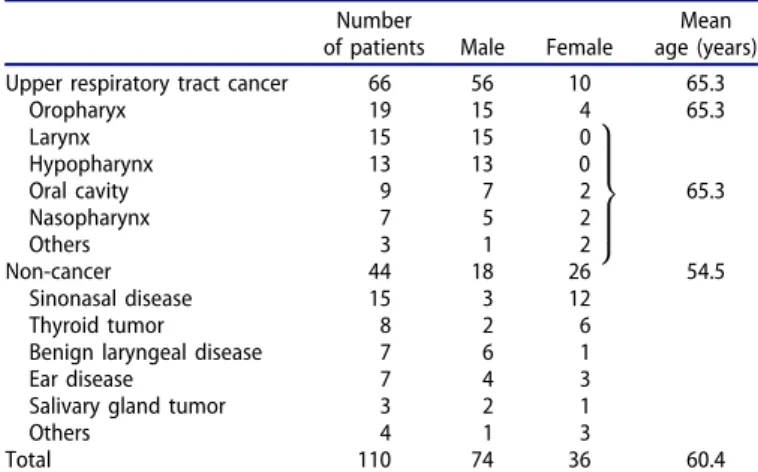
One hundred and ten patients with various head and neck diseases including 19 OPCs treated in Kanazawa University Hospital (Kanazawa, Japan) and Ishikawa Prefectural Central Hospital (Kanazawa, Japan) were enrolled in this study. The clinical characteristics of the patients are shown in Table 1. This study was approved by the Ethics Committee of Kanazawa University School of Medicine, and Ishikawa Prefectural Central Hospital, and written consent was obtained from all enrolled patients.
Sample collection
With respect to the oral rinse sample, 20 mL of normal saline gargled for 20 s was collected and centrifuged at 3000 rpm for 10 min. The pellet was suspended in 2.0 mL of preservative solution for liquid-based cytology (Medical &
Biological Laboratories, Nagoya, Japan) and stored at 4
C until use. With respect to the tonsillar swab sample, scratch- ing of the palatine tonsil using a swab for uterine cervix
CONTACTShigeyuki Murono [email protected] Department of Otolaryngology, Fukushima Medical University, 1 Hikarigaoka, Fukushima 960-1295, Japan ß2017 Acta Oto-Laryngologica AB (Ltd)
ACTA OTO-LARYNGOLOGICA, 2017 VOL. 137, NO. 7, 773–777
http://dx.doi.org/10.1080/00016489.2016.1274426
cytodiagnosis was performed, and the sample was stored at 4
C until use after being suspended in a preservative solution.
HPV-DNA detection
DNA was extracted from all samples before treatment using a DNA extraction kit (G&G Science, Fukushima, Japan) according to the manufacturer ’ s protocol. b -Globin was first amplified by polymerase chain reaction (PCR) to confirm the adequacy of the extracted DNA in all samples. In b-globin-positive samples, PCR using GP5 þ and GP6 þ primers, which is widely used for the detection of a broad spectrum of mucosotropic HPV genotypes, was per- formed [5–7]. This generates an 140-bp-long fragment from the HPV L1 structural gene.
Samples that were negative in the first GP5þ/
GP6 þ PCR were re-amplified using the same set of pri- mers, which was defined as auto-nested PCR in this study.
Thirty-six cycles and 20 cycles of amplification were per- formed in the first and second PCR, respectively. Each cycle consisted of 20 s at 94
C for denaturation, 30 s at 48
C for annealing, and 30 s at 72
C for extension. The amplified DNA was subjected to electrophoresis on 2%
agarose gel, and then visualized by ultraviolet illumination using ethidium bromide. The negative control included all reagents except for DNA, while the p1203 PML2d HPV-16 plasmid, which was a gift from Peter Howley (Addgene plasmid # 10869), was used as a positive control.
HPV genotyping
HPV genotyping using HPV Geno Array Test Kits (Hybribio, Hong Kong, China) was performed according to the manufacturer’s protocol for HPV-DNA-positive samples [8]. This can detect 37 HPV genotypes, consisting of 15 high-risk types (16, 18, 31, 33, 35, 39, 45, 51, 52, 53, 56, 58, 59, 66, and 68), six low-risk types (6, 11, 42, 43, 44, and CP8304), and 16 probably low-risk types (26, 34, 40, 54, 55, 57, 61, 67, 69, 70, 71, 72, 73, 82, 83, and 84), by flow- through hybridization using HPV-DNA amplified by PCR.
Immunohistochemistry
Immunostaining for p16 was also performed in 19 OPC cases as described previously [9]. A mouse monoclonal anti- body against human p16 (Santa Cruz Biotechnology, Dallas, TX) was used at a dilution of 1:200. Immunohistological p16 positivity was evaluated as described previously [10].
Statistical analysis
Differences in the age and HPV detection between p16-posi- tive and -negative OPC were analyzed using the Mann- Whitney U-test. The sex, smoking status, T stage, N stage, and subsite were analyzed using two-tailed Fisher ’ s exact test. All analyses were carried out using SPSS19.0 software (SPSS Inc., Chicago, IL). In all tests, p < 0.05 was considered significant.
Results
HPV-DNA detection in head and neck diseases
b-Globin was detected in all oral rinse and tonsillar swab samples. With respect to HPV-DNA detection, adding the second PCR clearly increased the detection threshold, as shown in Figure 1. In accordance, most HPV-DNA-positive samples were identified using the second PCR in both oral rinse and tonsillar swab in the present study, as shown in case 2 of Figure 2.
One hundred and ten cases were divided into three groups: OPC, upper respiratory tract cancer other than OPC (URTC), and non-cancer. In oral rinse, HPV-DNA was detected in nine of 19 cases (47.4%) with OPC, eight of 47 cases (17.0%) with URTC, and seven of 44 cases (15.9%) with non-cancer (Table 2). In tonsillar swab, HPV-DNA was
Figure 1. Detection of HPV-DNA in the first and second PCR using gradient concentrations of the positive control.
Table 1. Patient characteristics.
Number
of patients Male Female
Mean age (years)
Upper respiratory tract cancer 66 56 10 65.3
Oropharyx 19 15 4 65.3
Larynx 15 15 0
Hypopharynx 13 13 0
9>
>>
>=
>>
>>
;
Oral cavity 9 7 2 65.3
Nasopharynx 7 5 2
Others 3 1 2
Non-cancer 44 18 26 54.5
Sinonasal disease 15 3 12
Thyroid tumor 8 2 6
Benign laryngeal disease 7 6 1
Ear disease 7 4 3
Salivary gland tumor 3 2 1
Others 4 1 3
Total 110 74 36 60.4
774 H. YOSHIDA ET AL.
detected in 10 of 19 cases (52.6%) with OPC, four of 47 cases (8.5%) with URTC, and four of 44 cases (9.1%) with non-cancer (Table 2). The concordance of HPV-DNA detec- tion between oral rinse and tonsillar swab was 94.7% in OPC, 85.1% in URTC, and 79.5% in non-cancer.
HPV-DNA detection and p16 immunohistochemistry in oropharyngeal cancer
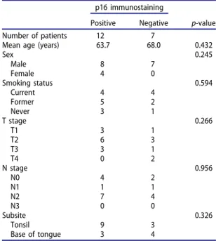
Because almost half of the cases with OPC were positive for HPV-DNA in both oral rinse and tonsillar swab, we next investigated the association with p16 immunostaining in OPC. The clinical characteristics of OPC patients are shown in Table 3. Twelve of the 19 OPC cases were positive for p16, while the remaining seven were negative. There were no significant differences with respect to the sex, smoking status, T stage, N stage, or tumor sub-site between p16-posi- tive and -negative cases.
In addition, we also performed PCR using GP5þ/
GP6 þ primers to detect HPV-DNA in samples obtained from formalin-fixed paraffin-embedded tissue of OPC, and demonstrated that all p16-positive cases were also positive for HPV-DNA. Therefore, there was no p16-positive case who was negative for HPV in the present study.
In oral rinse, HPV-DNA was detected in nine of 12 p16- positive cases, while none of the seven p16-negative cases demonstrated HPV-DNA, showing a significant difference (p ¼ 0.003) (Table 4, Figure 2). Similarly, in tonsillar swab, HPV-DNA was detected in 10 of 12 cases, while none of the seven p16-negative cases demonstrated HPV-DNA, showing a significant difference (p ¼ 0.001) (Table 4, Figure 2). Based on p16 immunostaining, the sensitivity, specificity, positive predictive value, and negative predictive value of HPV-DNA detection in oral rinse and tonsillar swab were 75, 100, 100, and 70%, and 83, 100, 100, and 78%, respectively.
HPV-DNA detection in oral rinse after treatment in oropharyngeal cancer
Eight of nine OPC patients positive for HPV-DNA in oral rinse at diagnosis were further investigated after treatment (Table 5). In these eight, oral rinse samples were collected 5–15 months (mean ¼ 9.6 months) after the completion
of treatment. As shown in Table 5, HPV-DNA was undetectable in oral rinse in seven of eight cases after treat- ment, while the remaining case was positive for HPV-DNA.
Four were treated with chemoradiotherapy, and four were
Figure 2. HPV-DNA detection in oropharyngeal cancer cases. Cases 1 and 2 are p16-positive, while case 3 is p16-negative. PC: positive control; OR: oral rinse; NC:
negative control; NS: non-specific band.
Table 2. Positive rate of HPV-DNA detection in oral rinse and tonsillar swab.
Oral rinse Tonsillar swab
Oropharyngeal cancer 9/19 (47.4%) 10/19 (52.6%)
Non-oropharyngeal upper respiratory tract cancer
8/47 (17.0%) 4/47 (8.5%)
Non-cancer 7/44 (15.9%) 4/44 (9.1%)
Table 3. Characteristics of oropharyngeal cancer patients.
p16 immunostaining
Positive Negative p-value
Number of patients 12 7
Mean age (years) 63.7 68.0 0.432
Sex 0.245
Male 8 7
Female 4 0
Smoking status 0.594
Current 4 4
Former 5 2
Never 3 1
T stage 0.266
T1 3 1
T2 6 3
T3 3 1
T4 0 2
N stage 0.956
N0 4 2
N1 1 1
N2 7 4
N3 0 0
Subsite 0.326
Tonsil 9 3
Base of tongue 3 4
Table 4. Comparison between HPV-DNA detec- tion and p16 immunostaining.
p16 immunostaining
HPV-DNA Positive Negative p-value
Oral rinse 0.003
Positive 9 0
Negative 3 7
Tonsillar swab 0.001
Positive 10 0
Negative 2 7
ACTA OTO-LARYNGOLOGICA 775
treated with surgery. All patients achieved a complete response after treatment, and were without any evidence of disease at sample collection.
HPV genotyping
HPV genotyping was further performed in 42 samples with HPV-DNA detection. A certain HPV genotype was detected in only 14 (35.0%) samples, while no types of HPV were detected in the remaining samples. In p16-positive OPC, HPV type 16 (HPV16) was detected in four of nine oral rinse samples, and four of 10 tonsillar samples, while HPV type 58 was detected in one tonsillar swab sample. On the other hand, HPV genotyping from tissues of OPC patients identified HPV16 in 11 of 12 p16-positive OPC cases and HPV58 in the remaining case. In non-oropharyngeal URTC samples, HPV16 was detected in one of eight cases which were HPV-positive in oral rinse samples, and one of four tonsillar swab samples. In non-cancer samples, HPV16 was detected in three of 15 cases which were HPV-positive in oral rinse samples, and three of eight tonsillar swab samples.
Only a single sub-type of HPV could be detected in all evaluable samples.
Discussion
The prevalence of oral HPV infection among healthy persons was reported to be 5.7–8.3% in the US [2]. On the other hand, the HPV-positive rate in oral rinse in non-cancer patients was 15.9% in the present study, being higher than in previous reports. Adding a second PCR may increase the detection threshold, while a difference in the patient back- ground may affect the positive rate. Accordingly, the avail- ability of auto-nested GP5 þ /GP6 þ PCR to detect HPV-DNA was described by Remmerbache et al. [7]. Furthermore, the increased sensitivity of auto-nested GP5þ/GP6 þ PCR com- pared with conventional single-round GP5þ/GP6 þ PCR was demonstrated [11]. Thus, auto-nested PCR may be a useful method to investigate samples with low viral loads [11].
This was also supported by the fact that only one third of the samples with HPV-DNA detection identified certain HPV subtypes in the present study, which suggests the higher detection sensitivity of auto-nested PCR than the regular gen- otyping kit.
There have been several reports with respect to HPV- DNA detection in oral rinse, showing positive rates from 39 – 54% in HPV-related OPC [12 – 14]. In comparison with these reports, the present study demonstrated a higher posi- tive rate of 75% in p16-positive OPC. This is in accordance with the favorable result of auto-nested PCR as described above.
HPV-DNA was not detected in oral rinse in three p16- positive OPC cases. Among them, two involved OPC arising at the base of the tongue, and showing a submucosal appearance. Thus, the HPV-DNA detection rate in p16-posi- tive OPC was 89% (eight of nine) at sub-sites of the tonsil, and 33% (one of three) at the base of the tongue. Therefore, it is considered that HPV-DNA in oral rinse could be more detectable in OPC in the tonsil than at the base of the tongue. Wang et al. [14] similarly reported a higher detec- tion rate in HPV-related OPC of the tonsil (67%) than at the base of the tongue (24%).
In the present study, the concordance rate of HPV-DNA detection between oral rinse and tonsillar swab was 94.7%
in OPC, 85.1% in URTC, and 79.5% in non-cancer. In add- ition, based on p16 immunostaining, the sensitivity and spe- cificity of HPV-DNA detection were 75% (nine of 12) and 100% (seven of seven) in oral rinse, and 83.3% (10 of 12) and 100% (seven of seven) in tonsillar swab, respectively. A high concordance rate suggests the usefulness of unobstruc- tive sample collection, namely oral rinse, being in accord- ance with a study comparing oral rinse and cytology brush sampling [15].
Interestingly, HPV-DNA in oral rinse after treatment became negative in seven of eight evaluable cases. Although we did not observe any recurrent cases among these eight patients, HPV-DNA in oral rinse after treatment could be a biomarker of the disease status. Rettig et al. [12] demon- strated that persistent HPV16 DNA in oral rinse after treat- ment was associated with poorer disease-free survival and overall survival. Further research with respect to the preva- lence of HPV-DNA in oral rinse after treatment detected with our strategy is warranted.
One of the limitations of the present study is the small number of cases of OPC. However, we could at least dem- onstrate the superiority of our method to detect HPV- DNA in oral rinse. Another limitation is the relatively short period of post-treatment observation. Actually, it is unclear whether persistent HPV-DNA in oral rinse after treatment detected with our procedure is associated with persistent disease, or whether HPV-DNA will be re-detected in cases of disease recurrence at the primary site. Another limitation is the low rate of HPV genotyping in oral rinse samples. The HPV genotyping procedure included one round of PCR consisting of 40 cycles accord- ing to the manufacturer ’ s protocol, while the second PCR was added after the first PCR to identify HPV-DNA in oral rinse samples in the present study. Therefore, we con- sider that the procedural difference between genotyping and auto-nested PCR was a major reason for the low rate of HPV genotyping in oral rinse samples. Further study is planned to answer these limitations.
Table 5.Comparison of HPV-DNA detection in oral rinse before and after treatment in p16-positive oropharyngeal cancer.
HPV-DNA detection in oral rinse
Case Subsite TNM Before After State
1 Tonsil T3N2cM0 þ CR
2 Tonsil T1N2bM0 þ CR
3 Tonsil T3N0M0 þ CR
4 Tonsil T1N2bM0 þ CR
5 Tonsil T2N2cM0 þ þ CR
6 Tonsil T2N2bM0 þ CR
7 Tonsil T2N0M0 þ CR
8 Tonsil T2N2bM0 þ CR
CR: Complete response.
776 H. YOSHIDA ET AL.
Conclusion
In conclusion, HPV-DNA detection in oral rinse is useful as a biomarker of HPV-related OPC. Auto-nested GP5þ/
GP6 þ PCR was a helpful method for this purpose.
Acknowledgments
We thank Dr P. Howley for providing p1203 PML2d HPV16. The authors would like to thank Dr M. Namiki and Dr H. Yaegashi for technical advice and helpful discussion. This study was supported in part by a research grant from the Daiwa Securities Health Foundation [42-17].
Disclosure statement
The authors report no conflicts of interest. The authors alone are responsible for the content and writing of the paper.
Funding
This study was funded in part by the Daiwa Securities Health Foundation [42-17].
References
[1] Chaiwongkot A, Pientong C, Ekalaksananan T, Kongyingyoes B, Thinkhamrop J, Yuenyao P, Sriamporn S.
Evaluation of primers and PCR performance on HPV DNA screening in normal and low grade abnormal cervical cells.
Asian Pac J Cancer Prev 2007;8:279
–82.
[2] Gillison ML, Broutian T, Pickard RK, Tong ZY, Xiao W, Kahle L, et al. Prevalence of oral HPV infection in the United States, 2009
–2010. JAMA 2012;307:693
–703.
[3] Chaturvedi AK, Engels EA, Pfeiffer RM, Hernandez BY, Xiao W, Kim E, et al. Human papillomavirus and rising oro- pharyngeal cancer incidence in the United States. J Clin Oncol 2011;29:4294
–301.
[4] Edelstein ZR, Schwartz SM, Hawes S, Hughes JP, Feng Q, Stern ME, et al. Rates and determinants of oral human papillomavi- rus infection in young men. Sex Transm Dis 2012;39:860
–7.
[5] Jacobs MV, de Roda Husman AM, van den Brule AJ, Snijers PJ, Meijer CJ, Walboomers JM. Group-specific differentiation between high- and low-risk human papillomavirus genotypes by
general primer-mediated PCR and two cocktails of oligonucleo- tide probes. J Clin Microbiol 1995;33:901
–5.
[6] Harwood CA, Spink PJ, Surentheran T, Leigh IM, de Villiers EM, McGregor JM, et al. Degenerate and nested PCR: a highly sensitive and specific method for detection of human papillo- mavirus infection in cutaneous warts. J Clin Microbiol 1999;37:3545
–55.
[7] Remmerbach TW, Brinckmann UG, Hemprich A, Chekol M, K
€uhndel K, Liebert UG. PCR detection of human papillomavi- rus of the mucosa: comparison between MY09/11 and GP5
þ/ 6
þprimer sets. J Clin Virol 2004;30:302
–8.
[8] Nakashima K, Shigehara K, Kawaguchi S, Wakatsuki A, Kobori Y, Nakashima K, et al. Prevalence of human papilloma- virus infection in the oropharynx and urine among sexually active men: a comparative study of infection by papillomavirus and other organisms, including
Neisseria gonorrhoeae, Chlamydia trachomatis, Mycoplasma spp., and Ureaplasma spp.BMC Infect Dis 2014;14:43.
[9] Wakisaka N, Yoshida S, Kondo S, Kita M, Endo K, Tsuji A, et al. HPV status determines the efficacy of adjuvant chemo- therapy with S-1, an oral fluorouracil prodrug, in oropharyngeal cancer. Ann Otol Rhinol Laryngol 2015;124:400
–6.
[10] Loizou C, Laurell G, Lindquist D, Ofverman C, Stefansson K,
€Nylander K, Olofsson K. Incidence of tonsillar cancer in north- ern Sweden: Impact of human papilloma virus. Oncol Lett 2015;10:3565
–72.
[11] Souto Damin AP, Guedes Frazzon AP, de Carvalho Damin D, Beck Biehl H, Abruzzi de Oliveira L, Auler R, et al Detection of human papillomavirus DNA in squamous cell carcinoma of the esophagus by auto-nested PCR. Dis Esophagus 2006;19:64
–8.
[12] Rettig EM, Wentz A, Posner MR, Gross ND, Haddad RI, Gillison ML, et al. Prognostic implication of persistent human papillomavirus type 16 DNA detection in oral rinses for human papillomavirus-related oropharyngeal carcinoma. JAMA Oncol 2015;1:907
–15.
[13] Dang J, Feng Q, Eaton KD, Jang H, Kiviat NB. Detection of HPV in oral rinse samples from OPSCC and non-OPSCC patients. BMC Oral Health 2015;15:126.
[14] Wang Y, Springer S, Mulvey CL, Silliman N, Schaefer J, Sausen M, et al. Detection of somatic mutations and HPV in the saliva and plasma of patients with head and neck squamous cell carcinomas. Sci Transl Med 2015;7:293ra104.
[15] Steinau M, Reddy D, Sumbry A, Reznik D, Gunthel CJ, Del Rio C, et al. Oral sampling and human papillomavirus gen- otyping in HIV-infected patients. J Oral Pathol Med 2012;41:288
–91.
ACTA OTO-LARYNGOLOGICA 777