Detection of Serum ƒ¿-fetoprotein levels in Rats Bearing Hepatocellular Carcinoma Induced by Diethylnitrosamine
Tomohiko SETOGUCHI, Tetsuro KANDA, Hideo TERAO, Hiroto YAMASHITA, and Hideyo ITAKURA
D
epartment of Pathology, Institute for Tropical Medicine, Nagasaki University, Nagasaki, Japan
(Chief: Hideyo ITAKURA)
ABSTRACT: Serum ƒ¿-Fetoprotein (AFP) levels of hepatocellular carcinoma-bearing Wistar rats treated continuously with 50 ppm of Diethylnitrosamine (DEN) in drinking water were examined. Various types of tumorous changes in the liver were observed succeedingly as follows; 1) area of hyperplasia appearing after 2 months of treatment, 2) nodular hyper-
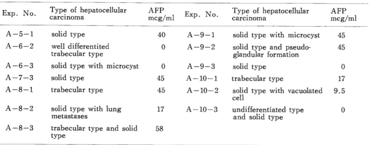
plasia after 3 months of treatment, 3) area of basophilic cells after 2.5 months of treatment, 4) nodular basophilic cells after 3.5 months of treatment and 5) hepatocellular carcinoma including trabecular type, solid type, tubular type, undifferentiated type and mixed type of these patterns after 3.5 months of treatment. The AFP appeared suddenly at 3.5 months from commencementand the number of cases which had hepatocellular carcinoma showing positive serum AFP was 9 out of 13 rats. The titer of serum AFP varied from 9.5 mcg/ml to 58
mcg/ml by single radial immunodiffusion (S. R. I. D.) method. Although any association of serum AFP with non-cancerous or pre-cancerous lesion of the liver within 3.5 months was not revealed, the correlation between serum AFP level and degree of differentiation of hepatocellu- lar carcinoma are discussed.
whatever mechanism of AFP synthesis may concern, the detection of AFP in the pati‑
ent′s serum has a marked diagnostic value for primary hepatocellular carcinoma and embryonal
teratoma (Avelev, 1968). Many authors reported that a small amount o土DEN induced
various types of hepatic lesion and hepatocellular carcinoma in the rat (Magee, 1967;
scherener, 1972; and Takayama, 1973)・ The association of AFP with hepatocellular carcinoma induced by DEN has been confirmed in some species of mammal other than rat
(Stranislawski‑Birencwajg, 1967 ; Scherener, 1972 ; and Takayama, 1973)二
The correlation between serum AFP levels and progressive tumorous changes of the rat liver induced by DEN were examined immunologically and histologically, and a short analysis
of experimental data are given in this report・
Contribution No・ 728 from the Institute for Tropical Medicine,Nagasaki University・
Received for publication, June 13, 1975
MATERIALS AND METHODS
Animals
40 male rats of Wistar strain (30 for experiment and 10 for control) weighing 240 g on
average were used・Treatment
50 ppm of DEN (Tokyo Kasei Co・ Tokyo) in drinking water and synthetic diet (Funabashi Nojyo, Chiba) was given freely・
Experimental group
Rats were divided into 10 groups. Each group composed of 4 rats including one control was sacrificed every half or one month until1 6 months as shown in Fig. 1・
Preparation of anti‑Serum
Anti rat‑AFP horse serum was prepared by Department of Biochemistry, School o土
Medicine, Hokkaido University. Specific rabbit anti‑serum against rat AFP was prepared by immunizing a rabbit with fetal rat serum. The anti‑serum obtained was absorbed with
adult rat serum. Horse二antiserum to AFP was obtained after immunization with antigen‑
antibody complex produced by mixing newborn rat serum with monospecific rabbit serum.
Antibody titer was 1: 250 in C‑F test.
Standard serum
Purified AFP‑ from ascites of hepatoma rat by affinity chromatography was used for standard sample・ AFP concentration was 200 meg/ml・
Immunodiffusion method
S. R. I. D. method (Mancini et al., 1965) was used for detection of AFP.
Histopathological Examination
Section material of rat liver were fixed in 20% formalin and embedded in paraffin.
Each section material was cutted in 4μ in thickness・ All sections were stained with Hemato‑
xylin二Eosin, Periodic Acid Schiff reaction, Heidenhain′s Aniline Blue Method and Silver
lmpregnation for reticulum.
RESULTS
Succeeding appearances of tumorous changes of the liver are the following (Fig. 1) 1 ) Area of hyperplasia showing slight nuclear pleomorphism and clear abundant eosinophilic cytoplasm, appeared after 2 months. 2) Nodular hyperplasia showing moderate pleomor‑
phism o土nuclei, nodular proliferation and moderate irregular arrangement of hepatic cell cord,
appeared after 3 months・ 3) Area of basophilic cells showing moderate pleomorphism of nuclei with basophilic cytoplasm and irregular arrangement of hepatic cell cords, appeared
after 2.5 months. 4 ) Nodular basophilic cells showing advanced pleomorphism of nuclei and irregular arrangement of hepatic cell cords, appeared after 3.5 months. 5) Hepatocellular
carcinoma showing several kinds of histological appearance such as trabecular type, solid type, undifferentiated type, and pseudoglandular type were observed after 3.5 months.The number of animals which had hepatocellular carcinoma was thirteen in total・
A‑10 A‑2
No. of animal
30
Group A一l
15
0
良
fc c<‑.
1,二, J8
● A‑6
8
O normal
(g¥ area of hyperplasia
㊨ nodular hyperplasia
①area 。f has〇phllie cells
①㌔。dular has。ph土Iie cells
Ahepatocellular careln〇ma
・
A‑7 g
A‑8
A‑9
1 2 3 4 5 6 Month
Fig. 1. Histological findings of tumorous lesions of each rat treated with DEN ・
60
50
■・・■
巳 iZ!
。1 e
P1 3
* 30
∈ コ h lt
∽
苫 20
「‑1 こ)
>
)?
子」
10
● :;;1三〇n;三三Iular‑
o non hepat℃cellular‑
carc in℃ma
・
●
e
● ○
●
o ooo oopoop OOP OO <BォO OO ●
1 2 3 4 5 6 month durati℃n ℃f trea亡inent
Fig. 2・ Association of serum AFP with DEN・
Their histological types and the level of serum AFP of each case are showing in Table 1.
The serum AFP appeared after 3・5 months and could be detected in nine cases out of the above thirteen rats which had hepatocellular carcinoma (Fig・ 2)・ In cases of precancerous tumorous changes such as area of hyperplasia, nodular hyperplasia, area of basophilic cells and nodular basophilic cells other than hepatocellular carcinoma, no serum AFP could be revealed・
Table 1. Histological type of tumor and level of serum AFP
Exp・No・と"ypeof
arcin。m聖epatocellular孟FP eg/mlExp・No.Typ?‑
carcin。茂聖epatocellular急FP eg/ml
A二5二1
A‑6‑2 A‑6‑3 A‑7‑3
A‑8二1
A二8‑2
A‑8‑3
solid type well differentited trabecular type
solid type with microcyst solid type
trabecular type
solid type with lung metastases
trabecular type and solid type
40 A二9‑1
0 A二9‑2
A‑9‑3 45 A‑10‑1
45 A二10‑2
17 A‑10‑3 58
solid type with microcyst 45 solid type and pseudo‑ 45 glandular formation
solid type
trabecular type 17 solid type with vacuolated ・5 cell
undifferentiated type and solid type
0
DISCUSSION
Serum AFP appeared abruptly at 3.5 months士ollowing commencement of the experiment
Thereafter there was relati、rely high incidence of AFP appearance in rats bearing hepatocellular carcinoma. Kitagawa (1972) described that area of hyperplasia and nodular hyperplasia did not seem to produce AFP・ No appearance of AFP was noted within 3・5 months and any association of AFP with preneoplastic changes could not be seen in this work・ The sudden occurrence of AFP in the sera of rats with induced hepatocellular carcinoma is one evidence in favor of the suggestion that the appearance o士this protein involved a mechanism of malignant transformation and dose not concern with precancerous lesion.
Although two phase appearance of AFP had been reported in DAB carcinogenesis (Watabe, 1972), only one phase of appearance of AFP was observed in this study. Trans‑
formation of normal liver cells in precancerous stage in DAB carcinogenesis has been proved by some experiments (Onoe, 1972 and lnaoka, 1967) and it was suggested that early appearance of AFP was produced from their ovall cells・
We could not observe ovall cells in preneoplastic changes of the liver and probably that is the reason why two phase appearance of serum AFP was not seen.
The correlation between the level of AFP and the histological type of hepatocellular carcinoma had been discussed by some authors. Abelev (1968) described in his hypothesis that there might be correlation of degree of malignancy with AFP production・ In other words, rapidly growing hepatocellular carcinoma showed high level of AFP, but slowly growing nepatocellular carcinoma showed negative AFP・ Our experiments of DEN could not find out the correlation between, the AFP production and type of hepatocellular carcinoma.
Serum AFP was not found in all hepatocellular carcinoma‑bearing rats. In some cases of poorly
differentiated tumors showed relatively low level of AFP. Even in some of them no production
of AFP was noted. Stanislawski‑Birencwajg (1967) described similarly that he had not clear
evidence of association of the type of hepatocellular carcinoma with the AFP production・
we may suppose that hepatocellular carcinoma is composed of various types of AFP producing
cells, and therefore, it is very difficult to prove the relationship between the atypism of tumor tissue and the production of AFP. In view of this, further investigation of the problem appears necessary.
AcKNOWLEGEMENTS
we would like to thank professor Hidematsu Hirai and his staff of Department of llioche‑
mistry, School of Medicine, Hokkaido University, for their excellent instruction and valuable
suggestion of detection of serum AFP二 We also express our appreciation to Mr. Masachika
Senba for his valuable assistance.
REFERENCES
1) Abelev, G. I. (1968): Production of Embryonal serum alpha‑fetoglobulin by hepatoma : Review of experimental and clinical data. Cancer Res. , 28, 1344‑1350.
2) Inaoka, Y・ (1967) : Significance of so‑called ovall cell proliferation during azo二dye hepatocarcinogenesis
GANN, 58, 355‑366.
3) Kitagawa, T., et al. (1972): Alpha‑fetoprotein and hepatocarcinogenesis in rat fed 3'‑methyl‑4‑
(dimethylamino) azobenzen or N‑2‑fluorenylacetamide. Int. J. Cancer, 10, 368‑381.
4) Mancini, G., Carbonara, A・ O., and Heremans, J・ F・ (1965): Immunochemical quantitation of
antigen by single radial immunodiffusion二Immunochemistry, 2, 235‑254.
5) Magee, P. N. et al. (1967): Carcinogenic nitroso compound. Advance Cancer Res., 10, 163‑247, Academic Press.
6) Ouchterony, O・ (1953): Antigen‑antibody reaction in Gels IV types of reaction in cordinated system
of diffusion. Acta・ Path二 Microbiol・ Scand・, 32, 231‑240.
7) Onoe, T., (1972): Experimental hepatocarcinogenesis. Kanzo, Recent advances series, 77‑5 二(in
Japanese)I
8) Stranislawski‑Birencwajg, M. (1967) : Association of embryonic antigens with experimentally induced hepatic lesions in rat. Cancer Res., 31, 1192‑1194.
9) Scherener, E. et al. (1972): Quantitative study on foci of altered liver cells induced in the rat by a single dose of DEN and partial hepatectomy・ J・ Natl. Cancer lnst., 49, 93‑106・
10) Takayama, S. (1973) : Hepatocarcinogenesis by diethylnitrosamine. Igaku‑no‑Ayumi, 86, 854‑858 (in Japanese)・
ll) Watabe, H. (1971): Early appearance of embryonic alpha‑globulin in rat serum during carcinogenesis with 4‑dimethylaminoazobenzen・ Cancer Res・ , 31, 1192‑1194.
12) Watabe, H. (1974): Purification and chemical characterization of alphaィetoprotein from rat and
mouse. Int. J二 Cancer, 13, 377‑388.
Diethylnitrosamine (DEN)誘発肝癌に於ける血中AFPの測定
瀬戸口智彦,神田哲郎,寺尾英夫,山下裕人,板倉英世(長崎大学熱帯医学研究所病理学部門)
50 ppmのDENを, Wistar系ラットに6か月間継続投与し,1か月又は半月毎に,肝の発癌過程
を,病理組織学的に検討するとともに各時間におけるラット血中AFP濃度を測定した.腫瘍性病変は
次の様な結果となった.1) Area of hyperplasia: DEN投与開始后2か月目で出現,2) Nodular hyperplasia: DEN投与后3か月目で出現,3) Area of basophilic cells: 2.5か月目で出現,4) Nodular basophilic cells: 3.5か月目で出現,5) Hepatocellular carcinoma: 3.5か月目で出現,i) Trabecular type,ii) Solid type,iii) Tubular type,iv) Undifferentiated type,v) Mixed typeに 分類された.ラット血中AFP濃度はManciniの一元免疫拡散法によって半定量的に測定し,肝癌13 例中9例に陽性を認め,9.5mg/mlから58mg/mlの範囲にあった.血中AFPはDEN投与后3.5か月目 に突然検出され,謂ゆる前癌状態においては血中に証明することはできなかった.さらに,肝癌の分 化度とAFP濃度との関連性については,Solid typeには最高58mg/mlを始めとして高濃度のAFPが 検出されたにもかかわらずUndifferentiated typeを示す肝癌血中には,AFPは検出できなかった.
一応諸家の報告と一致する様であるが今後この機能についてはさらに検索をすすめねばならない.
熱帯医学 第17巻 第2号 65‑72頁, 1975年8月
窺
二== :二二二4̲‑二i ‑‑二… ‑I‑二二
Photo. 1. Solid type of hepatocellular car‑
cinoma in A‑5‑1 after 3.5 M
treatment. (H・ & E , ×214)
Photo. 3二 Solid type with microcyst in A‑
6‑3 after 4 M treatment. (H. &
E., ×214)
二1二二=:I,る‑二=‑ =
Photo. 5. Trabecular type in A‑8‑1 after 5 M treatment. (H.
&E , × 90)
、
・. ′
///
璽聖域室
&aHど真義済
∴・1 ・ ・・廿. .常4B虫 ㌔撃x y<photo. 2. Trabecular type in A‑6‑2 after
4M treatment・ (H・ & E・, ×90)
‑ ・・二=4一茂二l1 i二二)二二=
photo・ 4. Solid type in A‑7‑3 after 4.5
M treatment. (H・ &E., ×214)
. 壽・′ ノ・
JW l茂‑=
‑ # 号肴
#J /
亀子 x潜 鱒 ‑ 整ぜ義◇
fl
‑、† l‑
β ■′■}
:.I
′
静潜漣
*ォ
、
農 が 沖
藩#管だ機
・
好磨
感
挙 ヽシ
㌔:‑t二・J二.i
夢亀 ‑準豊第肴‑,砂篭凝 欝石 窟
・/妄 t
〜 / Jd
薫蒸
'津警
y静 熱◇
轡
I
;/
′・
鞠展長
号/
.・j
厳
●
■
義
一、,I ォn 海 =◇ る'
・ .〜
ヽ **
二+
〜
物 丁二〜・
‡帯
優
サ
・.t子/
葡 4*
逮義遥
●
感‑潜
∴ , ・・ . ‑ ,
Photo. 6. Solid type in A‑8‑2 after 5 M treatment. (H. & E.,
× 214)
Photo. 7・ trabecular and solid type in
A‑8‑3 after 5 M treatment・
(H.&E・, ×90)
/ //
Bl
/
hT{;5
∵ .
・∴′ ■
//
考 ◇
Photo・ 9・ Solid type with pseudoglandular
type in A‑9‑2 after 5.5 M
treatment. (H・ & E・, × 90)
謬誓*:‑が
ン.. S.
:J * l■
準▲撃藩表曜
報。 tx 藩
′・:
薫蒸
オ.・ +/
港
室砂
芸才
/
i// *
・〓v '4̀
祝さ I/)/ //・
‑//■ 一.・!
・ !
◇ベ魯
・;/
l::館 ∴
..る.‑
.>/負.:r
〜/ み
∴∴鷲 瀞
サ 書誌 y≡姿
■・・
■̀ナ
5'藁
‑ ′.
・///
//// ,
=∴
鞍
巻
′′
疾{ 祥 換
′
// /・
◇海∨
′
郡 安駕
::r/,:
戟
∴ 祭 物' 鴨◇∧
帝,. 1■
i/ち
∴▲
▼ 鷲 ///
′車
◇yx 半
: I
妻ギ
//t
▲
鴻誕
/てる,/
遠海 //サ/4
事二/・二 ;/;
染,好戦悪心 ∧莞<竃選
阜 ∴ 沖//
..:... 二
■ ■. ■■
ヽ
禦 櫛)‑ー演芸諸 宗
‑常 務緊 漕 ◇管#〉x る
嘩
・>奉.・
・ 芝淋
?/
1
・/・
//
義教 袈
"&.'‑ト ♯ /
ii //
{/
>
S/, / 婆 ,Wf*,
…義 警賀
/ ・
〜‑<
蛋 )・'
li
■ゎ・ ・.
∴ 「
・・ ・・ . / .÷
藻芸≡,賢
Photo・ ll. Solid type with vacuolar cells in A‑10‑2 after 6 M treatment.
(H.&E・, ×90)
Photo・ Solid type with microcyst in A‑9‑1 after 5・5 M treatment・
(H・&E二, ×90)
Photo・ 10・ Solid type in A‑9‑3 after
5二5 M treatment. (H・ & E・,
×90)
藩撃
Photo二 12・ Undifferentiated type in A‑
10‑3 after 6 M treatment二
(H・&E., ×90)