Dental Implant Surface Roughness and Topography
89 J. Gifu. Dent. Soc
Vol.35, No.3,89〜95 February,2009
Review Article
Dental Implant Surface Roughness and Topography: A Review of the Literature
ISABELde MONSERRATOSORIOBERNAL1), ITORISA2), KATAGIHIROKI2), TSUBOIKEN-ICHIRO2), YAMADANAOKO2), TANABETOSHI-ICHIRO2), NAGAHARAKUNITERU2)and MORIMASAHIKO3)
INTRODUCTION
Dental implant quality depends on the chemical, physical, me- chanical, and topographic characteristics of the surface1). These different properties interact and determine the activity of the at- tached cells that are close against the dental implant surface.
Dental implants have been designed to provide textures and shapes that may enhance cellular activity and direct bone apposi- tion(osseointegration)2). Osteogenesis at the implant surface is influenced by several mechanisms. A series of coordinated events, including cell proliferation, transformation of osteoblasts and bone tissue formation might be affected by different surface topographies3). There is a clinical impression that the amount of bone-to-implant contact(BIC)is an important determinant in the long-term success of dental implants. Consequently, maxi- mizing the BIC and osseointegration has become a goal of treat- ment, which is enhanced by implant surface roughness4).
The present literature review aimed to elucidate implant sur- face topography and to obtain a future perspective regarding the topography of the implant surface which could be beneficial to implant surgery when implemented in practice.
MATERIALS
A systematic online review of the main database and a manual search of relevant articles from refereed journals were per- formed. Thirty articles about surface roughness were reviewed, of which only22had the necessary information to carry out sta- tistical analysis. The categories were classified into normal bone tissue and augmented bone tissue. Of the22articles reviewed,
15belonged to the category of normal bone tissue and7to the category of augmented bone tissue.
1.Normal Bone Tissue
The influence of surface roughness on implant osseointegra- tion in normal bone tissue has been studied by various research- ers for several years, including several in vivo and in vitro stud- ies(Tables1,2and3). Buser et al.5)evaluated the influence of different surface characteristics on the bone integration of tita- nium implants and the highest extent of BIC was observed in sandblasted acid-etched surfaces with mean values of50―60%
and with an average roughness of18―23μm. Wong et al.6)found an excellent correlation between the average roughness of the implant surface and pushout failure load. In1999, it was sug- gested that the interface shear strength of titanium implants is significantly influenced by their surface characteristics7). Also, it has been suggested that only a very specific surface topography with a Ravalue(arithmetic average of absolute values of all pro- file points)1,3)between1and1.5μm provides an optimal surface for bone integration8).
Studies have shown that cells, including blood monocytes/
macrophages, are amongst the first cells to come into contact with the implant surface after its insertion. Monocytes/macro- phages have the potential to secrete a range of cytokines and growth factors, which have the capability of initiating both tissue destruction as well as healing or reparative responses. Soskolne et al.4)examined monocyte adherence to titanium discs with four different degrees of surface roughness and plastic surfaces. The results indicated that the number of monocytes attached to blasted titanium surfaces was significantly greater than to ma- The present review deals with the future perspectives regarding the topography of the implant surface which could be beneficial to implant surgery when implemented in practice. A systematic online review of the main database and a manual search of relevant articles from refereed journals were performed. Thirty articles about surface roughness were found, of which only22had information necessary to carry out statistical analysis. The categories were sepa- rated into normal bone tissue(15articles)and augmented bone tissue(7articles).
The category of normal bone tissue was subdivided into three groups: in vivo animal studies(8articles), in vivo hu- man studies(3articles), and in vitro studies(4articles). All articles that belonged to the augmented bone tissue category were in vivo studies carried out in humans. A total of423patients received1350implants, of which725 implants had accurate data.
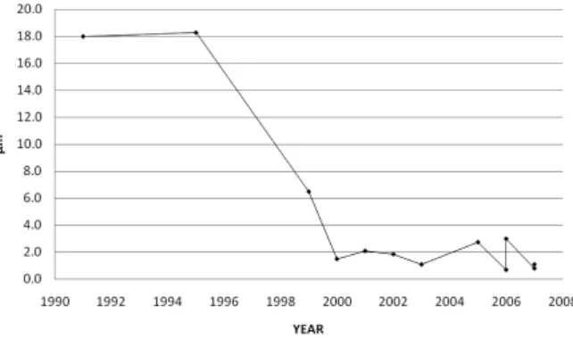
Since1991, several average degrees of roughness have been suggested to enhance implant osseointegration as fol- lows:18-23μm in1991and1995,6.5μm in1999, and <_2.7μm in2000-2007; however, these averages vary considerably, which might reflect the different types of measurements and techniques used by each author. As a result, there is currently no consensus in the degree of surface roughness that is optimum for bone cell attachment.
Key words: Dental implant, Surface roughness, Surface topography, Osseointegration
1)Department of Orthodontics, Autonomous University of State of Mexico
2)Department of Implantology, Division of Oral Pathogenesis and Disease Control, Asahi University School of Dentistry
3)Asahi University School of Dentistry Hozumi1851, Mizuho, Gifu501―0296,Japan
(Accepted November26,2009)
90 chined titanium surfaces, demonstrating that the characteristics of surfaces with which human blood monocytes interact affect the ability of macrophages to adhere to those surfaces as well as their ability to secrete various inflammatory mediators9).
On the other hand, Lekholm and Zarb10)have described four
qualities of the jawbone: Type I is composed of homogenous compact bone; Type II exhibits a thick layer of compact bone surrounding a core of dense trabecular bone; Type III exhibits a thin layer of cortical bone surrounding dense trabecular bone of favorable strength; and Type IV exhibits a thin layer of cortical Zm=average profile height(Martin)26)
Ra=mean height of roughness(Schwartz27), Sammons11), Marinucci9)) Sa=average height deviation(Soskolne4), Ivanoff24))
Table2In vivo studies in animals
NA=no available data
Ra=average roughness(Buser5,7), Wong6)) Sa=mean deviation of the surface(Gahlert28))
Table3In vivo studies in humans
NA=no available data
Scx=average wavelength crossing the mean plane(Ivanoff24)) Sa=average height deviation(Ivanoff24))
Ra=arithmetic average of absolute values of all profile points(Grassi1), Shibli3))
Rz=average value of the absolute heights of the five highest peaks and the depths of the five deepest valleys(Grassi1), Shibli3))
Dental Implant Surface Roughness and Topography
91 bone surrounding a core of low density trabecular bone. It has been shown that the survival rate of oral implants placed into Type IV bone is markedly decreased compared to other bone qualities. Type IV bone, which is common in the posterior max- illa, presents a considerable challenge to successful implant treatment in this location. In this regard, Huang et al.2)evaluated local bone formation and osseointegration at titanium porous ox- ide(TPO)-modified implants in Type IV bone. Bone density reflected the nature of Type IV bone in the posterior maxilla showing limited bone mass with large marrow spaces. The dif- ference in density between bone inside and immediately outside the threads was statistically significant and may be a reflection of remodeling processes in the immediate osteotomy site. The results suggested that the TPO surface possesses considerable osteoconductive potential in promoting a high level of implant osseointegration in Type IV bone of the posterior maxilla. Mean peri-implant bone density ranged from32%within the threads of the implant to37%immediately outside the threaded area.
Unfortunately, there is no available data about the average roughness of the implants used in this study.
Sammons et al.11)compared the interaction between rat calvar- ial bone osteoblasts and titanium dental implants with different microstructured surfaces, which include plasma-sprayed, grit- blasted and/or acid-etched, smooth-machined and anodized tita- nium. They concluded that a rough surface of the porous micro- structure may enhance the rate of cell spreading, although differ- entiation and calcification occurred on the surface of both rough and smooth microstructures. Furthermore, they found that cell spreading, morphology and alignment were influenced by sur- face microstructures in both suspensions and pocket cultures. In the latter, osteoblasts migrated from bone fragments onto all sur- faces, and cells proliferated to form multicellular layers overly- ing the microstructures with extracellular matrix both between layers and on implant surfaces.
With regard to bone remodeling around the implant rough sur- face, a systematic review of this topic has been described by Shalabi et al.12)who searched the literature from1953to2003 with the following criteria for inclusion:1)abstracts of animal studies investigating implant surface roughness and bone heal- ing;2)observations of three-month bone healing, surface topog- raphy measurements, and biomechanical tests; and3)provision of data on surface roughness, BIC, and biomechanical test val- ues. The literature search revealed5966abstracts;470,23, and 14articles included the first, second and third selection steps, re- spectively. Only14studies remained for data analysis, all of which investigated the relation between surface roughness and BIC. They concluded that statistical analysis on the available data provided supportive evidence of a positive relationship be- tween BIC and surface roughness. At present, the consensus is that the implant-bone response is influenced by the topographic surface of the implant.
Roughness not only provides better mechanical stability be- tween bone tissue and the implant surface, but is also a configu- ration that retains blood clots completely and stimulates the bone-healing process3). In vitro, cultured osteoblasts from hu- man mandibular bone and three titanium surfaces were studied:
machined titanium, micro-sandblasted titanium(average surface roughness of0.5μm)and macro-sandblasted titanium(average surface roughness of3μm). Cell morphology was estimated by scanning electron microscope(SEM)analysis and cell prolifera-
tion by measuring the amount of3H-thymidine incorporation into DNA. mRNA expression of osteonectin, osteopontin, bone sialoprotein(BSP)and Runx2, which are markers of osteoblas- tic phenotype, were determined by reverse trasncriptase polym- erase chain reaction(RT-PCR)analysis. Compared with a ma- chined titanium surface, micro- and macro-sandblasted surface increased the secretion of TGFβ2,(growth factor involved in os- teoblast proliferation and differentiation), expression of Runx2 Type II, mRNA(which regulates the expression of osteoblast genes that are key players in mineralized phenotype develop- ment), BSP, and osteopontin, but not osteonectin. Osteonectin is mostly expressed late in osteogenesis, and BSP and osteopon- tin are highly expressed in the early stage of bone maturation, suggesting that osteoblast differentiation on rough surfaces oc- curs in the early stage. Moreover, the results indicated that the macro-sandblasted(3μm)titanium surface facilitated the in- creased expression of BSPs and growth factors more than the micro-sandblasted(0.5μm)surface, which favor osteoblast dif- ferentiation9).
Several investigators have demonstrated higher removal torque values and the percentage of bone-to-implant contact
(BIC%)for rough dental implant surfaces compared to ma- chined surfaces. Furthermore, histological studies suggest that the sandblasted acid-etched(SLA)surface provides a better hu- man bone tissue response than machined implants under un- loaded conditions after a healing period of2months. An impor- tant feature was that bone density in a500-μm-wide zone lateral to the implant surface around the SLA implants did not differ be- tween the maxilla and mandible, suggesting that this surface to- pography may enhance bone quality close to dental implants placed in soft bone1).
Also, the influence of surface morphology on the osseointe- gration of zirconia has been studied. Studies suggest that zirco- nia implants with a sandblasted surface(rough, ZrO2r)with a roughness value of Sa=0.56μm can achieve higher stability in bone than zirconia implants with a machined surface(ZrO2m)
with a roughness value of Sa=0.13μm. Roughening the sur- faces of zirconia implants enhances bone apposition and has a beneficial effect on interfacial shear strength; however, the mean removal torque values were higher for titanium SLA implants
(Sa=1.15μm)in comparison with the two zirconia implants13). The state of the bone-implant interface at modified zirconia im- plants was evaluated after removal torque(RTQ)testing and showed a strong bone tissue response to surface-modified zirco- nia implants after6weeks of healing in rabbit bone. The modi- fied zirconia implants showed resistance to torque forces similar to that of oxidized implants and a four to fivefold increase com- pared with machined zirconia implants14,15).
2.Augmented Bone Tissue
Roughened implants have been associated with higher sur- vival rates than machined implants in grafted sinuses13,16). Only a few controlled longitudinal studies have assessed the impact of rough surfaces versus machined surfaces on long-term implant success in conjunction with the sinus augmentation technique
(Table4). The studies have demonstrated that placement of roughened implants in augmented maxillary sinus has a higher BIC12,16―19).
Studies have shown that the higher the percentage of BIC, the faster and firmer the bone integration, but the development of BIC is dependent on the implant surface, bone density, and heal-
92 ing time. Clinical studies have assessed how BIC is influenced by different implant surfaces in augmented bone. In2006, To- disco and Trisi examined the BIC and osteoconductive capacity
(OC)of the surface of6different implant surfaces after early loading in humans, which included a microtextured surface with an average surface roughness(Sa)value of3.30±0.22μm; tita- nium plasma sprayed(TPS)with a reported Savalue from3.60
±0.30μm to9.90±1.06μm; an oxidized surface with a re- ported Savalue of3.14±0.11μm; sandblasted and acid-etched surface with a reported Savalue of3.32±0.22μm; acid-etched surface with a reported Sa value of1.82±0.08μm; and hy- droxyapatite treatment with an average surface roughness(aver- age peak height)value of4.9μm. Two implants with different surfaces were placed side-by-side in the grafted(n=5)and non- grafted(n=1)sinuses of3volunteers, restorations were deliv- ered60days later and after6months of full occlusal loading the implants were retrieved in block sections. Highest BIC and OC values were exhibited by the microtextured surface, and lowest values were exhibited by the TPS surface. All other surfaces showed excellent BIC(>50%)but varied widely in surface os- teoconductivity(range=17.55%-28.62%)20).
In addition, implants with a rough surface in their whole length(FR)have been compared with implants with a2mm coronal machined portion(PR)when used in association with the sinus-lift procedure, which yielded no significant differences in terms of the clinical and radiographical characteristics or sur- vival between both groups21).
RESULTS
The category of normal bone tissue was subdivided into three groups: in vivo studies carried out in animals, in vivo studies car- ried out in humans, and in vitro studies. According to the sam- ple size,420implants were analyzed in in vivo studies in ani- mals,159implants were analyzed in in vivo studies in humans, and193samples were analyzed in vitro(Fig.1).
NA=no available data
Sa=average surface roughness(Todisco20)) TR=totally rough
PR=partially rough
*No significant differences were found between partially rough implants compared with totally rough implants
Fig.1. Samples(in%)divided into in vivo studies in animals, in vivo studies in humans and in vitro studies, which were carried out in normal bone tissue studies by different authors from1991to 2007, according with the data obtained from the articles.
Dental Implant Surface Roughness and Topography
93 The average roughness in normal bone tissue varied widely according to the author and to the unit of measurement used in each study; in general, it was observed that the range has de- creased since1991, when Buser performed one of the first stud- ies on this subject(Fig.2).
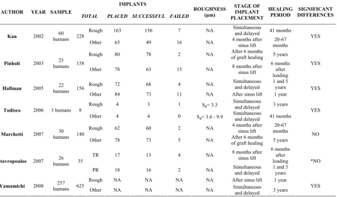
In respect to the second category(augmented bone tissue), all were in vivo studies carried out in humans. A total of423pa- tients participated in the studies, underwent surgical procedures for sinus lift, and received1350implants(corresponding to7 different researches), of which725implants had accurate data
(corresponding to6different researches). The seven studies were carried out from2002to2008and most authors agreed that significant differences exist between rough and machined im- plants in augmented bone tissue for implant osseointegration
(Fig.3). Some of these studies evaluated differences between implants placed at same time as the sinus lift procedure and im- plants that were placed in stage2(after sinus lift).7,19―22). The average healing period was40months, ranging from6to67 months, and during this time the implants remained functional.
Only one author specified the average roughness of implants used in his study; thus, we were not able to analyze the average roughness for augmented bone tissue.
DISCUSSION
Some studies that have evaluated osseointegration on ma- chined surfaces inserted into human jaws showed that the per- centage of BIC ranged between9%and13%after a5-6-month healing period1,3,23,24); however, these values were lower than the results presented by Grassi et al.1), which yielded a mean of
42.83%in SLA-surface implants after a2-month healing pe- riod; and Shibli et al.3)suggested that the oxidized surface with an Ravalue of0.87±0.14μm had a higher BIC rate(39.04%)
than machined surfaces(21.71%)with an Ravalue of0.32±
0.03μm under unloaded conditions with a healing period of two months1,3,7). Consequently, it has been suggested that rough sur- face implants can be loaded at an earlier time than machined sur- faces3,25).
Since1991, several average degrees of roughness have been suggested to enhance implant osseointegration(Fig.4)1,3―5,8,9,11,
24,26―28)
. Wennerberg and Albrektsson8)suggested that only a very specific surface topography with an Ravalue between1and1.5 μm provides an optimal surface for bone integration. In addi- tion, Marinucci et al.9)demonstrated that an average surface roughness of3μm is more suitable than0.5μm for osteoblast differentiation in vitro. Todisco and Trisi20)compared six differ- ent implant surfaces after early loading in humans, in which a microtextured surface with a reported Savalue of3.30±0.22μ m achieved the highest BIC and OC values(94.08%and 34.31%, respectively)in grafted bone; however, these averages varied considerably, which might reflect the different types of measurements and techniques used by each author. As there is currently no consensus on the degree of surface roughness that is optimum for bone cell attachment; further research is needed in this field.
REFERENCES
1)Grassi S, Piatelli A, de Figueredo LC, de Melo L, Iezzi G, Alba RC Jr and Shibli JA. Histologic evaluation of early human bone response to different implant surfaces. J Periodontol. 2006; 77:
1736-1743.
2)Huang YH, Xiropaidis AV, Albabdar RG, Hall J and Wikesjo UME. Bone formation at titanium porous oxide(TiUniteTM)
oral implants in Type!bone. Clin Oral Impl Res. 2005; 16:
105-111.
3)Shibli JA, Grassi S, de Figueiredo LC, Feres M, Marcantonio E Jr, Iezzi G and Piattelli A. Influence of implant surface topogra- phy on early osseointegration: a histological study in human jaws. J Biomed Mater Res B Appl Biomater. 2007; 80: 377-385.
4)Soskolne W, Cohen S, Sennerby L, Wennerberg A and Shapira L. The effect of titanium surface roughness on the adhesion of monocytes and their secretion of TNF-αand PGE 2. Clin Oral Impl Res. 2002; 13: 86-93.
5)Buser D, Schenk RK, Steinemann S, Fiorellini JP, Fox CH and Stich H. Influence of surface characteristics on bone integration of titanium implants. A histomorphometric study in miniature Fig.2. Average roughness suggested by different authors from1991
to2007in different studies in normal bone tissue, including in vivo in animals, in vivo in humans and in vitro studies.
Fig.3. Percentages of failure and success of rough surface implants and implants with other surfaces, which were used in aug- mented bone tissue in vivo studies in humans from2002to 2007.
Fig.4. Average roughness suggested by different authors in in vivo and in vitro studies in normal bone tissue from1991to2007. A marked decrease of the degree can be seen.
94 6)Wong M, Eulenberger J, Schenk R and Hunziker E. Effect of
surface topology on the osseointegration of implant materials in trabecular bone. J Biomed Mater Res. 1995; 29: 1567-1575.
7)Buser D, Nydegger T, Oxland T, Cochran DL, Schenk RK, Hirt HP, Snétivy D and Nolte LP. Interface shear strength of titanium implants with a sandblasted and acid-etched surface: a biome- chanical study in the maxilla of miniature pigs. J Biomed Mater Res. 1999; 45: 75-83.
8)Wennerberg A and Albrektsson T. Suggested guidelines for the topographic evaluation of implant surfaces. Int J Oral Maxillofac Implants. 2000; 15: 331-344.
9)Marinucci L, Balloni S, Becchetti E, Belcastro S, Guerra M, Calvitti M, Lilli C, Maria E and Locci P. Effect of titanium sur- face roughness on human osteoblast proliferation and gene ex- pression in vitro. Int J Oral Maxillofac Impl. 2006; 21: 719-725.
10)Lekholm U and Zarb GA; Zarb GA and Albrektsson T, eds. Tis- sue Integrated Prosthesis. Chicago: Quintessence Publishing Co;
1985: 199-209.
11)Sammons RL, Lumbikanonda N, Gross M and Cantzler P. Com- parison of osteoblast spreading on microstructured dental implant surfaces and cell behaviour in an explant model of osseointegra- tion. A scanning electron microscopic study. Clin Oral Imp Res.
2005; 16: 657-664.
12)Shalabi MM, Gortemaker A, Van’t Hof MA, Jansen JA and Creugers NHJ. Implant surface roughness and bone healing: a systematic review. J Dent Res. 2006; 85: 496-500.
13)Marchetti C, Pieri F, Trasarti S, Corinaldesi G and Degidi M.
Impact of implant surface and grafting protocol on clinical out- comes of endosseous implants. Int Oral Maxillofac Implants.
2007; 22: 399-407.
14)Sennerby L, Dasmah A, Larsson B and Iverhed M. Bone tissue responses to surface-modified zirconia implants: A histo- morphometric and removal torque study in the rabbit. Clin Im- plant Dent Relat Res. 2005; 7 Suppl 1: S 13-20.
15)Schreiner U, Schroeder-Boersch H, Schwarz M and Scheller G.
Improvement of osseointegration of bio-inert ceramics by modi- fication of the surface-results of an animal experiment. Biomed Tech(Berl). 2002; 47: 164-168.
16)Wallace SS and Froum SJ. Effect of maxillary sinus augmenta- tion on the survival of endosseous dental implants. A systematic review. Ann Periodontol. 2003; 8: 328-343.
17)Pinholt EM. Branermark and ITI dental implants in the human bone-grafted maxilla: a comparative evaluation. Clin Oral Im- plants Res. 2003; 14: 584-592.
CJ. Factors affecting the survival of implants placed in grafted maxillary sinuses: a clinical report. J Prosthet Dent. 2002; 87:
485-489.
19)Yamamichi N, Itose T, Neiva R and Wang HL. Long-term evalu- ation of implant survival in augmented sinuses: a case series. Int J Periodontics Dent. 2008; 28: 163-169.
20)Todisco M and Trisi P. Histomorphometric evaluation of six den- tal implant surfaces after early loading augmented human sinuses.
J Oral Impl. 2006; 32: 153-166.
21)Stavropoulos A, Karring T and Kostopoulos L. Fully vs. partially rough implants in maxillary sinus floor augmentation: a randomized- controlled clinical trial. Clin Oral Impl Res. 2007; 18: 95-102.
22)Hallman M, Mordenfeld A and Strandkvist T. A retrospective 5- year follow-up study of two different titanium implant surfaces used after interpositional bone grafting for reconstruction of the atrophic edentulous maxilla. Clin Implant Dent Relat Res. 2005;
7: 121-126.
23)Ivanoff CJ, Widmark G, Johansson C and Wennerberg A. His- tologic evaluation of bone response to oxidized and turned tita- nium microimplants in human jawbone. Int J Oral Maxillofac Implants. 2003; 18: 341-348.
24)Ivanoff CJ, Hallgren C, Widmark G, Sennerby L and Wenner- berg A. Histologic evaluation of the bone integration of TiO2 blasted and turned titanium microimplants in humans. Clin Oral Impl Res. 2001; 12: 128-134.
25)Trisi P, Lazzara R, Rebaudi A, Rao W, Testori T and Porter SS.
Bone-implant contact on machined and dual acid-etched surfaces after 2 months of healing in the human maxilla. J Periodontol.
2003; 74: 945-956.
26)Martin JY, Schwartz Z, Hummert TW, Schraub DM, Simpson J, Lankford J Jr, Dean DD, Cochran DL and Boyan BD. Effect of titanium surface roughness on proliferation, differentiation, and protein synthesis of human osteoblast-like cells(MG 63). J Bio- med Mater Res. 1995; 29: 389-401.
27)Schwartz Z, Lohmann CH, Oefinger J, Bonewald LF, Dean DD and Boyan BD. Implant surface characteristics modulate differ- entiation behavior of cells in the osteoblastic lineage. Adv Dent Res. 1999; 13: 38-48.
28)Gahlert M, Gudehus T, Eichhorn S, Steinhauser E, Kniha H and Erhardt W. Biomechanical and histomorphometric comparison between zirconia implants with varying surface textures and a ti- tanium implant in the maxilla of miniature pigs. Clin Oral Impl Res. 2007; 18: 662-668.
Dental Implant Surface Roughness and Topography
95 岐 歯 学 誌
35巻3号 89〜95 2009年2月
インプラント体の表面粗さ及び形状:論文的総説
Isabel de Monerrat Osorio Bernal
1) 伊 藤 理 妙2) 片 木 紘 樹2)坪 井 健一郎2) 山 田 尚 子2) 田 辺 俊一朗2)
永 原 國 央2) 森 昌 彦3)
本総説では,臨床的にインプラント手術において埋入されたインプラント体に対して有利に働くと考えら れるインプラント体表面の粗さに関し,どの様な結論が現在得られているのか,また,将来に向けての展望 はどうかを追求することを目的としている.
インターネットにてPub Med検索において散見し得た文献をもとにインプラント体の表面粗さに関する 論文で,本総説の主旨である統計的分析に対応しうる22論文を用いた.
また,通常の骨組織内での検索は15論文,骨造成を行った部位での論文は7であった.さらに,通常の骨 組織での論文は,動物実験8,臨床論文3で残りの4論文はin vitroであった.骨造成を行った部位での論 文はすべて臨床報告であった.これらすべての論文での患者数は423人,インプラント体数は1350で,その 内725本に関してはすべての検索データーが得られた.
1991年からインプラント体の表面性状が骨接合に重要であることが報告され,その粗さは,1991年:18〜
23μm,1999年:6.5μm,その後2000年から2007年までに2.7μmからそれ以下に変化している.このように 年代により表面粗さにかなりの違いがあり,それが論文の著者が用いた測定方法と手技の違いと考えられて いる.そのため,今日においてもその粗さの詳細な程度には一定の結論がなく,さらなる検索が必要と考え る.
キーワード:歯科インプラント治療,表面粗さ,表面形状,骨接合
1)メキシコ州立自治大学歯学部歯科矯正学講座
2)朝日大学歯学部口腔病態医療学講座インプラント学分野
3)朝日大学歯学部
501―0296 岐阜県瑞穂市穂積1851
(2008年11月26日受理)