Volume 2011, Article ID 151596,9pages doi:10.1155/2011/151596
Research Article
Comparative Effects of α -, β -, and γ -Carbolines on Platelet Aggregation and Lipid Membranes
Hironori Tsuchiya
Department of Dental Basic Education, Asahi University School of Dentistry, 1851-1 Hozumi, Mizuho, Gifu 501-0296, Japan
Correspondence should be addressed to Hironori Tsuchiya,
[email protected]Received 20 February 2011; Revised 23 April 2011; Accepted 2 June 2011
Academic Editor: M. Firoze Khan
Copyright © 2011 Hironori Tsuchiya. This is an open access article distributed under the Creative Commons Attribution License, which permits unrestricted use, distribution, and reproduction in any medium, provided the original work is properly cited.
Cigarette smoking and alcohol consumption possibly affect platelet functions. To verify the hypothesis that some
α-,β-, andγ-carboline components in cigarette smoke and alcoholic beverages may change platelet aggregability, their effects on human platelets were determined by aggregometry together with investigating their membrane effects by turbidimetry. Carbolines inhibited platelet aggregation induced by five agents with the potency being 3-amino-1,4-dimethyl-5H-pyrido[4,3-b]indole
>3-amino- 1-methyl-5H-pyrido[4,3-b]indole
>1-methyl-9H-pyrido[3,4-b]indole. The most potent 3-amino-1,4-dimethyl-5H-pyrido[4,3- b]indole showed 50% aggregation-inhibitory concentrations of 6–172
μM. Bothγ-carbolines interacted with phosphatidylcholinemembranes to lower the lipid phase transition temperature with the potency correlating to the antiplatelet activity, suggesting that the interaction with platelet membranes to increase their fluidity underlies antiplatelet effects. Given their possible concentration and accumulation in platelets,
γ- andβ-carbolines would provide cigarette smokers and alcohol drinkers with reduced plateletaggregability, and they may be responsible for the occurrence of hemorrhagic diseases associated with heavy smoking and alco- holics.
1. Introduction
Cigarette smoking has the possibility to affect the functions of platelets. Platelet aggregation stimulated by collagen is di- minished in smokers compared with nonsmokers, suggesting that habitual smoking reduces platelet aggregability [1].
Smokers are more susceptible to aspirin in collagen- and adenosine 5
-diphosphate- (ADP-) induced platelet aggrega- tion than nonsmokers [2]. Smoking also promotes the plate- let inhibition mediated by an antiplatelet prodrug clopido- grel [3]. Cigarette smoking and alcohol drinking are often characterized by concurrent use, and alcohol drinking is gen- erally accompanied by an increase in smoking. Platelet de- fects are also noted in alcoholics [4]. Alcohol consumption is inversely related to platelet aggregation in response to colla- gen, epinephrine, and ADP [5], and it reduces platelet aggre- gability [6, 7]. However, the influences of cigarette smoking and alcohol consumption on platelet aggregability appear to be conflicting, with some studies indicating a decrease and others an increase of aggregation. Platelet aggregation induced by epinephrine and ADP is increased in habitual
smokers [8, 9]. The platelets of alcoholics are initially hy- poaggregable but become hyperaggragable after the cessation of drinking [10]. ADP-induced platelet aggregation is in- creased in alcoholics [11].
Cigarette smoke contains a variety of bioactive com- pounds [9]. Alcoholic beverages are not consumed as pure ethanol. Therefore, some components in cigarette smoke and alcoholic beverage are assumed to influence the properties of platelets. The objective of this study was to determine antiplatelet e ff ects of the assumed compounds and to relate cigarette smoking and alcohol consumption to hypoaggre- gability. In addition, their e ff ects on lipid membranes were investigated to address one possible mode of antiplatelet action. From the viewpoint of this mechanistic membrane interaction, an opposite phenomenon, hyperaggregability, in smokers and alcoholics was also discussed.
Cigarette smoking and alcohol drinking have been re- ferred to as risk factors for intracerebral hemorrhage, suba- rachnoid hemorrhage, and hemorrhagic stroke [12–16].
These factors correlate with each other. Reduced platelet
aggregability is involved in the development of hemorrhagic
complications [17, 18]. The occurrence of hemorrhage is in- creased by heavy cigarette smoking and binge alcohol drink- ing [12, 19]. A graded increase in risk of intracerebral hemo- rrhage and hemorrhagic stroke depends on how many ciga- rettes are smoked [13], and several chemicals in smoke are likely to relate to the increased risk of intracranial hemo- rrhage [20]. Heavy alcohol drinking, but not light-to-moder- ate alcohol intake, increases the risk of intracerebral and sub- arachnoid hemorrhage [12, 14]. These suggest that certain antiplatelet components in cigarette smoke and alcoholic beverages are possibly associated with serious hemorrhagic diseases. This study aimed at discussing such a possibility by determining their e ff ects on human platelets.
A class of compounds with the pyridoindole structure, α-, β-, and γ-carbolines, are contained in cigarette smoke and alcoholic beverages. Their levels in mainstream smoke con- densates are 2.01–10.3 ng/cigarette for α-carboline, 0.25–
2.53 μg/cigarette for β-carbolines, and 0.29–1.10 ng/cigarette for γ-carbolines [21, 22]. The concentrations of α- and γ-carbolines in combustion smoke samples are 0–1.96 and 0.33–0.74 ng/g in mainstream smoke, respectively, but 5.00–6.51 and 0.27–0.37 ng/g in sidestream smoke, respec- tively [23]. β-carbolines show higher levels of 2.10–
8.99 μg/cigarette in sidestream smoke compared with 0.36–
4.24 μg/cigarette in mainstream smoke [24]. Various alco- holic beverages contain nM–μM levels of β-carbolines depending on brands [25]. Therefore, it is reasonable to assume that the in vivo concentrations of carboline com- pounds may be increased by smoking cigarettes and con- suming alcoholic beverages. The basal concentrations of one of β-carbolines, harmane, in plasma (47.7
±41.2 pM) and platelets (0.060
±0.108 pmol/10
9platelets) of smokers are respectively, two- and four-times higher compared with those (22.5
±14.3 pM and 0.015
±0.008 pmol/10
9platelets) of nonsmokers, and harmane concentrations increase to 150.4
±110.8 pM and 0.142
±0.324 pmol/10
9platelets after smoking [26, 27]. Another β-carboline, norharmane, also shows plasma and platelet concentrations (114.1
±115.3 pM and 0.176
±0.078 pmol/10
9platelets) in smokers higher than those (56.5
±29.7 pM and 0.043
±0.027 pmol/10
9platelets) in nonsmokers, and norharmane concentrations increase to 1.06
±0.52 nM and 0.245
±0.086 pmol/10
9platelets after smoking [26]. Different γ-carbolines are contained in plasma with the concentrations of 18.8
±5.0 pM to 68.3
±24.0 pM, which would markedly increase by smoking cigarettes [28, 29]. The plasma levels of norharmane are higher in alcoholics (591.5
±154.6 pM) compared with a control group (159.9
±63.6 pM) [25]. Harmane in plasma is increased following alcohol ingestion [30]. These elevated concentrations appear to result from inhaling cigarette smoke and drinking alco- holic beverages [31, 32].
α-, β-, and γ-carbolines exhibit a wide range of bioactiv- ities such as neuropsychiatric, hallucinogenic, and carcino- genic, which have been exclusively studied [33–35]. In addi- tion to these known bioactivities, α-, β-, and γ-carbolines may exert some e ff ects at the periphery, because they are found in human blood and platelets. Certain carbolines such as harmane, harmine, and harmol were recently reported to show the inhibitory e ff ects on collagen-induced platelet
aggregation [36]. The structure and antiplatelet activity rela- tionship suggested that the presence of a methyl group in the pyrido moiety is important for carboline compounds to inhibit platelet aggregation [37].
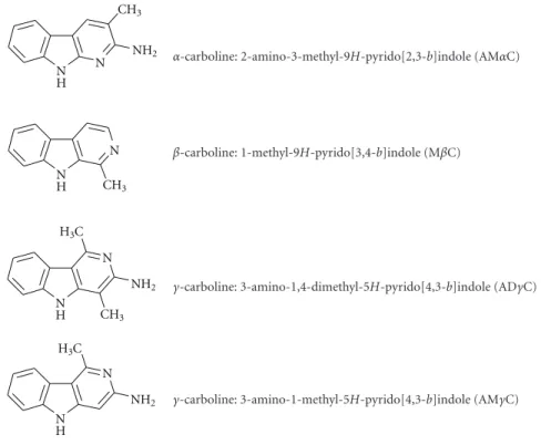
In this study, the hypothesis that α-, β-, and γ-carbo- lines may a ff ect the functions of human platelets was ver- ified. 2-Amino-3-methyl-9H-pyrido[2,3-b]indole (AMαC), 1-methyl-9H-pyrido[3,4-b]indole (MβC), 3-amino-1,4-di- methyl-5H-pyrido[4,3-b]indole (ADγC), and 3-amino-1- methyl-5H-pyrido[4,3-b]indole (AMγC) (see Figure 1 for their structures) were chosen as tested compounds because they have a methyl substituent as the antiplatelet activity determinant in the common pyrido structure and their con- centrations in cigarette smoke and/or alcoholic beverages are relatively high [21–25]. Their effects on platelet aggregation induced by collagen, epinephrine, platelet-activating factor (PAF), ADP, and thrombin were comparatively determined.
Consequently, γ-carbolines were found to inhibit platelet aggregation in response to all of the tested inducers which have different aggregation mechanisms, suggesting that the site of antiplatelet action of γ-carbolines is not confined to receptors or enzymes for individual aggregation agonists.
The physicochemical property of biomembranes plays a crucial role in signal transduction and influences the activity of platelets. Changes in membrane fluidity induce the inhi- bition or the promotion of platelet aggregation stimulated by various aggregants [38–41]. The e ff ects of γ-carbolines on lipid membranes were studied to get a clue to one of possible antiplatelet mechanisms.
2. Materials and Methods
2.1. Chemicals. AMαC, MβC, ADγC (commercially referred to as Trp-P-1), and AMγC (commercially referred to as Trp-P-2) were purchased from Wako Pure Chemicals (Osaka, Japan). Collagen (MC Medical, Tokyo, Japan), PAF (Funakoshi, Osaka, Japan), epinephrine (Daiichi Sankyo, Tokyo, Japan), ADP (MC Medical) and thrombin (Sigma- Aldrich, St. Louis, Mo, USA) were used for inducing platelet aggregation. 1,2-Dipalmitoylphosphatidylcholine (DPPC) was obtained from Avanti Polar Lipids (Alabaster, Ala, USA).
Dimethyl sulfoxide (DMSO) of spectroscopic grade (Kishida, Osaka, Japan) and water of liquid chromatographic grade (Kishida) were used for preparing sample solutions. All other reagents were of the highest grade available.
2.2. Platelet Aggregation Assay. The experiments were desig-
ned and performed according to the guidelines of the Japa-
nese Pharmacological Society. Human platelet-rich plasma
(PRP) and platelet-poor plasma (PPP) were prepared with
citrated blood which was obtained from healthy male
donors aged 38–48 years (n
=6) of nonsmokers and
nonalcoholics without taking any drugs at least for one
month according to the previous method [42]. Only male
subjects were employed as blood donors because platelet
aggregation induced by collagen, PAF and ADP was different
between males and females in the study which evaluated
the comparative e ff ects of the smoking habit and the act of
smoking on platelet aggregability [43]. Sharp increases of
N H
N
NH N N H
N N H
N CH3
CH3
CH3 H3C
H3C
NH2
NH2
NH2
α-carboline: 2-amino-3-methyl-9H-pyrido[2,3-b]indole (AMαC)
β-carboline: 1-methyl-9H-pyrido[3,4-b]indole (MβC)
γ-carboline: 3-amino-1,4-dimethyl-5H-pyrido[4,3-b]indole (ADγC)
γ-carboline: 3-amino-1-methyl-5H-pyrido[4,3-b]indole (AMγC)
Figure 1: The structures ofα-,β-, andγ-carbolines tested in this study.
the carboline concentrations in plasma and platelets were found in nonsmokers after 5 minutes following smoking one cigarette [25–27] and in nonalcoholics 30 minutes after drinking alcoholic beverages [32]. Therefore, PRP and PPP were prepared from nonsmokers and nonalcoholics in order to determine the inherent effects of α-, β-, and γ-carbolines on platelet aggregation without exposing platelets to carbo- line compounds. The platelet count of PRP was adjusted to be 3
×10
5platelets/μL by diluting PRP with PPP. Both PRP and PPP were used within 3 hours after preparation.
Platelet aggregation was assayed by the same method as the aggregometry using an antiplatelet reference aspirin which was recently reported from our laboratory [37]. Since its methodological validity had been confirmed [44, 45], this study was performed without using references and the obtained results were discussed with referring to our recent report [37]. The aggregation response of PRP was monitored using a HEMA Tracer 601 aggregometer (Niko Bioscience, Tokyo, Japan) by an increase of percent light transmission (%
T) at 660 nm as a function of time. PPP and unstimulated PRP were defined as 100% T and 0% T, respectively. The tested carbolines were dissolved in and diluted with DMSO and/or water. After adjustment of % T to 0, 20 μL of the sample solutions were added to 170 μL of PRP. Vehicle alone was added for controls. The volume of DMSO was kept less than 0.1%–1.0% (v/v) of the total volume so as not to affect platelet aggregability and aggregation inducers. After treating PRP with each carboline for 1 minute, platelet aggregation was induced by adding 10 μL of the aqueous solutions of collagen (50 μg/mL), PAF (5 μM), epinephrine (40 μg/mL), ADP (60 μM) or thrombin (5 units/mL). The time of their
addition was defined as 0 minute. A maximal % T of aggre- gation response (T
max), an area under curve of aggregation response (AUC, from 0 to 5 minutes) and a single slope of aggregation response were measured in platelet aggrega- tion induced by collagen, PAF, ADP, and thrombin. Since epinephrine showed the biphasic aggregation, first T
max(at 1 minute) and second T
max(at 5 minutes), first AUC (from 0 to 1.5 minutes) and second AUC (from 1.5 to 5 minutes), and slopes of the first (% T at 45 seconds) and second (% T at 4 minutes) phases were measured. The tested compounds were assayed at various concentrations (1–1000 μM). Their % inhibitions were plotted against their concentrations to prepare concentration-inhibition curves, from which 50%
inhibitory concentrations were calculated [46].
2.3. Membrane Interaction Assay. The changes in turbidity of liposomal membrane suspensions reflect the thermotropic phase transition of membrane-constituting phospholipids from the gel to the liquid crystalline phase and the phase transition temperatures are altered by membrane-interacting compounds [47]. The membrane interactions of antiplatelet ADγC and AMγC were assayed as reported previously [48]. Briefly, DPPC liposomes were prepared by hydrating the dry film of DPPC (final concentration of 0.4 mM) with water, and then treated with ADγC and AMγC (0, 0.5, 1 and 1.5 mM for each). The absorption of DPPC liposome suspensions was measured at 450 nm by a UV-260 spectrophotometer equipped with a CPS thermocontroller (Shimadzu, Kyoto, Japan) as the temperature was increased.
The transition between gel and liquid crystalline phases in
phospholipid liposomes dispersed in the aqueous medium
Tmax
AUC Slope
N N N N N N
AMαC MβC
AMγC 0 ADγC
100 200 300 400 500
50%inhibitoryconcentration(μM)
Figure 2: Concentrations of carbolines to produce 50% inhibition
of the platelet aggregation responses (T
max, AUC, and slope) to collagen. N: Not inhibited by 50% at the indicated concentrations.
Data are presented as mean
±SE (n
=7).
is accompanied by a sudden change in turbidity [49]. The phase transition temperatures defined as the mid-point of the abrupt step in absorbance were calculated.
2.4. Statistical Analysis. All results are expressed as mean
±SE (n
=7 for antiplatelet experiments and n
=6 for membrane experiments). Data were analyzed by ANOVA, followed by post hoc Fisher’s PLSD test using StatView version 5.0 (SAS Institute, Cary, NC, USA). Values of P < .05 were considered statistically significant.
3. Results
3.1. Antiaggregatory E ff ect. Carboline compounds a ff ected human platelets with the potency di ff erent between sub- classes. Their inhibitory effects on platelet aggregation in- duced by collagen, PAF, epinephrine, ADP and thrombin are shown in Figures 2, 3, 4, 5, 6, and 7. The compar- isons showed that γ-carbolines were the most effective in inhibiting platelet aggregation, followed by less effective β- carboline, with α-carboline being almost ineffective at the tested concentrations (250 or 500 μM). AMαC showed a 50% inhibitory concentration of 499.6
±20.3 μM only for AUC of aggregation response to ADP. In contrast, ADγC and AMγC decreased T
maxand AUC of aggregation responses to all of collagen, PAF, epinephrine, ADP, and thrombin.
Both γ-carbolines also decreased the slopes of collagen- and epinephrine-induced platelet aggregation. When comparing 50% inhibitory concentrations, ADγC was more potent than AMγC. Even at 10 μM or less, ADγC inhibitorily influ- enced the aggregability of platelets stimulated by PAF and ep- inephrine.
3.2. Membrane Interaction. Antiplatelet γ-carbolines influ- enced the phase transition of membrane DPPC as shown in Figure 8. ADγC lowered the phase transition temperature
Tmax
AUC Slope
N N N N N
N N
AMαC MβC
AMγC 0 ADγC
100 200
50%inhibitoryconcentration(μM)
Figure 3: Concentrations of carbolines to produce 50% inhibition
of the platelet aggregation responses (T
max, AUC, and slope) to PAF.
N: Not inhibited by 50% at the indicated concentrations. Data are presented as mean
±SE (n
=7).
Tmax
AUC Slope
N N N N N N
AMαC MβC
AMγC 0 ADγC
100 200 300 400 500
50%inhibitoryconcentration(μM)
Figure 4: Concentrations of carbolines to produce 50% inhibition
of the first phase platelet aggregation responses (T
max, AUC, and slope) to epinephrine. N: Not inhibited by 50% at the indicated concentrations. Data are presented as mean
±SE (n
=7).
from a control value of 40.51
±0.02
◦C to 40.07
±0.03
◦C at 500 μM, 39.73
±0.03
◦C at 1 mM and 39.45
±0.02
◦C at 1.5 mM. AMγC shifted to 40.17
±0.03
◦C, 39.93
±0.03
◦C and 39.73
±0.02
◦C at 500 μM, 1 mM and 1.5 mM, respec- tively. ADγC was more effective in interacting with DPPC membranes than AMγC (P < .05 at 500 μM and P < .01 at 1 and 1.5 mM).
4. Discussion
The comparative studies have revealed that β- and γ-carbo-
lines, but not α-carboline, inhibit platelet aggregation in-
duced by five di ff erent agents with the potency being
ADγC > AMγC > MβC. The relation between structure and
Tmax
AUC Slope
N N N
AMαC MβC
AMγC 0 ADγC
100 200 300
50%inhibitoryconcentration(μM)
Figure 5: Concentrations of carbolines to produce 50% inhibition
of the second phase platelet aggregation responses (T
max, AUC, and slope) to epinephrine. N: Not inhibited by 50% at the indicated concentrations. Data are presented as mean
±SE (n
=7).
Tmax
AUC Slope
N
N
N N N
N N
AMαC MβC
AMγC 0 ADγC
100 200 300 400 500
50%inhibitoryconcentration(μM)
Figure 6: Concentrations of carbolines to produce 50% inhibition
of the platelet aggregation responses (T
max, AUC, and slope) to ADP.
N: Not inhibited by 50% at the indicated concentrations. Data are presented as mean
±SE (n
=7).
antiplatelet activity indicates that the basic structure of 5H- pyrido[4,3-b]indole (γ-carboline) is important for inhibit- ing platelet aggregation, followed by the structure of 9H- pyrido[3,4-b]indole (β-carboline). An additional methyl group in the pyrido moiety provides γ-carbolines with high- er activity. Since ADγC and AMγC themselves do not aggre- gate platelets despite partly resembling serotonin in structure [50], these γ-carbolines are referred to as potent antiplatelet compounds.
Antiplatelet γ-carbolines showed relatively low concen- trations to produce 50% inhibition of AUC of aggregation response. Since the antiplatelet effects of aspirin were recently reported by using the same method for platelet aggregation assay from our laboratory [37], its 50% AUC-inhibitory con- centrations are usable for comparing with the activity of
Tmax
AUC Slope
N N
N N N N N N
AMαC MβC
AMγC 0 ADγC
100 200 300 400 500
50%inhibitoryconcentration(μM)
Figure 7: Concentrations of carbolines to produce 50% inhibition
of the platelet aggregation responses (T
max, AUC, and slope) to thrombin. N: Not inhibited by 50% at the indicated concentrations.
Data are presented as mean
±SE (n
=7).
Control
500μM 1 mM 1.5 mM
44 43 42 41 40 39 38 37
Temperature (◦C) 0.2
0.3 0.4 0.5 0.6
Absorbanceat450nm
Figure 8: Effects of antiplateletγ-carboline on membrane DPPC
phase transition. The absorbance of DPPC liposome suspensions treated with 0, 0.5, 1, or 1.5 mM ADγC was measured at 450 nm with increasing the temperature. Typical traces in multiple mea- surements are shown.
γ-carbolines. Aspirin showed 116.1
±6.4 μM for collagen- induced, 71.3
±5.5 μM for epinephrine-induced first phase and 19.3
±3.6 μM for epinephrine-induced second phase response, although it did not inhibit PAF-, ADP-, and thrombin-induced responses by 50% at 500 μM. In this study, ADγC and AMγC have been found to show 50%
AUC-inhibitory concentrations of 84.0
±7.1 and 179.5
±20.7 μM, 106.1
±8.5 and 153.6
±8.5 μM, and 21.5
±1.9 and
117.2
±11.0 μM for collagen-induced, epinephrine-induced
first phase and epinephrine-induced second phase response,
respectively. ADγC and AMγC are also effective in inhibiting AUC of aggregation responses induced by PAF, ADP and thrombin to show 50% inhibitory concentrations of 6.01
±0.58 and 32.0
±5.8 μM, 65.2
±2.4 and 115.5
±8.8 μM, and 105.8
±19.5 and 230.9
±25.8 μM, respectively. With respect to platelet aggregation inhibition, γ-carbolines, especially ADγC, are comparable to or more potent than aspirin.
A question arises as to whether carboline components actually a ff ect the aggregability of platelets of cigarette smokers and alcohol drinkers. α-, β-, and γ-carbolines are contained in mainstream cigarette smoke (
≥22 ng/cigarette for AMαC,
≥2.2 μg/cigarette for MβC,
≥0.5 ng/cigarette for ADγC and
≥1.1 ng/cigarette for AMγC), and higher levels are found in sidestream cigarette smoke (
≥3.0 μg/cigarette for MβC) [21, 22, 24, 25]. β-carbolines are also present in various alcoholic beverages at the concentrations of
≥3.2 μM for MβC [25, 51]. Compared with nonsmokers (
≥23 pM), the plasma levels of MβC are higher in smokers (
≥48 pM) and increase to
≥165 pM by smoking a cigarette [25].
The platelet concentrations of MβC are
≥0.015 pmol/10
9platelets for nonsmokers but
≥0.060 pmol/10
9platelets for smokers, and MβC shows the increasing platelet concentra- tions to be
≥0.142 pmol/10
9platelets 13 minutes after smok- ing [26]. β-carbolines increase in blood rapidly following cigarette smoking and alcohol drinking, and their concen- trations in platelets are much higher than in plasma, indi- cating their significant concentration and accumulation in platelets. The accurate γ-carboline concentrations in plate- lets of smokers and alcoholics have been unknown. In the dosing experiment using rabbits, however, ADγC was present in blood, especially in red blood cells, for a long time after oral dosing [52]. Although aspirin is less potent in aggregation inhibition than ADγC, it has been frequently used as an antiplatelet drug. Considering that cigarette smoke- and alcoholic beverage-derived β- and γ-carbolines are concentrated and accumulated in platelets, the possibility for them to affect platelet aggregability is not necessarily excluded.
Although the platelet aggregation mechanisms for colla- gen, PAF, epinephrine, ADP, and thrombin differ, ADγC and AMγC were inhibitory on platelet aggregation induced by all of these aggregants, and MβC by two aggregants. These results indicate that γ- and β-carbolines influence the step common to different aggregation agonists in addition to their specific action at the receptor and enzyme levels. The physic- ochemical property of biomembranes, such as fluidity, plays a crucial role in signal transduction and a ff ects the activity of platelets. Membrane fluidity modulates platelet aggregability and membrane-fluidizing compounds attenuate collagen- and thrombin-induced platelet aggregation [53]. A change in platelet membrane fluidity is mechanistically related to various antiplatelet compounds [38, 54].
The membrane effects of toxins and drugs have been most frequently studied by measuring fluorescence polar- ization of liposomal and cellular membranes labeled with fluorescent probes [37–39, 54]. However, such a method was not applicable to antiplatelet γ-carbolines, because they are naturally fluorescent with the maximal excitation and emis- sion wavelengths almost similar to those of typical fluores-
cent probes. Therefore, turbidimetry was used for them. The changes in turbidity of liposome suspensions reflect the pro- motion of a gel to liquid crystalline transition of membrane phospholipids, indicating an increase of membrane fluidity [47, 48]. Consequently, ADγC and AMγC were found to lower the phase transition temperature of membrane DPPC at 500 μM–1.5 mM with the potency correlating to their relative antiplatelet e ff ects. Antiplatelet γ-carbolines appear to interact with lipid membranes and increase their fluidity at platelet aggregation-inhibitory concentrations.
The antiplatelet mechanisms previously reported for β- and γ-carbolines and their relating structures include the inhibition of aggregation-relevant enzymes and receptors, and the suppression of cytosolic calcium mobilization and arachidonic acid liberation. β- and γ-carbolines influence cy- clooxygenase activity and arachidonic acid metabolism to reduce the production of prostaglandins and thromboxane [55]. Antiplatelet γ-carbolines (ADγC and AMγC) and β-carbolines (MβC) are the potent inhibitors of mono- amine oxidase and serotonin uptake of platelets [33, 50].
β-carbolines like MβC inhibit phospholipase Cγ2 and pro- tein tyrosine phosphorylation [36]. Biological membranes require the lipid bilayer environments optimal for mem- brane-embedded enzymes, receptors, and transporter sys- tems. The fluidity changes of platelet membranes modify the activities of phospholipase, cyclooxygenase, and aggregation agonists’ receptors with the subsequent inhibition of phos- phoinositide breakdown, and of prostaglandin and throm- boxane formation [38, 39]. The increased membrane fluidity also implies that aggregation-relevant receptors on platelet membranes are less exposed to the external environment.
The present results suggest that platelet hypoaggregabil- ity might be induced in smokers and alcoholics by γ- and β-carbolines. However, the influences of cigarette smoking and alcohol consumption on platelet aggregation have been conflicting, with some studies reporting a reduction of aggre- gability [1–7] but others an enhancement of aggregability [8–11]. In contrast to fluid membranes (with increased fluidity) induced by ADγC and AMγC, rigid membranes (with decreased fluidity) show the enhanced platelet aggre- gability in response to epinephrine and thrombin [40, 41].
The decreased membrane fluidity renders platelet receptors
more exposed to the external environment and makes the
binding of agonists to the receptors more efficiently, result-
ing in an increase of platelet sensitivity to aggregants (hyper-
aggregability) [56, 57]. Varying lipid compositions modify
the fluidity of biomembranes and cholesterol is one of deter-
minants for decreasing membrane fluidity. While the en-
hanced platelet aggregability could be found in chronic
smokers, subjects who had smoked 10
±2 cigarettes per
day for 7–10 years showed the decreased fluidity of platelets,
which was due to an increase of cholesterol in platelet mem-
branes [58]. Platelet aggregability in response to ADP and
collagen was enhanced in alcoholics with increasing cho-
lesterol in platelet membranes [11]. The conflicting phenom-
ena, hypoaggregability and hyperaggregability, associated
with cigarette smoking and alcohol consumption appear to
be explained by the biphasic effects of membrane fluidity
changes.
Reduced platelet aggregability is important in the devel- opment of intracranial hemorrhage [17, 28]. Antiplatelet medication is known to increase the incidence of intracere- bral hemorrhage, hemorrhagic stroke and other hemorrhag- ic complication [59, 60] and also increase the recurrence risk of intracerebral hemorrhage [61]. Intracerebral hemorrhage, subarachnoid hemorrhage, and hemorrhagic stroke are closely associated with smoking and drinking habits [12–
16]. The present results suggest the possibility that γ- and β-carboline components in cigarette smoke and/or alcoholic beverages are pharmacotoxicologically relevant to these seri- ous hemorrhagic diseases as well as antiplatelet agents by de- creasing platelet aggregation responses to di ff erent aggre- gants.
Another etiological role of cigarette smoking and alcohol consumption has been indicated in cardiovascular events [62, 63] although it is conflicting similarly to their influence on platelet aggregability. Platelet aggregation is patho- logically related to coronary artery disease and coronary thrombosis leads to myocardial infarction. The antiplatelet effects of γ- and β-carbolines are in line with several studies that cigarette smoking and alcohol drinking protect against coronary heart disease and also lower the risk of coronary artery disease and myocardial infarction [63–65]. However, such e ff ects are inconsistent with other studies that cigarette smoking is a risk factor for coronary artery disease and binge alcohol drinking precipitates fatal myocardial infarction [62, 64]. Chronic smokers and heavy drinkers show an increase of cholesterol in platelet membranes [6, 58], by which the membrane fluidity of platelets is decreased. The aggregability should be enhanced in such rigid platelet membranes, in- creasing cardiovascular morbidity.
In conclusion, the reduced platelet aggregability found in cigarette smokers and alcoholic drinkers would be attribu- table to antiplatelet carboline components in cigarette smoke and alcoholic beverages. γ- and β-carbolines may be respon- sible for the occurrence of hemorrhagic diseases associated with heavy smoking and alcoholics by inhibiting platelet aggregation.
Abbreviations
ADγC: 3-amino-1,4-dimethyl-5H-pyrido[4,3-b]indole ADP: adenosine 5
-diphosphate
AMαC: 2-amino-3-methyl-9H-pyrido[2,3-b]indole AMγC: 3-amino-1-methyl-5H-pyrido[4,3-b]indole AUC: area under curve of aggregation response DMSO: dimethyl sulfoxide
DPPC: 1,2-dipalmitoylphosphatidylcholine MβC: 1-methyl-9H-pyrido[3,4-b]indole PAF: platelet-activating factor
PPP: platelet-poor plasma PRP: platelet-rich plasma
% T: percent light transmission
T
maxmaximal % T of aggregation response.
Conflict of Interests
The author declares that there is no conflict of interests.
Acknowledgments
This study was partly supported by a Grant-in-Aid for Scientific Research (C) 23593005 (to H. Tsuchiya) from the Japan Society for the Promotion of Science. Additional support was provided from the San-Ei Gen Foundation for Food Chemical Research.
References
[1] L. C. Foo, I. Roshidah, and M. B. Aimy, “Platelets of habitual smokers have reduced susceptibility to aggregating agent,”
Thrombosis and Haemostasis, vol. 65, no. 3, pp. 317–319, 1991.
[2] A. A. Weber, S. Liesener, A. Schanz, T. Hohlfeld, and K. Schr¨or,
“Habitual smoking causes an abnormality in platelet throm- boxane A2 metabolism and results in an altered susceptibility to aspirin effects,” Platelets, vol. 11, no. 3, pp. 177–182, 2000.
[3] T. Gremmel, S. Steiner, D. Seidinger, R. Koppensteiner, S.
Panzer, and C. W. Kopp, “Smoking promotes clopidogrel- mediated platelet inhibition in patients receiving dual anti- platelet therapy,” Thrombosis Research, vol. 124, no. 5, pp. 588–
591, 2009.
[4] R. Rubin, “Effect of ethanol on platelet function,” Alcoholism:
Clinical and Experimental Research, vol. 23, no. 6, pp. 1114–
1118, 1999.
[5] K. J. Mukamal, J. M. Massaro, K. A. Ault et al., “Alcohol consumption and platelet activation and aggregation among women and men: the Framingham Offspring Study,” Alco- holism: Clinical and Experimental Research, vol. 29, no. 10, pp.
1906–1912, 2005.
[6] S. C. Renaud and J. C. Ruf, “Effects of alcohol on platelet functions,” Clinica Chimica Acta, vol. 246, no. 1-2, pp. 77–89, 1996.
[7] Q. H. Zhang, K. Das, S. Siddiqui, and A. K. Myers, “Effects of acute, moderate ethanol consumption on human platelet aggregation in platelet-rich plasma and whole blood,” Alco- holism: Clinical and Experimental Research, vol. 24, no. 4, pp.
528–534, 2000.
[8] Y. Fusegawa and S. Handa, “Platelet aggregation induced by ADP or epinephrine is enhanced in habitual smokers,”
Thrombosis Research, vol. 97, no. 5, pp. 287–295, 2000.
[9] J. A. Ambrose and R. S. Barua, “The pathophysiology of cigarette smoking and cardiovascular disease: an update,”
Journal of the American College of Cardiology, vol. 43, no. 10, pp. 1731–1737, 2004.
[10] K. Desai, J. S. Owen, D. T. Wilson, and R. A. Hutton, “Platelet aggregation and plasma lipoproteins in alcoholics during alcohol withdrawal,” Thrombosis and Haemostasis, vol. 55, no.
2, pp. 173–177, 1986.
[11] M. Watanabe, K. Shiraishi, M. Itakura, and S. Matsuzaki,
“Relationship between platelet membrane lipid compositions and platelet aggregability in alcoholic liver disease,” Alco- holism: Clinical and Experimental Research, vol. 22, no. 3, supplement 1, pp. 97S–102S, 1998.
[12] W. T. Longstreth, L. M. Nelson, T. D. Koepsell, and G. van Belle, “Cigarette smoking, alcohol use, and subarachnoid hemorrhage,” Stroke, vol. 23, no. 9, pp. 1242–1249, 1992.
[13] T. Kurth, C. S. Kase, K. Berger, E. S. Schaeffner, J. E. Buring,
and J. M. Gaziano, “Smoking and the risk of hemorrhagic
stroke in men,” Stroke, vol. 34, no. 5, pp. 1151–1155, 2003.
[14] M. J. Ariesen, S. P. Claus, G. J. E. Rinkel, and A. Algra, “Risk factors for intracerebral hemorrhage in the general popula- tion: a systematic review,” Stroke, vol. 34, no. 8, pp. 2060–2065, 2003.
[15] S. Juvela, M. Hillbom, and H. Palom¨aki, “Risk factors for spontaneous intracerebral hemorrhage,” Stroke, vol. 26, no. 9, pp. 1558–1564, 1995.
[16] T. Kurth, C. S. Kase, K. Berger, J. M. Gaziano, N. R. Cook, and J. E. Buring, “Smoking and risk of hemorrhagic stroke in women,” Stroke, vol. 34, no. 12, pp. 2792–2795, 2003.
[17] J. Neiman, M. L. Rand, D. M. Jakowec, and M. A. Packham,
“Platelet responses to platelet-activating factor are inhibited in alcoholics undergoing alcohol withdrawal,” Thrombosis Research, vol. 56, no. 3, pp. 399–405, 1989.
[18] S. Juvela and M. Kaste, “Reduced platelet aggregability and thromboxane release after rebleeding in patients with sub- arachnoid hemorrhage,” Journal of Neurosurgery, vol. 74, no.
1, pp. 21–26, 1991.
[19] S. Juvela, M. Hillbom, H. Numminen, and P. Koskinen,
“Cigarette smoking and alcohol consumption as risk factors for aneurysmal subarachnoid hemorrhage,” Stroke, vol. 24, no.
5, pp. 639–646, 1993.
[20] L. O. D. Koskinen and P. C. Blomstedt, “Smoking and non- smoking tobacco as risk factors in subarachnoid haemor- rhage,” Acta Neurologica Scandinavica, vol. 114, no. 1, pp. 33–
37, 2006.
[21] S. Manabe, O. Wada, and Y. Kanai, “Simultaneous deter- mination of amino-α-carbolines and amino-γ-carbolines in cigarette smoke condensate by high-performance liquid chro- matography,” Journal of Chromatography—Biomedical Appli- cations, vol. 529, no. 1, pp. 125–133, 1990.
[22] C. J. Smith, X. Qian, Q. Zha, and S. C. Moldoveanu, “Analysis of
α- and β-carbolines in mainstream smoke of referencecigarettes by gas chromatography-mass spectrometry,” Journal of Chromatography A, vol. 1046, no. 1-2, pp. 211–216, 2004.
[23] H. Kataoka, K. Kijima, and G. Maruo, “Determination of mutagenic heterocyclic amines in combustion smoke sam- ples,” Bulletin of Environmental Contamination and Toxicology, vol. 60, no. 1, pp. 60–67, 1998.
[24] Y. Totsuka, H. Ushiyama, J. Ishihara et al., “Quantification of the co-mutagenic
β-carbolines, norharman and harman,in cigarette smoke condensates and cooked foods,” Cancer Letters, vol. 143, no. 2, pp. 139–143, 1999.
[25] W. Pfau and K. Skog, “Exposure to
β-carbolines norharmanand harman,” Journal of Chromatography B, vol. 802, no. 1, pp.
115–126, 2004.
[26] H. Rommelspacher, M. Meier-Henco, M. Smolka, and C.
Kloft, “The levels of norharman are high enough after smok- ing to affect monoamineoxidase B in platelets,” European Journal of Pharmacology, vol. 441, no. 1-2, pp. 115–125, 2002.
[27] R. Talhout, A. Opperhuizen, and J. G. C. van Amsterdam,
“Role of acetaldehyde in tobacco smoke addiction,” European Neuropsychopharmacology, vol. 17, no. 10, pp. 627–636, 2007.
[28] S. Manabe and O. Wada, “Analysis of human plasma as an exposure level monitor for carcinogenic tryptophan pyrolysis products,” Mutation Research, vol. 209, no. 1-2, pp. 33–38, 1988.
[29] S. Manabe and O. Wada, “Carcinogenic tryptophan pyrolysis products in cigarette smoke condensate and cigarette smoke- polluted indoor air,” Environmental Pollution, vol. 64, no. 2, pp. 121–132, 1990.
[30] H. Rommelspacher, H. Damm, S. Lutter et al., “Harman (1- methyl-β-carboline) in blood plasma and erythrocytes of nonalcoholics following ethanol loading,” Alcohol, vol. 7, no.
1, pp. 27–31, 1990.
[31] R. Spijkerman, R. van den Eijnden, D. van de Mheen, I.
Bongers, and D. Fekkes, “The impact of smoking and drinking on plasma levels of norharman,” European Neuropsychophar- macology, vol. 12, no. 1, pp. 61–71, 2002.
[32] H. Tsuchiya, K. Yamada, K. Tajima, and T. Hayashi, “Urinary excretion of tetrahydro-β-carbolines relating to ingestion of alcoholic beverages,” Alcohol and Alcoholism, vol. 31, no. 2, pp.
197–203, 1996.
[33] T. Herraiz, D. Gonz´alez, C. Anc´ın-Azpilicueta, V. J. Ar´an, and H. Guill´en, “β-Carboline alkaloids in Peganum harmala and inhibition of human monoamine oxidase (MAO),” Food and Chemical Toxicology, vol. 48, no. 3, pp. 839–845, 2010.
[34] B. Grella, M. Dukat, R. Young et al., “Investigation of hallucinogenic and related
β-carbolines,” Drug and AlcoholDependence, vol. 50, no. 2, pp. 99–107, 1998.
[35] W. Pfau and H. Marquardt, “Cell transformation in vitro by food-derived heterocyclic amines Trp-P-1, Trp-P-2 and N
2- OH-PhIP,” Toxicology, vol. 166, no. 1-2, pp. 25–30, 2001.
[36] J. H. Im, Y. R. Jin, J. J. Lee et al., “Antiplatelet activity of
β-carboline alkaloids from Perganum harmala: a possible mech- anism through inhibiting PLCγ2 phosphorylation,” Vascular Pharmacology, vol. 50, no. 5-6, pp. 147–152, 2009.
[37] H. Tsuchiya and S. Ohmoto, “Comparative effects of
β-carbolines on platelet aggregation and lipid membranes,”
Pharmacological Reports, vol. 62, no. 4, pp. 689–695, 2010.
[38] J. R. Sheu, G. Hsiao, H. N. Luk et al., “Mechanisms involved in the antiplatelet activity of midazolam in human platelets,”
Anesthesiology, vol. 96, no. 3, pp. 651–658, 2002.
[39] J. R. Sheu, Y. M. Lee, L. W. Lee, H. N. Luk, and M. H.
Yen, “Inhibitory mechanisms of naloxone on human platelets,”
Clinical and Experimental Pharmacology and Physiology, vol.
25, no. 7-8, pp. 585–591, 1998.
[40] K. Srivastava and D. Dash, “Altered membrane fluidity and sig- nal transduction in the platelets from patients of thrombotic stroke,” Molecular and Cellular Biochemistry, vol. 224, no. 1-2, pp. 143–149, 2001.
[41] K. Srivastava and D. Dash, “Changes in membrane microen- vironment and signal transduction in platelets from NIDDM patients—a pilot study,” Clinica Chimica Acta, vol. 317, no. 1- 2, pp. 213–220, 2002.
[42] H. Tsuchiya, M. Sato, and I. Watanabe, “Antiplatelet activity of soy sauce as functional seasoning,” Journal of Agricultural and Food Chemistry, vol. 47, no. 10, pp. 4167–4174, 1999.
[43] R. R. Taylor, M. Sturm, R. Vandongen, J. Strophair, and L. J.
Beilin, “Whole blood platelet aggregation is not affected by cigarette smoking but is sex-related,” Clinical and Experimen- tal Pharmacology and Physiology, vol. 14, no. 8, pp. 665–671, 1987.
[44] M. Furusawa, H. Tsuchiya, M. Nagayama, T. Tanaka, K.
Nakaya, and M. Iinuma, “Anti-platelet and membrane-rigid- ifying flavonoids in brownish scale of onion,” Journal of Health Science, vol. 49, no. 6, pp. 475–480, 2003.
[45] H. Tsuchiya, T. Tanaka, M. Nagayama, M. Oyama, and M.
Iinuma, “Membrane activity-guided isolation of antiprolifera- tive and antiplatelet constituent from Evodiopanax innovans,”
Natural Product Communications, vol. 3, no. 5, pp. 809–814, 2008.
[46] A. Odawara, K. Kikkawa, M. Katoh, H. Toryu, T. Shimazaki, and Y. Sasaki, “Inhibitory effects of TA-993, a new 1,5- benzothiazepine derivative, on platelet aggregation,” Circula- tion Research, vol. 78, no. 4, pp. 643–649, 1996.
[47] F. Korkmaz and F. Severcan, “Effect of progesterone on
DPPC membrane: evidence for lateral phase separation and
inverse action in lipid dynamics,” Archives of Biochemistry and
Biophysics, vol. 440, no. 2, pp. 141–147, 2005.
[48] M. Mizogami, H. Tsuchiya, T. Ueno, M. Kashimata, and K.
Takakura, “Stereospecific interaction of bupivacaine enan- tiomers with lipid membranes,” Regional Anesthesia and Pain Medicine, vol. 33, no. 4, pp. 304–311, 2008.
[49] I. Ueda, C. Tashiro, and K. Arakawa, “Depression of phase- transition temperature in a model cell membrane by local anesthetics,” Anesthesiology, vol. 46, no. 5, pp. 327–332, 1977.
[50] S. Manabe, Y. Kanai, S. Ishikawa, and O. Wada, “Carcino- genic tryptophan pyrolysis products potent inhibitors of type A monoamine oxidase and the platelet response to 5- hydroxytryptamine,” Journal of Clinical Chemistry and Clinical Biochemistry, vol. 26, no. 5, pp. 265–270, 1988.
[51] T. Herraiz, “Analysis of the bioactive alkaloids tetrahydro-β- carboline and
β-carboline in food,” Journal of ChromatographyA, vol. 881, no. 1-2, pp. 483–499, 2000.
[52] S. Manabe, Y. Kanai, and O. Wada, “Exposure level monitor of 3-amino-1,4-dimethyl-5H-pyrido[4,3-b]indole, a dietary carcinogen, in rabbits,” Environmental and Molecular Mutage- nesis, vol. 14, no. 1, pp. 34–41, 1989.
[53] N. Vlasic, M. S. Medow, S. M. Schwarz, K. A. Pritchard, and M.
B. Stemerman, “Lipid fluidity modulates platelet aggregation and agglutination in vitro,” Life Sciences, vol. 53, no. 13, pp.
1053–1060, 1993.
[54] H. F. Chiu, S. P. Yang, Y. L. Kuo, Y. S. Lai, and T. C. Chou,
“Mechanisms involved in the antiplatelet effect of C-phy- cocyanin,” British Journal of Nutrition, vol. 95, no. 2, pp. 435–
440, 2006.
[55] S. Ishikawa, S. Manabe, H. Yanagisawa, Y. Kitagawa, Y. Kanai, and O. Wada, “Inhibitory effects of tryptophan pyrolysis products on human platelet aggregation through inhibition of prostaglandin endoperoxide synthetase,” Food and Chemical Toxicology, vol. 25, no. 11, pp. 829–835, 1987.
[56] C. Watala, “May the alterations in lipid fluidity-mediated platelet hypersensitivity contribute to accelerated aging of platelets in diabetes mellitus?” Medical Hypotheses, vol. 36, no.
2, pp. 142–145, 1991.
[57] C. Watala, J. Gola ´nski, B. Walkowiak et al., “Does reduced membrane lipid fluidity underlie the altered thrombin- induced expression of integrin
αIIbβ3and PADGEM-140 in membranes of platelets from diabetic juveniles?” Platelets, vol.
7, no. 3, pp. 173–180, 1996.
[58] P. Padmavathi, V. D. Reddy, P. Maturu, and N.
Varadacharyulu, “Smoking-induced alterations in platelet membrane fluidity and Na
+/K
+-ATPase activity in chronic cigarette smokers,” Journal of Atherosclerosis and Thrombosis, vol. 17, no. 6, pp. 619–627, 2010.
[59] L. Bj¨orklund, M. A. Wallander, S. Johansson, and E. Les´en,
“Aspirin in cardiology—benefits and risks,” International Journal of Clinical Practice, vol. 63, no. 3, pp. 468–477, 2009.
[60] A. M. Naidech, B. R. Bendok, R. K. Garg et al., “Reduced platelet activity is associated with more intraventricular hemorrhage,” Neurosurgery, vol. 65, no. 4, pp. 684–688, 2009.
[61] A. Biffi, A. Halpin, A. Towfighi et al., “Aspirin and recurrent intracerebral hemorrhage in cerebral amyloid angiopathy,”
Neurology, vol. 75, no. 8, pp. 693–698, 2010.
[62] T. Inoue, “Cigarette smoking as a risk factor on coronary artery diseases and its effects on platelet function,” Tobacco Induced Diseases, vol. 2, no. 1, pp. 27–33, 2004.
[63] K. J. Mukamal, “The effects of smoking and drinking on cardiovascular disease and risk factors,” Alcohol Research and Health, vol. 29, no. 3, pp. 199–202, 2006.
[64] H. Numminen, M. Syrj¨al¨a, G. Benthin, M. Kaste, and M.
Hillbom, “The effect of acute ingestion of a large dose of alcohol on the hemostatic system and its circadian variation,”
Stroke, vol. 31, no. 6, pp. 1269–1273, 2000.
[65] Y. H. Jeong, J. H. Cho, M. K. Kang et al., “Smoking at least 10 cigarettes per day increases platelet inhibition by clopidogrel in patients with ST-segment-elevation myocardial infarction,”
Thrombosis Research, vol. 126, no. 4, pp. e334–e338, 2010.
Submit your manuscripts at http://www.hindawi.com
Pain
Research and TreatmentHindawi Publishing Corporation
http://www.hindawi.com Volume 2014
Hindawi Publishing Corporation
http://www.hindawi.com Volume 2014
Toxins
Journal of
Vaccines
Journal of
Hindawi Publishing Corporation
http://www.hindawi.com Volume 2014
Toxicology
Journal of
Hindawi Publishing Corporation
http://www.hindawi.com Volume 2014
Stroke
Research and TreatmentHindawi Publishing Corporation
http://www.hindawi.com Volume 2014
Drug Delivery
Journal ofHindawi Publishing Corporation
http://www.hindawi.com Volume 2014
Hindawi Publishing Corporation
http://www.hindawi.com Volume 2014
Advances in Pharmacological Sciences
Medicinal Chemistry
International Journal ofHindawi Publishing Corporation
http://www.hindawi.com Volume 2014
Addiction
Journal ofHindawi Publishing Corporation
http://www.hindawi.com Volume 2014
Hindawi Publishing Corporation
http://www.hindawi.com Volume 2014
BioMed
Research International Emergency Medicine International
Hindawi Publishing Corporation
http://www.hindawi.com Volume 2014
Hindawi Publishing Corporation
http://www.hindawi.com Volume 2014
Anesthesiology Research and Practice
Journal of
Hindawi Publishing Corporation
http://www.hindawi.com Volume 2014
Pharmaceutics
Hindawi Publishing Corporation
http://www.hindawi.com Volume 2014