Online edition : ISSN 2188-3610 Print edition : ISSN 2188-3602 Received : July 12, 2014 Accepted : August 15, 2014 Published online : September 30, 2014
Original Artcle
Masayuki Yagi 1), Lanny Parengkuan 1), Haruhi Sugimura 2), Nobuhiko Shioya 3), Yoichi Matsuura 4), Norihisa Nishida 4), Akifumi Nagatomo 4), Yoshikazu Yonei 1),
1) Anti-Aging Medical Research Center and Glycation Stress Research Center, Graduate School of Life and Medical Sciences, Doshisha University, Kyotanabe, Kyoto, Japan
2) Medical Corporation Shinkokai C'est La Vie Shinbashi Clinic, Minato-ku, Tokyo, Japan 3) KSO Co., Ltd., Minato-ku, Tokyo, Japan
4) Morishita Jintan Co., Ltd., Chuo-ku, Osaka, Japan
KEY WORDS: glycative stress, pomegranate (Punica granatum L.), advanced glycation end products (AGEs), 3-deoxyglucosone (3DG), Nε-(carboxymethyl)lysine (CML), pentosidine.
Abstract
Objective: The purpose of the present study is to elucidate the effect of pomegranate (Punica granatum L.) extracts on glycation stress reduction in an open clinical study without control subjects.
Methods: Subjects were 10 post-menopausal females (aged 58.5 ± 3.9) who received oral administration of 100mg/day pomegranate extract (PE) for 12 weeks. At 0, 8 and 12 weeks the following glycative stress markers were analyzed: blood glucose, HbA1c, glycoalbumin, 3-deoxyglucosone (3DG), Nε-(carboxymethyl)lysine (CML), pentosidine, and skin fluorescence from advanced glycation end products (AGEs).
Results: HbA1c significantly was decreased at 8 and 12 weeks after PE administration. Glycoalbumin, 3DG and pentosidine showed significant reduction at 8 weeks, but not at 12 weeks. Serum CML increased at 12 weeks. No changes were noted in skin fluorescence AGEs intensity and skin elasticity. No severe adverse effect was observed during the test period.
Conclusions: The 12-week oral administration of PE to healthy menopausal females showed a decrease in the glycative stress markers, indicating the potential effect of PE in reducing glycative stress.
Anti-glycation effect of pomegranate (Punica granatum L.) extract:
An open clinical study
Introduction
Glycation reaction, also called the Maillard reaction after the chemist who discovered the reaction, is a non-enzymatic irreversible reaction between reducing sugars and protein, finally generating advanced glycation end products (AGEs).
AGEs are associated with the progression of diabetes and aging, and have been recently considered as a risk factor for age- related diseases and aging 1, 2) .
In our previous study on the inhibitory effects of various fruits, a high anti-glycation activity was found in pomegranate (Punica granatum L.) extract (PE)3). Using the glycation model kit between collagen and glucose, the activity of PE was proven to be 3 fold that in other fruits which are reported to have anti-glycative activity 4). Since it will be important to examine the anti-glycation effect in humans, we undertook the present study to elucidate the anti-glycative effect of PE in an open clinical pilot study without control subjects.
Methods
Subjects
Subjects were 10 post-menopausal healthy females (aged 30 to 60 years old, 58.5 ± 3.9 years), with body mass index (BMI) of 22.3 ± 3.2 from who informed consent and written permission was obtained before participation. Persons with the following criteria were excluded:
1) Persons who have diabetes, renal dysfunction or a history of gastrointestinal surgery.
2) Persons taking anti-oxidative drugs or medications.
3) Persons not considered to be qualified by the responsible medical doctor.
Glycative Stress Research 2014; 1 (3): 60-67 Address correspondence to: Yoshikazu Yonei MD, PhD
Anti-Aging Medical Research Center/ Glycation Stress Research Center, Graduate School of Life and Medical Sciences Doshisha University 1-3 Tatara-Miyakodani, Kyotanabe, Kyoto 610-0321, Japan Tel & FAX: +81-774-65-6394 / E-mail: [email protected]
Co-authors: Yagi M, [email protected] Parengkuan L, [email protected]
Sugimura H, [email protected] Shioya N, [email protected] Matsuura Y, [email protected]
Study design
In this study, an open pilot study without control subjects, the subjects orally took 2 capsules per day, once a day for 12 weeks for a daily PE amount of 100 mg.
Before the study (0W), at 8 weeks (8W) and 12 weeks after the study (12W), the subjects underwent the following examinations: Anti-Aging QOL (quality of life) Common Questionnaire (AAQol) test, physical tests (blood pressure, pulse count, body weight and BMI), blood biochemistry tests [fasting plasma glucose (FPG), immune reactive insulin (IRI), HbA1c, glycoalbumin, 3-deoxyglucosone (3DG), Nε-(carboxymethyl)lysine (CML) and pentosidine], and measurement of skin fluorescence from AGEs.
For the evaluation of safety, the subjects underwent the following examinations: peripheral blood tests (white blood cell count, red blood cell count, hemoglobin, hematocrit, platelet count, MCV, MCH, MCHC and white blood cell profile) and serum biochemistry tests [total protein,albumin, AST, ALT, LDH, total bilirubin, ALP, γ-GTP, BUN, creatinine, uric acid, sodium, chloride, potassium, calcium, total cholesterol (TC), low-density-lipid-cholesterol (LDL-C), high-density- lipid-cholesterol (HDL-C), and triglyceride (TG)]. The safety analyses will be documented in a subsequent report.
The subjects recorded, written in notes, about their lifestyles, adverse effects, and compliance of test sample intake.
The study was conducted at Medical Corporation Shinkokai C'est La Vie Shinbashi Clinic (Minato-ku, Tokyo, Japan) during the period from November 1, 2012 to May 31, 2013.
Test product
The profile of the test product is presented in Table 1 and components in one capsule in Table 2. The test products were provided by Morishita Jintan Co., Ltd. (Chuo-ku, Osaka, Japan).
Examination items
Subjective symptoms and anthropometry
Physical and mental symptoms were recorded and rated on a 5-grade scale using the Anti-Aging QOL Common Questionnaire (AAQol) 5, 6). The following anthropometric parameters were recorded: height (cm), body weight (kg), blood pressure (systolic/diastolic: mmHg), pulse rate (pulse/
min).
Blood chemistry
The glycative stress markers measured by blood biochemistry included fasting plasma glucose (FPG), immune reactive insulin (IRI), HbA1c, glycoalbumin, 3-deoxyglucosone (3DG), CML and pentosidine. CML and 3DG were measured at Fushimi Pharmaceutical Co., Ltd.
(Marugame, Kagawa, Japan). The rests were measured at LSI Medience Corporation (Chiyoda-ku, Tokyo, Japan).
Skin AGEs auto-fluorescence
The amount of AGEs in a subject’s skin was measured using an AGE ReaderTM ( DiagnOptics, Groningen, Netherlands) 7, 8). This is a non-invasive method detecting auto- fluorescence (AF) specific for fluorescent AGEs accumulated
Test product Pomegranate extract-containing capsule Lot number
Production date
Active ingredients per capsule Active ingredients per day Type
Amount Reserve condition Date limit
Lot.No.20121017 2012/10/12
50 mg 100 mg/2capsile
Hard capsules 199.5 mg/capsule Room temperature
1 year
Ingredients
Total
Test product
Pomegranate extract Cellulose
Silicon dioxide Calcium stearate Gelatin Caramel color
50.0 94.0 3.0 3.0 48.0 1.5 199.5
25.1 47.1 1.5 1.5 24.1 0.7 100.0 Amount (mg/capsule) Ratio (%) Table 2. Ingredients in the test product per one capsule.
in the skin exited by ultra-violet ray9, 10). According to examinations of skin biopsy specimens from diabetic patients and hemodialysis patients with chronic renal failure, skin AF has been confirmed to be correlated with representative AGEs, i.e., pentosidine, a fluorescent AGE, and CML, a non- fluorescent AGE 9, 10). In the present study, subjects were asked to rest their elbow on the AGE Reader. Measurements were taken at the outer upper right arm, 10 cm away from the edge of the elbow. After wiping the measuring area using a cotton swaband alcohol, AF intensity was measured three times at the same point, and the results were expressed as the mean of three values.
Skin elasticity test
Skin elasticity was evaluated using a Cutometer (MPA580; Courage & Khazaka Electronic, GmbH, Cologne, Germany) 11,12). Briefly, a probe was placed on the skin surface of the inner side of the right arm at 10 cm from the elbow, and an area of skin was drawn up into the probe using negative pressure; the length of skin drawn into the probe was then measured using a glass prism. The R2 index is the ratio of skin length recovery after elongation and constriction (Ua1/Uf1), indicating improvement in elasticity as the ratio approaches 1.00; an ideal elastic material has an R2 value of 1.00, and normal skin has values in the range of 0.3 ~ 0.5. The R7 index is a ratio of skin elasticity during constriction (Ur1/Uf1); the most elastic skin has an R7 value close to 1.00. The R2 and R7 indices are the most reliable indices derived from a Cutometer;
a previous study found that these indices decrease with aging, with the curve shifted down and forward in patients with diabetes mellitus 13). Skin elasticity was measured four times to reduce error, with the highest and lowest values removed before averaging calculations.
Ethical standards
The trial followed the Japanese Ministry of Health and Welfare Ordinance No. 28 “Standards of implementation of the clinical trial of a pharmaceutical" (March 27, Heisei 9)”
and was completed at a third-party medical institution (Medical Corporation Shinkokai C'est La Vie Shinbashi Clinic, Minato- ku, Tokyo, Japan). The trial was approved by the Ethics Committee of the clinic and KSO Co., Ltd. (Minato-ku, Tokyo, Japan).
Statistical analysis
Test results are expressed as the mean ± standard deviation (SD) of absolute values. Differences in variables between the start (0W), and at eight (8W) and twelve weeks after (12W), were tested by Wilcoxon rank sum test or by Dunnett’s test.
Statistics were calculated with Dr. SPSSII (IBM Japan, Chuo- ku, Tokyo), and the level of significance was set at < 5% (two- tailed test).
*p < 0.05, vs.0W, n = 10, by Dunnet's Wilcoxon test. AAQol, Anti-Aging QOL Common Questionnaire; SD, standard deviation.
Table 3. Physical subjective symptom score in AAQol.
Tired eyes Blurry eyes Eye pain Stiff shoulders Muscular pain/stiffness Palpitations
Shortness of breath Tendency to gain weight Weight loss; thin Lethargy
No feeling of good health Thirst
Skin problems Anorexia Early satiety Epigastralgia Liable to catch colds Coughing and sputum Diarrhea
Constipation Hair loss Gray hair Headache Dizziness Tinnitus Hearing difficulty Lumbago Arthralgia Edematous
Easily breaking into a sweat Frequent urination Hot flash Cold skin
2.20 1.60 1.20 2.50 1.80 1.40 1.70 2.30 1.40 2.00 1.80 1.60 1.80 1.50 1.70 1.50 1.80 1.80 1.50 2.20 2.20 3.60 1.60 1.20 1.70 1.70 2.20 1.80 1.40 2.10 2.10 1.40 2.50
1.03 0.70 0.42 1.35 0.79 0.52 0.82 1.42 0.52 1.05 0.92 0.84 0.42 0.53 0.67 0.71 0.79 0.79 0.53 1.03 0.79 0.97 0.70 0.42 1.06 0.82 0.79 0.92 0.52 0.99 0.88 0.52 0.85
±
±
±
±
±
±
±
±
±
±
±
±
±
±
±
±
±
±
±
±
±
±
±
±
±
±
±
±
±
±
±
±
± mean
2.20 2.10 1.50 2.60 2.10 1.70 1.90 2.30 1.50 2.10 2.00 2.10 2.00 1.80 1.80 1.80 2.20 1.70 1.60 2.40 2.50 4.00 1.70 1.30 1.90 2.00 2.50 1.70 1.50 2.50 2.50 1.50 2.90 mean SD
±
0 W 8 W 12 W
0.79 0.99 0.53 1.35 0.99 0.48 0.57 1.42 0.53 0.99 0.47 0.99 0.67 0.63 0.79 0.63 0.79 0.82 0.52 0.97 1.08 0.82 0.67 0.48 1.29 0.82 1.35 0.67 0.71 1.08 0.97 0.53 1.37
±
±
±
±
±
±
±
±
±
±
±
±
±
±
±
±
±
±
±
±
±
±
±
±
±
±
±
±
±
±
±
±
± SD
1.000 0.313 0.250 1.000 0.500 0.250 0.750 1.000 1.000 1.000 0.727 0.250 0.500 0.375 1.000 0.250 0.125 1.000 1.000 0.500 0.250 0.125 1.000 1.000 0.625 0.250 0.531 1.000 1.000 0.500 0.289 1.000 0.313 p value
±
2.50 2.50 1.70 2.70 2.30 2.00 2.10 2.60 1.90 2.00 2.10 2.30 2.00 1.60 2.00 2.00 2.00 2.30 1.70 2.50 2.80 3.70 1.90 1.70 2.20 2.20 2.50 2.10 1.70 2.10 2.70 1.70 3.00 mean
0.71 0.53 0.48 1.34 0.95 0.47 0.57 1.17 0.99 0.67 0.88 0.82 0.82 0.52 0.94 0.67 0.67 0.82 0.48 0.97 1.03 0.82 0.57 0.48 1.32 0.92 0.85 0.88 0.67 0.99 0.95 0.48 1.25
±
±
±
±
±
±
±
±
±
±
±
±
±
±
±
±
±
±
±
±
±
±
±
±
±
±
±
±
±
±
±
±
± SD
0.563 0.016*
0.063 0.625 0.250 0.031*
0.344 0.375 0.375 1.000 0.375 0.109 0.688 1.000 0.250 0.063 0.500 0.125 0.500 0.375 0.148 1.000 0.250 0.063 0.063 0.125 0.375 0.453 0.375 1.000 0.109 0.250 0.180 p value Parameter ±
Results
Mental and physical subjective symptoms in AAQol
All 10 subjects were qualified for the test protocol; no subject dropped out, and all were analyzed. In 34 items of the physical symptoms, no score was significantly improved at 12 weeks after PE intake (Table 3). In contrast, the “blurry eyes” score changed from 0W; 1.6 ± 0.7 to 12w;2.5 ± 0.5 (p
= 0.016), “palpitation” score from 0W; 1.4 ± 0.5 to 12W; 2.0
± 0.5 (p = 0.031). Precise examination of the individual cases revealed that no subject claimed the symptoms of “blurry eye”
or “palpitation” as adverse effects. In the 21 items of mental symptoms, significant changes were not noted in the score (Table 4).
Blood biochemistry
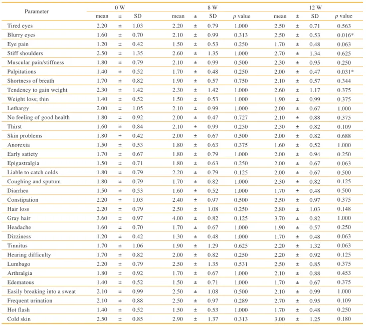
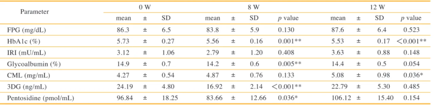
The results of blood glycative stress markers are presented in Table 5. During the 12-week observation period with oral PE
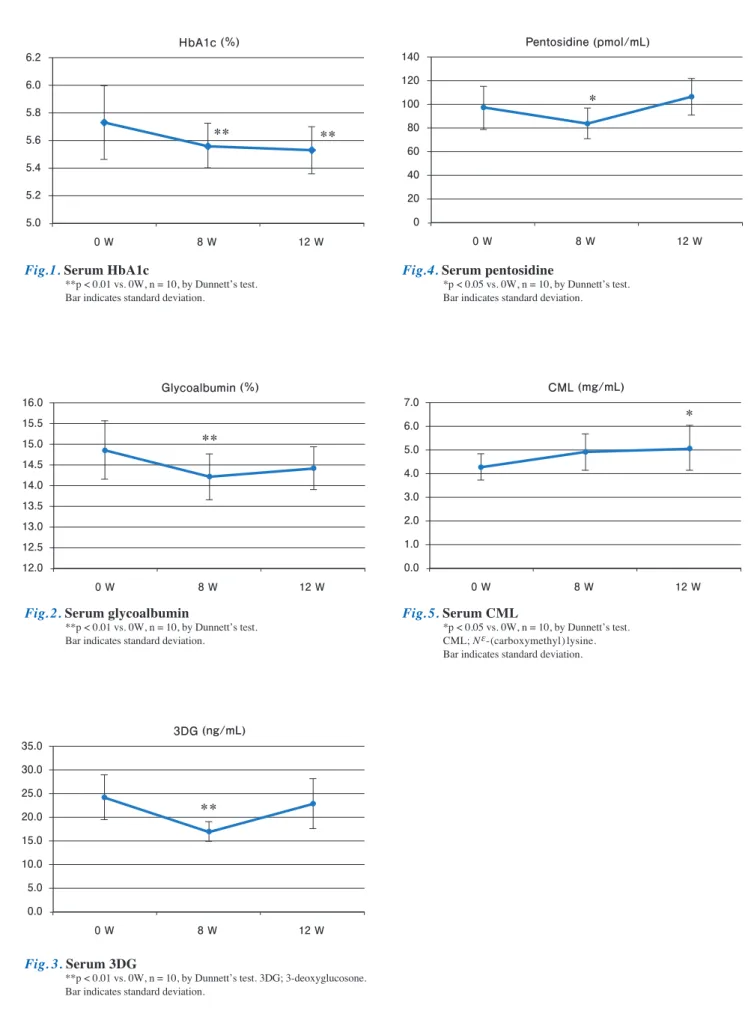
decreased from 0W; 5.73 ± 0.27%, to 8W; 5.56 ± 0.16% (p = 0.001) and remained low to 12W; 5.53 ± 0.17% (p = 0.001, Fig. 1). Glycoalbumin, 0W; 14.9 ± 0.7%, was significantly lower at 8W; 14.2 ± 0.6% (p = 0.005), however, it was not significant at 12W; 14.4 ± 0.5% (p = 0.054, Fig. 2). Similarly, 3DG, 0W; 24.19 ± 4.80 ng/mL, was significantly lower at 8W;
16.92 ± 2.14 ng/mL (p < 0.001), but not significant at 12W;
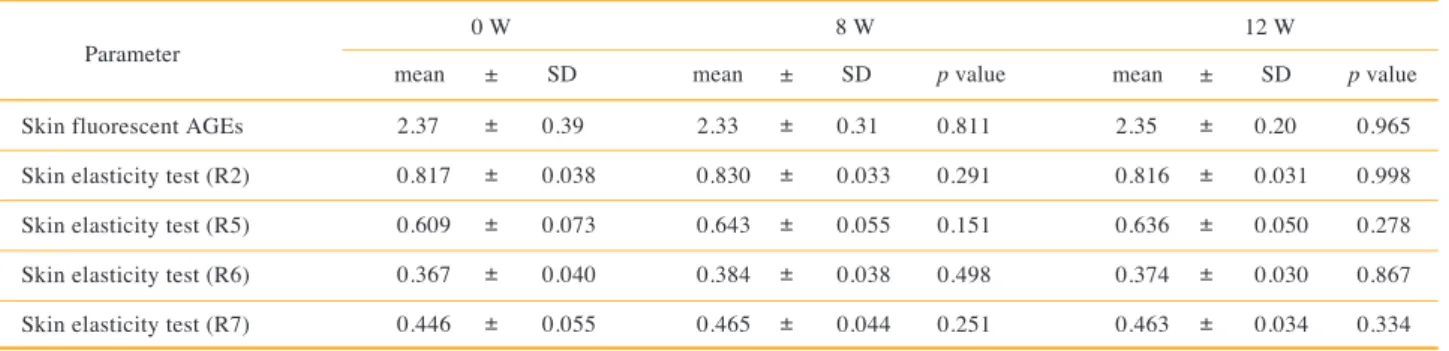
22.79 ± 5.30 ng/mL (p = 0.485, Fig. 3). Pentosidine showed 96.84 ± 18.25 pmol/mL at 0W and it was significantly lower at 8W; 83.66 ± 12.66 pmol/mL (p = 0.036), and then it returned to the baseline at 12W; 106.12 ± 15.40 pmol/mL (p = 0.154, Fig. 4). CML, 0W; 4.27 ± 0.54 μg/ml, was significantly higher
Skin AGEs auto-fluorescence
The amount of skin fluorescent AGEs was not significantly changed by PE intake during the 12-week observation period (Table 6).
Skin elasticity test
PE showed no significant effect on index R2, R5, R6 and R7 of the skin elasticity test during the observation period (Table 6).
p Value vs. 0W, n = 10,by Dunnett's Wilcoxon test. AAQol, Anti-Aging QOL Common Questionnaire; SD, standard deviation.
Table 4. Mental subjective symptom score in AAQol.
Irritability Easily angered Loss of motivation No feeling of happiness Nothing to look forward to in life Daily life is not enjoyable Lose confidence
Reluctance to talk with others Depressed
Feeling of uselessness Shallow sleep
Difficulty in falling asleep Pessimism
Lapse of memory Inability to concentrate Inability to solve problems Inability to make judgments readily Inability to sleep because of worries A sense of tension
Feeling of anxiety for no special reason Vague feeling of fear
1.90 1.60 1.80 1.50 1.50 1.70 1.70 1.50 1.40 1.70 2.30 1.50 1.70 2.60 1.80 1.80 2.10 1.90 2.10 1.20 1.10
0.74 0.52 0.42 0.53 0.53 0.48 0.48 0.71 0.52 0.67 1.25 0.71 0.67 0.70 0.63 0.42 0.74 0.74 0.99 0.42 0.32
±
±
±
±
±
±
±
±
±
±
±
±
±
±
±
±
±
±
±
±
± mean
2.00 1.90 1.80 1.50 1.50 1.40 1.60 1.70 1.50 1.80 2.30 2.00 2.10 2.80 2.30 2.10 2.20 2.20 1.80 1.70 1.20 mean SD
±
0 W 8 W 12 W
0.82 0.57 0.63 0.71 0.53 0.52 0.70 0.95 0.71 0.42 0.82 1.05 0.99 0.63 0.48 0.74 0.79 0.79 0.63 0.48 0.42
±
±
±
±
±
±
±
±
±
±
±
±
±
±
±
±
±
±
±
±
± SD
1.000 0.375 1.000 1.000 1.000 0.250 1.000 0.500 1.000 1.000 1.000 0.063 0.219 0.625 0.125 0.375 1.000 0.375 0.500 0.063 1.000 p value
±
2.00 2.00 2.00 1.60 1.80 1.50 1.80 1.80 1.70 1.80 2.40 2.20 2.30 3.00 2.40 2.20 2.30 2.00 2.20 1.50 1.30 mean
0.67 0.67 0.94 0.70 0.92 0.53 0.63 0.63 0.67 0.42 0.84 1.23 0.67 0.82 0.52 0.63 0.82 0.67 0.63 0.53 0.48
±
±
±
±
±
±
±
±
±
±
±
±
±
±
±
±
±
±
±
±
± SD
1.000 0.313 0.766 1.000 0.750 0.625 1.000 0.250 0.250 1.000 0.984 0.031 0.063 0.313 0.125 0.250 0.625 1.000 1.000 0.250 0.500 p value Parameter ±
*p < 0.05, **p < 0.01 vs. 0W, n = 10, by Dunnett’s test.
FPG, fasting plasma glucose; IRI; immune reactive insulin; 3DG, 3-deoxyglucosone; CML, Nε-(carboxymethyl)lysine; SD, standard deviation.
Table 5. Blood glycative stress markers.
FPG (mg/dL) HbA1c (%) IRI (mU/mL) Glycoalbumin (%) CML (mg/mL) 3DG (ng/mL) Pentosidine (pmol/mL)
86.3 5.73 3.12 14.9 4.27 24.19 96.84
6.5 0.27 1.06 0.7 0.54 4.80 18.25
±
±
±
±
±
±
± mean
83.8 5.56 2.79 14.2 4.87 16.92 83.66 mean SD
±
0 W 8 W 12 W
5.9 0.16 1.20 0.6 0.76 2.14 12.66
±
±
±
±
±
±
± SD
0.130 0.001**
0.408 0.005**
0.133
<0.001**
0.036*
p value
±
87.6 5.53 3.63 14.4 5.08 22.79 106.12 mean
6.4 0.17 0.88 0.5 0.98 5.30 15.40
±
±
±
±
±
±
± SD
0.523
<0.001**
0.148 0.054 0.036*
0.485 0.154 p value Parameter ±
Fig.1. Serum HbA1c
**p < 0.01 vs. 0W, n = 10, by Dunnett’s test.
Bar indicates standard deviation.
Fig.4. Serum pentosidine
* p < 0.05 vs. 0W, n = 10, by Dunnett’s test.
Bar indicates standard deviation.
Fig.2. Serum glycoalbumin
**p < 0.01 vs. 0W, n = 10, by Dunnett’s test.
Bar indicates standard deviation.
Fig.5. Serum CML
*p < 0.05 vs. 0W, n = 10, by Dunnett’s test.
CML; Nε-(carboxymethyl)lysine.
Bar indicates standard deviation.
Fig. 3. Serum 3DG
**p < 0.01 vs. 0W, n = 10, by Dunnett’s test. 3DG; 3-deoxyglucosone.
Bar indicates standard deviation.
** **
HbA1c (%) 6.2
6.0 5.8 5.6 5.4 5.2 5.0
0 W 8 W 12 W
*
Pentosidine (pmol/mL) 140
120 100 80 60 40 20 0
0 W 8 W 12 W
**
Glycoalbumin (%) 16.0
15.5 15.0 14.5 14.0 13.5 13.0 12.5 12.0
0 W 8 W 12 W
* CML (mg/mL)
7.0 6.0 5.0 4.0 3.0 2.0 1.0 0.0
0 W 8 W 12 W
**
3DG (ng/mL) 35.0
30.0 25.0 20.0 15.0 10.0 5.0 0.0
0 W 8 W 12 W
p Value vs. 0W, n = 10, by Dunnett’s test. AGEs, advanced glycation end products; SD, standard deviation.
Table 6. Skin fluorescent AGEs and elasticity index.
Skin fluorescent AGEs Skin elasticity test (R2) Skin elasticity test (R5) Skin elasticity test (R6) Skin elasticity test (R7)
2.37 0.817 0.609 0.367 0.446
0.39 0.038 0.073 0.040 0.055
±
±
±
±
± mean
2.33 0.830 0.643 0.384 0.465 mean SD
±
0 W 8 W 12 W
0.31 0.033 0.055 0.038 0.044
±
±
±
±
± SD
0.811 0.291 0.151 0.498 0.251 p value
±
2.35 0.816 0.636 0.374 0.463 mean
0.20 0.031 0.050 0.030 0.034
±
±
±
±
± SD
0.965 0.998 0.278 0.867 0.334 p value Parameter ±
Discussion
Effects of pomegranate extract (PE)
The active ingredient of test products is PE. The extracts of pomegranate seed, rind, flower are reported, in in vitro studies, experimental animals and culture cells, to show various activities including anti-oxidant capacity14, 15), anti-inflammation
16-19), anti-bacteria 20-23), anti-tumor 24-27), and anti-diabetes 28-30). PE shows anti-bacterial actions against Listeria monocytogenes
21), Pseudomonas stutzeri 22), and Staphylococcus aureus 23). Anti-tumor effect was shown against cell lines derived from mammalian gland cancer 24), ovarian cancer 24), endometrium cancer 24), osteosarcoma 25), colon cancer 26) and prostate cancer
27). Furthermore, in rats PE is reported, to show a preventive effect against carvacrol on methotrexate-induced bone marrow toxicity 31), renal ischemia-reperfusion injury 32), hepatotoxicity induced by diethylnitrosamine and phenobarbital 33), and bleomycin-induced pulmonary fibrosis 34).
PE seems favorable towards sugar metabolism showing anti-diabetic actions 28 - 30). Recent research has focused on the mechanism of PE actions and experiments using diabetic rats have demonstrated the stimulation of insulin secretion
35) and inhibition of α-amylase and α-glucosidase 36). PPARγ activation by PE 37) may be involved in these actions. As a result, PE is expected to be a possible treatment for fatty liver
38) and hypertension care 39), wound healing enhancement 40, 41) and cognitive function improvement 42).
Recent studies on anti-glycation activity by PE have shown that PE inhibits albumin glycation 43) and AGEs generation
3, 44, 45). In these findings on anti-diabetic actions, the basi of PE actions may be its anti-glycation effect.
Data interpretation
In the present study, after the 12-week PE administration, glycative stress markers, FPG and IRI did not change. HbA1c, glycoalbumin, 3DG and pentosidine were significantly reduced at 8 weeks, and HbA1c stayed significantly low at 12 weeks.
PE, as in the previously reported experiments, may fulfill its anti-glycation effect, thus reducing AGEs generation in the body. The reason why CML tended to increase still remains unclear. In order to obtain new findings, we have a study plan in the next trial to check skin corneal CML by the tape stripping method 46).
Since the purpose is focused on confirming safety and effect as a pilot open study, the subjects were limited only to
healthy persons, those who include both persons with high glycation stress and persons with normal glycation stress. In the latter, markers such as skin fluorescent AGEs may be low or normal, and there would be no room for reducing these markers by PE intake. It is necessary to modify the design in a subsequent study, for an example, to include subjects with high skin fluorescent AGEs as assessed by screening.
In a previous clinical study of type 2 diabetic patients and healthy subjects, serum malondialdehyde (MDA) and hydroxynonenal (HN) were reduced only in the diabetic patients; they were not changed in the healthy subjects 47). The reason may be that MDA and HN were almost normal in the healthy persons and there was no room for improvement.
Similarly, in the present study the subjects were healthy persons and skin fluorescent AGEs were not reduced for the same reason.
There were no adverse effects claimed in this study, indicating that PE can be considered a safe product. The evaluation of safety will be described in the next report.
Conclusions
In this open study without control subjects to elucidate the anti-glycation activity of PE, the subjects were 10 post- menopausal females who received oral administration of PE 100 mg/day for 12 weeks, and as a result, some glycative stress markers were reduced. This finding indicates an equivocal effect of PE on glycative stress improvement. Further studies are necessary to elucidate the anti-glycation effect of PE, such as a double-blind randomized controlled trial with subjects with high glycation stress with skin fluorescent AGEs selected by screening.
Conflict of interest statement
The authors are indeted to the Fund “Research and development support program for regional revitalization in agriculture, forestry, fisheries and in the food industry sector’’
of the New Business and Intellectual Property Division, Food Industry Affairs Bureau, from the Ministry of Agriculture, Forestry and Fisheries of Japan, 2013.
Mr. Matsuura, Mr. Nishida, and Mr. Nagatomo are employees of Morishta Jintan Co., Ltd., and were not involved in the data analysis.
References
1) Nagai R, Mori T, Yamamoto Y, et al. Significant of advanced glycation end products in aging-related disease. Anti-Aging Medicine. 2010; 7: 112-119.
2) Ichihashi M, Yagi M, Nomoto K, et al. Glycation stress and photo-aging in skin. Anti-Aging Medicine. 2011; 8: 23-29.
3) Parengkuan L, Yagi M, Matsushima M, et al. Anti-glycation activity of various fruits. Anti-Aging Medicine. 2013; 10: 70- 4) Morishita Jintan Co., Ltd. Patent: Maillard reaction inhibitor. 76.
WO 2013100105 A1. 2013 July 4.
5) Yonei Y, Mizuno Y, Togari H, et al. Muscular resistance training using applied pressure and its effects on the promotion of growth hormone secretion. Anti-Aging Medical Research. 2004; 1:13-27.
6) Yonei Y, Takahashi Y, Watanabe M, et al. A double- blind, randomized controlled trial (RCT) of L-carnitine and conjugated linoleic acid-based health food with health claims. Anti-Aging Medicine. 2007; 4:19-27.
7) Nomoto K, Yagi M, Arita S, et al. A survey of fluorescence derived from advanced glycation end products in the skin of Japanese: Differences with age and measurement location.
Anti-Aging Medicine. 2012; 9: 119-124.
8) Nomoto K, Yagi M, Arita S, Ogura M, Yonei Y. Skin accumulation of advanced glycation end products and lifestyle behaviors in Japanese. Anti-Aging Medicine. 2012;
9: 165-173.
9) Meerwaldt R, Graaff R, Oomen PH, et al. Simple non- invasive assessment of advanced glycationendproduct accumulation. Diabetologia. 2004; 47: 1324-1330.
10) Meerwaldt R, Hartog JW, Graaff R, et al. Skin autofluorescence, a measure of cumulative metabolic stress and advanced glycation end products, predicts mortality in hemodialysis patients. J Am Soc Nephrol. 2005; 16: 3687- 3693.
11) Enomoto DN, Mekkes JR, Bossuyt PM, et al. Quantification of cutaneous sclerosis with a skin-elasticity meter in patients with generalized scleroderma. J Am Acad Dermatol. 1996;
35: 381-387.
12) Rennekampff HO, Rabbels J, Pfau M, et al. Evaluating scar development with objective computer-assisted viscoelastic measurement. Kongressbd Dtsch Ges Chir Kongr. 2002; 119:
749-755. (in German)
13) Kubo M, Yagi M, Kawai H, et al. Anti-glycation effects of mixed-herb-extracts in diabetes and pre-diabetes. J Clin Biochem Nutr. 2008; 43(Suppl 1): 66-69.
14) Cerdá B, Espín JC, Parra S, et al. The potent in vitro antioxidant ellagitannins from pomegranate juice are metabolised into bioavailable but poor antioxidant hydroxy- 6H-dibenzopyran-6-one derivatives by the colonic microflora of healthy humans. Eur J Nutr. 2004; 43: 205-220.
15) Henning SM, Zhang Y, Rontoyanni VG, et al. Variability in the antioxidant activity of dietary supplements from pomegranate, milk thistle, green tea, grape seed, goji, and acai: Effects of in vitro digestion. J Agric Food Chem.
2014 Apr 29. [Epub ahead of print]
16) Khan N, Syed DN, Pal HC, et al. Pomegranate fruit extract inhibits UVB-induced inflammation and proliferation by modulating NF-κB and MAPK signaling pathways in mouse skin. Photochem Photobiol. 2012; 88: 1126-1134.
17) Rosillo MA, Sánchez-Hidalgo M, Cárdeno A, et al. Dietary supplementation of an ellagic acid-enriched pomegranate extract attenuates chronic colonic inflammation in rats.
Pharmacol Res. 2012; 66: 235-242.
18) Ismail T, Sestili P, Akhtar S. Pomegranate peel and fruit extracts: A review of potential anti-inflammatory and anti- infective effects. J Ethnopharmacol. 2012; 143: 397-405.
19) Winand J, Schneider YJ. The anti-inflammatory effect of a pomegranate husk extract on inflamed adipocytes and macrophages cultivated independently, but not on the inflammatory vicious cycle between adipocytes and macrophages. Food Funct. 2014; 5: 310-318.
20) Al-Zoreky NS. Antimicrobial activity of pomegranate (Punica granatum L.) fruit peels. Int J Food Microbiol.
2009; 134: 244-248.
21) Li G, Xu Y, Wang X, et al. Tannin-rich fraction from pomegranate rind damages membrane of Listeria monocytogenes. Foodborne Pathog Dis. 2014; 11: 313- 22) 319.Devatkal SK, Jaiswal P, Jha SN, et al. Antibacterial
activity of aqueous extract of pomegranate peel against Pseudomonas stutzeri isolated from poultry meat. J Food Sci Technol. 2013; 50: 555-560.
23) Su X, Howell AB, D'Souza DH. Antibacterial effects of plant-derived extracts on methicillin-resistant Staphylococcus aureus. Foodborne Pathog Dis. 2012; 9:
573-578.
24) Sreekumar S, Sithul H, Muraleedharan P, et al.
Pomegranate fruit as a rich source of biologically active compounds. Biomed Res Int. 2014;2014:686921. Epub 2014 Apr 10.
25) Li J, Zhang F, Wang S. A polysaccharide from pomegranate peels induces the apoptosis of human osteosarcoma cells via the mitochondrial apoptotic pathway. Tumour Biol. 2014 May 2. [Epub ahead of print]
26) Jaganathan SK, Vellayappan MV, Narasimhan G, et al.
Role of pomegranate and citrus fruit juices in colon cancer prevention. World J Gastroenterol. 2014; 20: 4618-4625.
27) Ming DS, Pham S, Deb S, et al. Pomegranate extracts impact the androgen biosynthesis pathways in prostate cancer models in vitro and in vivo. J Steroid Biochem Mol Biol. 2014 Feb 22;143C:19-28. doi: 10.1016/
j.jsbmb.2014.02.006. [Epub ahead of print]
28) Das AK, Mandal SC, Banerjee SK, et al. Studies on the hypoglycaemic activity of Punica granatum seed in streptozotocin induced diabetic rats. Phytother Res. 2001;
15: 628-629.
29) Bagri P, Ali M, Aeri V, et al. Antidiabetic effect of Punica granatum flowers: effect on hyperlipidemia, pancreatic cells lipid peroxidation and antioxidant enzymes in experimental diabetes. Food Chem Toxicol. 2009; 47: 50- 30) Parmar HS, Kar A. Antidiabetic potential of Citrus sinensis 54.
and Punica granatum peel extracts in alloxan treated male mice. Biofactors. 2007; 31: 17-24.
31) Şen V, Bozkurt M, Söker S, et al. The effects of pomegranate and carvacrol on methotrexate-induced bone marrow toxicity in rats. Clin Invest Med. 2014; 37:
E93-E101.
32) Sancaktutar AA, Bodakci MN, Hatipoglu NK, et al. The protective effects of pomegranate extracts against renal ischemia-reperfusion injury in male rats. Urol Ann. 2014; 6:
46-50.
33) Shaban NZ, El-Kersh MA, Bader-Eldin MM, et al. Effect of Punica granatum (pomegranate) juice extract on healthy liver and hepatotoxicity induced by diethylnitrosamine and phenobarbital in male rats. J Med Food. 2014; 17: 339- 349.
34) Hemmati AA, Rezaie A, Darabpour P. Preventive effects of pomegranate seed extract on bleomycin-induced pulmonary fibrosis in rat. Jundishapur J Nat Pharm Prod.
2013; 8: 76-80.
35) Nekooeian AA, Eftekhari MH, Adibi S, et al. Effects of pomegranate seed oil on insulin release in rats with type 2 diabetes. Iran J Med Sci. 2014; 39: 130-135.
36) Kam A, Li KM, Razmovski-Naumovski V, et al. A comparative study on the inhibitory effects of different parts and chemical constituents of pomegranate on α-amylase and α-glucosidase. Phytother Res. 2013; 27:
1614-1620.
37) Li Y, Qi Y, Huang TH, et al. Pomegranate flower: a unique traditional antidiabetic medicine with dual PPAR-alpha/- gamma activator properties. Diabetes Obes Metab. 2008;
10: 10-17.
38) Xu KZ, Zhu C, Kim MS, et al. Pomegranate flower ameliorates fatty liver in an animal model of type 2 diabetes and obesity. J Ethnopharmacol. 2009; 123: 280- 39) Mohan M, Waghulde H, Kasture S. Effect of pomegranate 287.
juice on angiotensin II-induced hypertension in diabetic Wistar rats. Phytother Res. 2010; 24(Suppl 2): S196-203.
40) Yan H, Peng KJ, Wang QL, et al. Effect of pomegranate peel polyphenol gel on cutaneous wound healing in alloxan-induced diabetic rats. Chin Med J (Engl). 2013;
126: 1700-1706.
41) Pirbalouti AG, Azizi S, Koohpayeh A, et al. Wound healing activity of Malva sylvestris and Punica granatum in alloxan-induced diabetic rats. Acta Pol Pharm. 2010; 67:
511-516.
42) Cambay Z, Baydas G, Tuzcu M, et al. Pomegranate (Punica granatum L.) flower improves learning and memory performances impaired by diabetes mellitus in rats. Acta Physiol Hung. 2011; 98: 409-420.
43) Dorsey PG, Greenspan P. Inhibition of nonenzymatic protein glycation by pomegranate and other fruit juices. J Med Food. 2014; 17: 447-454.
44) Mesías M, Navarro M, Gökmen V, et al. Antiglycative effect of fruit and vegetable seed extracts: Inhibition of AGE formation and carbonyl-trapping abilities. J Sci Food Agric. 2013; 93: 2037-2044.
45) Ito H, Li P, Koreishi M, Nagatomo A, et al. Ellagitannin oligomers and a neolignan from pomegranate arils and their inhibitory effects on the formation of advanced glycation end products. Food Chem. 2014; 152: 323-330.
46) Kamitani Y, Yagi M, Nomoto K, et al. Non-invasive collection of stratum corneum samples by a tape-stripping technique. Anti-Aging Medicine. 2013; 10: 55-59.
47) Basu A, Newman ED, Bryant AL, et al. Pomegranate polyphenols lower lipid peroxidation in adults with type 2 diabetes but have no effects in healthy volunteers:
A pilot study. J Nutr Metab. 2013;2013:708381. doi:
10.1155/2013/708381. Epub 2013 Jul 10.