Introduction
Diaphragmatic tumors are rare, and those reported previously have included malignant fibrous histiocytoma1) and gastrointestinal stromal tumor2). However, cases of hepatocellular carcinoma (HCC) invasive to the diaphragam have also sometimes been documented3-6). However, in cases of HCC requiring combined resection of the diaphragm, the area of the diaphragm resected is usually small and primary closure is feasible. On
the other hand, when the defect of the diaphragm is wide, reconstruction of the diaphragm becomes problematic. Although this can be done using an artificial prosthesis, such as mesh, living material may be superior because it minimizes the possibility of infectious complications.
Reconstruction of the diaphragm for congenital diaphragmatic defects has often been reported7, 8). Here we report a case of spontaneous rupture of a HCC for which we performed wide resection of the
A case of spontaneous rupture of hepatocellular carcinoma mimicking a diaphragm tumor:
Reconstruction of the diaphragm using a latissimus dorsi muscle flap
Ichiro Hirai*, Wataru Kimura*, Hiroyuki Oizumi**, Hiroto Fujimoto*, Toshihiro Watanabe*, Koji Tezuka*, Taketo Yamagishi*,
Shuichiro Sugawara*, Naoki Kanauchi**, Satoshi Shiono**, Tadanori Minagawa**, Mitsuaki Sadahiro**
*First Department of Surgery, Yamagata University
**Second Department of Surgery, Yamagata University
(Accepted August 18, 2013)
ABSTRACT
Spontaneous rupture of hepatocellular carcinoma (HCC) is sometimes encountered clinically. We experienced a case of spontaneous rupture of a HCC, which was surgically resected along with the diaphragm.
A 74-year old woman presented with anemia. Preoperative imaging suggested a tumor arising on the diaphragm. After thoracotomy and laparotomy, the intraoperative diagnosis was changed to spontaneous rupture of a HCC with invasion to the diaphragm and right lower lung. Wide resection of the diaphragm including an area measuring 13 × 10 cm, partial resection of the right lower lung and right hepatectomy were performed. The defect in the diaphragm was reconstructed by suturing with a reversed latissimus dorsi muscle flap.
There was no recurrence of HCC 7 years after surgery. Furthermore, the patient had no muscle weakness in the right arm during daily living activities.
In conclusion, a good outcome may be obtained by combined resection of a ruptured HCC along with the diaphragm, even if the tumor has infiltrated the diaphragm. A latissimus dorsi muscle flap is considered to have a better antibacterial effect than an artificial mesh prosthesis, and is suitable for patients requiring wide resection of the diaphragm.
Key words: hepatocellular carcinoma, spontaneous rupture, diaphragm, latissimus dorsi muscle flap.
diaphragm, with reconstruction using a latissimus dorsi muscle flap.
Case report
A 74-year-old woman presented with giddiness, a n d w a s f o u n d t o b e a n e m i c w i t h a s e r u m hemoglobin level of 7.8 g/dl. She was diagnosed as having a liver abscess and treated initially with antibiotics. However, her condition did not improve and she was referred to our hospital.
The abdomen was soft and flat, and neither the liver nor any abdominal tumor was palpable.
Laboratory data indicated anemia (red blood cells 288×104/μl, hemoglobin 8.0 g/dl). The serum total bilirubin level was 0.6 mg/dl, aspartate aminotransferase (AST) was 15 U/l, alanine aminotransferase (ALT) was 9 U/l, hepatitis B antigen, antibody, and hepatitis C antibody were all negative. The indocyanine green retention rate at 15 minutes after injection was 6%. Uptake determined by 99mTc-GSA SPECT scintigraphy w a s 9 3 % , i n d i c a t i n g g o o d l i v e r f u n c t i o n . Tumor markers were within normal limits, including alpha-fetoprotein (AFP) at 2.5 ng/ml, carcinoembryonic antigen (CEA) at 3.09 ng/ml, and protein induced by vitamin K absence or antagonist -II (PIVKA II) at <10 mAU/ml.
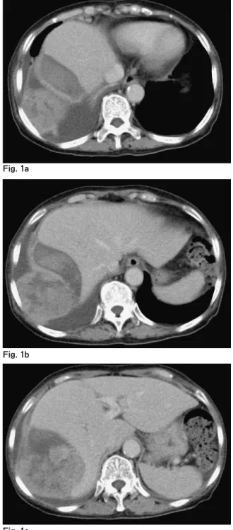
CT scan revealed a tumor located on the diaphragm, measuring 69×61 mm on the pleural (cranial) side and 49×57 mm on the abdominal (caudal) side (Fig. 1a-c). The tumor was iso-dense with the liver on non-enhanced CT. A dynamic CT study showed that the tumor was enhanced from the periphery through the early to delayed phase.
These CT findings were different from a typical HCC.
The tumor was located close to the 9th and 1 0t h r i b s . M R I e x a m i n a t i o n a l s o s h o w e d a diaphragmatic tumor rather than HCC because of a diaphragmatic compression pattern (Fig.
2a, b). Ultrasonography revealed a tumor with a snowman-like profile with the diaphragm located caudal to it.
On the basis of these findings we diagnosed this case as a tumor of diaphragmatic origin, such as a fibrosarcoma or rhabdomyosarcoma. We
planned to use a latissimus dorsi muscle flap for reconstruction of the diaphragm, anticipating that wide resection of the diaphragmatic tumor might be necessary. Gore-Tex mesh was also prepared for reconstruction of the diaphragm and chest
Fig. 1a-c The preoperative diagnosis was a tumor of diaphragm origin, because the tumor appeared to be located on both the cranial and caudal sides of the diaphragm.
Fig. 1a
Fig. 1b
Fig. 1c
wall. We also made preparations for hepatectomy because of the tumor’s location near the liver.
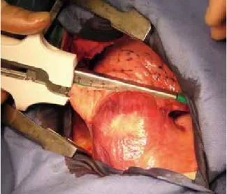
A skin incision was made in the 8th inter-costal space. The latissimus dorsi muscle was widely exposed (Fig. 3a), and its cranial side was divided with electric forceps. After thoracotomy, the tumor was detected on the diaphragm, and found to be attached to the right lower lobe of the lung.
Partial resection of the right lung was performed with autosuturing (Fig. 3b). Upon dissection of the diaphragm, old blood was detected in the abdominal cavity. Because the main tumor was located in the liver, laparotomy was added. This revealed that the right lobe of the liver had spontaneously divided, indicating that this case was one of HCC rupture
(Fig. 3c). The liver surface of the lateral sector was normal. However, the visceral peritoneum of the medial sector and right liver was thickened and white.
After cholecystectomy, the right lobe was mobilized and the short hepatic veins were ligated and cut. Right hepatectomy was performed by the Pean fracture method under the Pringle maneuver.
The right lobe and a wide area of the diaphragm were removed (Fig. 3d). The resected specimen weighed 689 g, and the area of the diaphragm resected was 13×10 cm.
The latissimus dorsi muscle flap was inserted into the pleural cavity through the 10th intercostal space. The flap and remnant diaphragm were
Fig. 2a, b We diagnosed this case as diaphragmatic sarcoma in view of the manner in which it compressed the diaphragm.
Fig. 3a Skin and subcutaneous tissue was detached widely from the latissimus dorsi muscle.
Fig. 3b The lower part of the right lung was dissected with autosuturing.
Fig. 2a
Fig. 2b
attached by horizontal mattress sutures using felt and absorbable strings (Fig. 3e). The wound was closed after placement of pleural and abdominal drainage tubes.
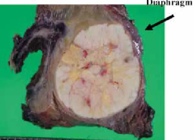
T h e c u t s u r f a c e o f t h e f o r m a l i n - f i x e d
resected specimen was 6.6 cm in diameter (Fig.
4a). Pathological examination showed that a moderately differentiated HCC had invaded the diaphragm (Fig. 4b), without infiltration into the lung.
Artificial ventilation was necessary after surgery, but the patient was extubated on postoperative day (POD) 1. The thoracic drainage tube was removed on POD 3, and the patient was discharged on 17 POD. Pleural effusion disappeared at 2 months after surgery. There was no recurrence of the HCC 7 years after surgery, and no pleural effusion. The patient has shown no muscle weakness in the right arm for daily living activities, and the latissimus dorsi flap has shown no signs of atrophy during outpatient follow-up (Fig. 5).
Fig. 3c The right lobe of the liver showed spontaneous rupture.
Fig. 3d Photograph after resection of the right liver lobe and partial resection of the right lung.
Fig. 3e Suturing between the remnant diaphragm and the latissimus dorsi muscle flap.
Fig. 4b Moderately differentiated hepatocellular carcinoma invasive into the diaphragm was confirmed pathologically.(This is the part of Fig. 4a indicated by an arrow)
Fig. 4a Cut surface of the formalin-fixed surgical specimen. The diaphragm is indicated with an arrow.
Discussion
We misdiagnosed this case as a tumor of diaphragmatic origin because of the enhanced pattern on CT and the diaphragmatic compression pattern. Enhanced ultrasonography was not performed. If enhanced ultrasonography had been carried out, a preoperative diagnosis of HCC might have been obtained.
HCC often ruptures spontaneously. One of the suggested causes of HCC rupture is vascular injury to small arteries9). The long-term outcome of liver resection for HCC, with or without rupture, is still uncertain10). One report has indicated that HCC rupture is associated with advanced disease and has a poor prognosis11). Shock and the need for blood transfusion have been reported as independent factors associated with early mortality in patients with ruptured HCC12). Although transarterial embolization (TAE) is usually performed for hemostasis of ruptured HCC13), our patient’s
blood pressure was stable despite the presence of anemia. Therefore, elective surgery was possible in this case. Ruptured HCC often exacerbates the risk of peritoneal dissemination and is usually difficult to resect completely14). On the other hand, HCC rupture itself is reported to have a negative impact on patient survival15). Among patients with ruptured HCC, those for whom hepatectomy was considered feasible after TAE had fairly good outcomes16). Our patient with ruptured HCC also showed a good outcome, as surgical removal of the whole tumor was possible, despite the erroneous preoperative diagnosis due to protrusion of the HCC into the pleural cavity.
It has been reported that en bloc resection of the diaphragm is justified for patients with gross diaphragmatic involvement of HCC, as there are no significant differences in short- or long-term surgical outcome3-6). It has also been reported that simultaneous resection of the diaphragm during partial hepatectomy for metastasic liver tumors does not significantly influence perioperative morbidity or mortality17). Reconstruction of the diaphragm is necessary when wide resection is performed. Materials used for reconstruction can include artificial prostheses or living material7, 18, 19). Shimamura et al. have reported diaphragm repair using an external oblique muscle flap20).
The latissimus dorsi muscle arises from the 9th- 12th ribs and is attached to the humerus. Tsukada
Fig. 4d Pathological appearance of the HCC part ( × 40).
Fig. 5 The latissimus dorsi muscle flap showed no atrophy. There was no pleural effusion and no recurrence of HCC during outpatient follow-up.
Fig. 4c Pathological appearance of the HCC part ( × 4).
et al. reported that there was no ischemic necrosis of the proximal part of a latissimus dorsi flap even if the tendinous part of the muscle is cut near the humeral insertion21). The branches of the thoracodorsal nerve usually run parallel to the branches of the thoracodorsal arteries. It has been reported that a latissimus dorsi muscle flap does not become atrophic8).
The distal portion of a latissimus dorsi flap is brought into the chest through an aperture obtained by resection of the tenth (or ninth) rib22). Arm motion is not compromised in any way because of anchorage of the still-functional proximal portion of the muscle and compensation by other muscle groups23). Seroma formation is the commonest complication after diaphragm reconstruction24). Our patient also had pleural effusion for 2 months after surgery, but atelectasis or pneumonia did not occur.
In conclusion, a good outcome may be obtained b y c o m b i n e d r e s e c t i o n o f a r u p t u r e d H C C along with the diaphragm, even if the tumor has infiltrated the diaphragm. A latissimus dorsi muscle flap is considered to have a better antibacterial effect than an artificial mesh prosthesis, and is suitable for patients requiring wide resection of the diaphragm.
References
1. Perelas A, Papantoni E, Sakorafas GH, Vassileiou P, Arkadopoulos N, Smyrniotis V. Malignant fibrous histiocytoma of the diaphragm. Am Surg. 2013; 79:
e12-3.
2. Yeung CK, Yuen CH, Chan IK, Chu RW. Malignant extra-gastrointestinal stromal tumour of diaphragm.
ANZ J Surg 2008; 78: 923-924.
3. Jeng KS, Chen BF, Lin HJ. En bloc resection for extensive hepatocellular carcinoma: is it advisable?
World J Surg 1994; 18: 834-839.
4. Leung KF, Chui AK, Leung KL, Lai PB, Liew CT, Lau WY. Clinicopathological study of hepatocellular carcinoma with diaphragmatic involvement. Br J Surg. 2001; 88: 681-682.
5. Lin MC, Wu CC, Chen JT, Lin CC, Liu TJ. Surgical results of hepatic resection for hepatocellular carcinoma with gross diaphragmatic invasion.
Hepatogastroenterology. 2005; 52: 1497-1501.
6. Yamashita Y, Morita K, Iguchi T, Tsujita E, Soejima Y, Taketomi A, Maehara Y. Surgical impacts of an en bloc resection of the diaphragm for hepatocellular carcinoma with gross diaphragmatic involvement. Surg Today. 2011; 41: 101-106.
7. S a m a r a k k o d y U , K l a s s e n M , H a m i l t o n N . R e c o n s t r u c t i o n o f c o n g e n i t a l a g e n e s i s o f hemidiaphragm by combined reverse lattisimus dorsi and serratus anterior muscle flaps. J Pediat Surg 2001;
36: 1637-1640.
8. L e e S , P o u l o s N D , G r e e n h o l z S K . S t a g e d reconstruction of large congenital diaphragmatic defects with synthetic patch followed by reverse latissimus dorsi muscle. J Pediat Surg 2002; 37: 367-370.
9. Zhu LX, Meng XL, Fan ST. Elasticity of small artery in patient with spontaneous rupture of hepatocellular carcinoma. Hepatol Res 2004; 29: 13-17.
10. L a i E C , L a u W Y . S p o n t a n e o u s r u p t u r e o f hepatocellular carcinoma: a systematic review. Arch Surg. 2006; 141: 191-198.
11. M i y o s h i A , K i t a h a r a K , K o h y a N , N o s h i r o H , M i y a z a k i K . O u t c o m e s o f p a t i e n t s w i t h spontaneous rupture of hepatocellular carcinoma.
Hepatogastroenterology 2011; 58: 99-102.
12. Tan FL, Tan YM, Chung AY, Cheow PC, Chow PK, Ooi LL. Factors affecting early mortality in spontaneous rupture of hepatocellular carcinoma. ANZ J Surg. 2006 ; 76: 448-452.
13. Li WH, Cheuk EC, Kowk PC, Cheung MT. Survival after transarterial embolization for spontaneous ruptured hepatocellular carcinoma. J Hepatobiliary Pancreat Surg. 2009; 16: 508-512.
14. Kaido T, Arii S, Shiota M, Imamura M. Repeated r e s e c t i o n f o r e x t r a h e p a t i c r e c u r r e n c e s a f t e r hepatectomy for ruptured hepatocellular carcinoma. J Hepatobiliary Pancreat Surg. 2004; 11: 149-152.
15. Aoki T, Kokudo N, Matsuyama Y, Izumi N, Ichida T, Kudo M, Ku Y, Sakamoto M, Nakashima O, Matsui O, Makuuchi M; for the Liver Cancer Study Group of Japan. Prognostic Impact of Spontaneous Tumor Rupture in Patients With Hepatocellular Carcinoma:
An Analysis of 1160 Cases From a Nationwide Survey.
Ann Surg. 2013
16. T a k e b a y a s h i T , K o n d o S , A m b o Y , H i r a n o S , O m i M , M o r i k a w a T , O k u s h i b a S , K a t o h
H . S t a g e d h e p a t e c t o m y f o l l o w i n g a r t e r i a l embolization for ruptured hepatocellular carcinoma.
Hepatogastroenterology. 2002; 49: 1074-1076.
17. Li GZ, Turley RS, Lidsky ME, Barbas AS, Reddy SK, Clary BM. Impact of simultaneous diaphragm resection during hepatectomy for treatment of metastatic colorectal cancer. J Gastrointest Surg. 2012; 16: 1508- 1515.
18. Whetzel TP, Stokes RB, Greenholz SK, Saunders CJ. Reconstruction of the toddler diaphragm in severe anterolateral congenital diaphragmatic hernia with the reverse latissimus dorsi flap. Ann Plast Surg 1997; 39:
615-619.
19. Sinna R, De Dominicis F, Quassemyar Q, Fuks D, Perignon D, Regimbeau JM, Berna P. Diaphragmatic reconstruction using combined reversed extended latissimus dorsi and serratus anterior fascia flaps. Ann Thorac Surg 2011; 91: 922-924.
20. Shimamura Y, Gunveb P, Ichii M, Taniguchi H, Kitai S, Endo Y, Okajima K, Shima Y, Sugai S, Takahashi A, et al. Repair of the diaphragm with an external oblique muscle flap. Surg Gynecol Obstet 1989; 169: 159-169.
21. T s u k a d a H , O s a d a H . R e c o n s t r u c t i o n o f t h e hemidiaphragm and hemipericardium using combined reversed latissimus dorsi and serratus anterior muscle flaps. Surg Today 2006; 36: 291-293.
22. Bedani AV, Andreani SM, Muscolino G. Latissimus dorsi reverse flap to substitute the diaphragm after extrapleural pnemonectomy. Ann Thorac Surg 2000; 69:
986-988.
23. Bedini A, Valente M, Andreani S, Ravasi G. Reverse flap of distal dorsi for diaphragm reconstruction in the adult: specification of the technical procedure and report on six cases. J Thoracic Cardiovasc Surg 1997;
114: 846-848.
24. McConkey MO, Temple CLF, Mcfadden S, Temple WJ. Autologous diaphragm reconstruction with the pedicled latissimus dorsi flap. J Surg Oncol 2006; 94:
248-251.