1
Anatomical Study of the Position and Orientation of the Coracoclavicular Ligaments:
1
Differences in Bone Tunnel Position by Sex
2
3
4
5
Terufumi Shibata
1, Teruaki Izaki
1, Satoshi Miyake
1, Nobunao Doi
1, Yozo Shibata
2,
6
Yutaka Irie
3, Katsuro Tachibana
3, Takuaki Yamamoto
17
8
1
Department of Orthopaedic Surgery, Fukuoka University Faculty of Medicine,
9
7-45-1 Nanakuma, Jonan-ku, Fukuoka 814-0180, Japan
10
11
2
Department of Orthopaedic Surgery, Fukuoka University Chikushi Hospital, Fukuoka
12
1-1-1 Zokumyoin, Chikushino, Fukuoka 818-8502, Japan
13
14
3
Department of Anatomy, Fukuoka University Faculty of Medicine,
15
7-45-1 Nanakuma, Jonan-ku, Fukuoka 814-0180, Japan
16
17
18
Corresponding author: Terufumi Shibata, MD
19
2
Department of Orthopaedic Surgery, Faculty of Medicine, Fukuoka University
20
7-45-1 Nanakuma, Jonan-ku, Fukuoka 814-0180, Japan
21
Tel: +81-92-801-1011 ext. 3465; FAX: +81-92-864-9055
22
E-mail: [email protected]
23
24
https://doi.org/10.1016/j.otsr.2018.10.020
25
3
Abstract
26
Background
27
Reconstructing both coracoclavicular ligaments following acromioclavicular
28
dislocation has recently been reported to restore the function of the acromioclavicular
29
joint better than traditional procedures. Knowing the appropriate position and
30
orientation of the bone tunnels and the potential risks of neurovascular injuries leads to
31
safe reconstruction. We aimed to answer the following questions: what is the difference
32
in the accurate clavicular bone tunnel positions (BTPs) during coracoclavicular ligament
33
reconstruction between sex, and what are the potential risks for neurovascular injuries?
34
Hypothesis
35
The BTPs differ by sex at the site of coracoclavicular ligament reconstruction.
36
Patients and methods
37
We introduced two Kirschner wires into 25 cadaver shoulders (17 male, 8 female), one
38
through the insertion center of the trapezoid ligament and one through the conoid
39
ligament, and measured the distance from the respective Kirschner wire insertion points
40
to the bony landmarks of the clavicle and the oblique angle of each Kirschner wire. The
41
shortest distance from the insertion point of each Kirschner wire to the suprascapular
42
nerve and artery was also measured.
43
Results
44
While the distance from the acromioclavicular joint to the respective Kirschner wire
45
insertion points tended to be longer in males, the ratio of these insertion points to total
46
clavicle length was constant. Other measurements for respective Kirschner wire
47
insertions to the bony landmarks and neurovascular structures were comparable, as were
48
abduction and retroversion angles. The distance from the suprascapular nerve to the
49
4
insertion point of the conoid ligament at the coracoid process was 13.8±4.0 mm, while
50
the distance from the suprascapular artery was 7.1±3.3 mm.
51
Discussion
52
Appropriate position and orientation of the bone tunnels
,and the ratio of the BTPs to the
53
total clavicular length, aid surgeons in performing the reconstruction. The conoid
54
ligament insertion on the coracoid was just proximal to the suprascapular artery, so
55
surgeons should be careful with conoid insertion .
56
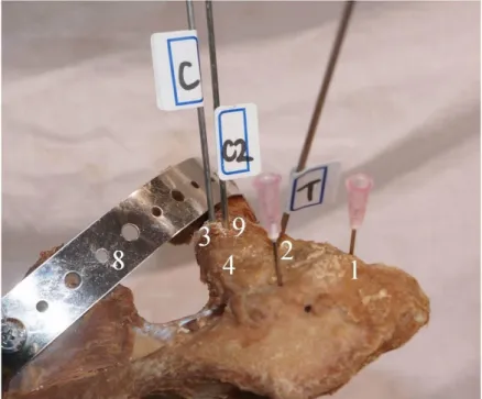
Level of evidence: Level V, cadaver study
57
Keywords:
58
Acromioclavicular joint; Instability; Blood vessels; Neuroanatomy
59
5
1. Introduction
60
Reconstruction of the coracoclavicular ligaments as separate anatomical structures has
61
been investigated to correct acromioclavicular dislocation because of their distinct
62
function. The conoid ligament is responsible for the restraint against anterior and
63
superior loading, whereas the trapezoid ligament provides resistance to posterior
64
loading [1]. Two-bundle stabilization of the acromioclavicular joint is biomechanically
65
superior and better restores joint function compared with single-bundle stabilization [2–
66
4]. Inappropriate tunnel positioning in both the clavicle and coracoid process increases
67
fracture risk [5]. Therefore, correct tunnel position and orientation is important to obtain
68
better clinical results. Several authors reported that a malpositioned insertion point
69
during bone tunnel drilling increases the risk of damaging the suprascapular nerve and
70
artery around the coracoid process; [6,7] however, the distance from the exit point has
71
not been investigated. Because the suprascapular artery and nerve pass just medial to the
72
coracoid process, the exit point of the drill on the coracoid process is crucial.
73
Zhu et al [8] studied the location and orientation of the coracoclavicular ligaments by
74
drilling through the center of the trapezoid and the conoid tuberosities. Although these
75
tuberosities are bony landmarks that are easily verified, the center of each tuberosity
76
does not necessarily match the center of the coracoclavicular ligament attachment. The
77
6
trapezoid ligament’s attachment extends medial to this tuberosity [9]. The center of the
78
conoid ligament is not also located on the most prominent aspect of the conoid
79
tuberosity, and the conoid tuberosity is broad and the exact center is not always distinct
80
[10]. Therefore, additional data on the bone tunnel position (BTP) and orientation that
81
passes through the insertion center of these ligaments are needed. The differences
82
between sex in the coracoclavicular ligament attachment on the clavicular undersurface
83
have been previously reported [10,11]. Thus, a difference in the BTP on the clavicular
84
surface between sex has been suspected. However, Zhu et al [8] did not report the sex of
85
the cadavers. For these reasons, a more accurate position and orientation of the
86
coracoclavicular ligament is needed.
87
The purpose of this study was to answer the following questions: (1) what is the
88
difference in the accurate clavicular BTPs for coracoclavicular ligament reconstruction
89
between sex; and (2) what are the potential risks of neurovascular injuries during
90
coracoclavicular ligament reconstruction? We hypothesized that accurate BTPs for
91
anatomical reconstruction of the acromioclavicular joint differ by sex.
92
2. Patients and Methods
93
2.1 Specimen demographics
94
This study included 25 shoulders from 17 male and 8 female cadavers donated to our
95
7
institution. We obtained a single shoulder from each cadaver, and the remainder of each
96
body was used for medical education. This study was approved by our Institutional
97
Review Board. Donors were preserved in formalin-based dilution, and none of the
98
cadavers had obvious congenital abnormalities, findings of trauma, or previous surgery
99
to the shoulder.
100
2.2 Specimen preparation
101
To expose the lateral clavicle, coracoid process, and coracoclavicular ligament, we
102
carefully removed all periscapular and clavicular muscles. To disconnect the
103
glenohumeral joint, we resected the rotator cuff and capsule at the proximal humerus.
104
Before each specimen was disarticulated at the scapulothoracic joint, the proximal
105
clavicle and scapular spine were fixed using a metal plate and screws to maintain the
106
position between the clavicle and the coracoid process. The screws did not damage the
107
area of interest. After dissection, the scapula was fixed using a bench vise, which we
108
mounted on a free-standing camera platform so that the specimen’s alignment could be
109
freely changed. We positioned the scapular body vertically and the upper surface of the
110
clavicle horizontally. Therefore, we were able to observe the orientation of the
111
coracoclavicular ligament from all directions, which allowed us to drill through the
112
insertion center of the coracoclavicular ligaments (conoid and trapezoid) along the
113
8
directions of these ligaments.
114
These ligamentous attachments were observed under the clavicle, and one 2-mm
115
Kirchner wire (K-wire) was introduced through the insertion center of the trapezoid (wire-
116
T) and another through the conoid ligament (wire-C) directed as accurately as possible to
117
the coracoid origins of the coracoclavicular ligaments, along their trajectory. We then
118
measured the distance from the respective K-wire insertion point to the lateral edge and
119
anterior or posterior border of the distal clavicle (Fig. 1A).
120
Both K-wires were evaluated for abduction and retroversion angles. To set the horizontal
121
baseline, we placed a 2-mm K-wire on the longitudinal axis of the clavicle, which passed
122
the anteroposterior midpoint of the distal and proximal clavicles as reference K-wire X.
123
To set the anteroposterior baseline, another 2-mm K-wire was drilled in the
124
anteroposterior direction horizontally and perpendicular to K-wire X as reference K-wire
125
Y (horizontal baseline). A digital camera was then placed at the same height as the clavicle
126
so that each reference K-wire (X, Y) was seen as a dot on the coronal or sagittal view of
127
the scapula, and photographs of the shoulder were taken in both planes (Fig. 1B and C).
128
Using the photographs, we measured the oblique angle of wire-T or wire-C to the
129
reference K-wire (X or Y) using Image J image analyzing software (National Institutes of
130
Health, Bethesda, MD, USA).
131
9
Blowout of the posterior wall of the medial clavicular tunnel leads to a loss of reduction,
132
as the clavicular attachment area of the conoid ligament was located around the conoid
133
tubercle posteriorly [12]. Lower bone density and the thinnest dorsal cortex in the lateral
134
clavicle also contributes to blowout [13]. To place the bone tunnel at a more robust
135
position, we drilled another 2-mm K-wire (C2 conoid K-wire, wire-C2) at the midpoint
136
of the anteroposterior diameter of the clavicle perpendicular to reference wire-X, which
137
was inserted parallel to wire-C (Fig. 1D). We performed the same measurements for wire-
138
C2 as wire-C.
139
Next, we measured the location of each K-wire penetrating the medial cortex of the
140
coracoid process (Fig. 2A). The proximal parts of the suprascapular artery and nerve
141
were resected when dissecting the scapula from the trunk, so the shortest distance from
142
each K-wire insertion point to the suprascapular artery and nerve was measured at the
143
superior transverse ligament level.
144
The footprints of the insertion areas for each coracoclavicular ligament attached to the
145
coracoid were clearly marked circumferentially with India ink after each ligament was
146
incised, leaving a short stump. Because the clavicle was an obstruction to the following
147
measurements, the clavicle was removed. The closest distance from the respective K-wire
148
insertion point on the superior surface of the coracoid process to the suprascapular artery
149
10
and nerve was then measured (Fig. 2B). Finally, we completely removed the ligament
150
stump and confirmed K-wire placement near the center of the coracoclavicular ligament
151
insertion on the clavicular undersurface and the coracoid surface.
152
2.3 Measuring the distances
153
All geometric measurements were recorded with calipers accurate to 0.1 mm, and the
154
photographs were taken at the same time to confirm the tip of the caliper fit the
155
measurement points accurately. Measurements were performed by a single investigator.
156
We then calculated the ratio of the distance between the lateral edge of the clavicle and
157
the K-wire insertion point in the trapezoid and the conoid divided by the clavicular
158
length.
159
2.4 Statistical analysis
160
Statistical analyses were performed using IBM SPSS, version 21 (IBM, Armonk, NY,
161
USA). Data are presented as means ± standard deviations (range). We used a paired t-test
162
for normally distributed data or the Mann–Whitney U test for non-normally distributed
163
data to compare sex differences. We also investigated whether the distance from the
164
acromioclavicular joint to the respective K-wire insertion point and the clavicular length
165
correlated with height. Pearson's correlation was performed to evaluate the strength of the
166
relationship between corresponding variables. p < .05 was considered statistically
167
11
significant.
168
3. Results
169
The cadaver demographic characteristics and the measurements of each K-wire at the
170
clavicular surface appear in Table 1. We found significant differences between male and
171
female height, total clavicular length, and distance from the lateral edge of the clavicle to
172
the K-wire insertion in the conoid ligament. The distance from the lateral edge of the
173
clavicle to the wire-T and wire-C2 insertion points tended to be longer in males. The
174
distance from the posterior border of the clavicle to the wire-C2 insertion was longer than
175
that of wire-C (p < .001). Other measurements for respective K-wire insertion points to
176
the clavicular bony landmarks were comparable between sex, as were abduction and
177
retroversion angles. We found high positive correlations between clavicular length and
178
cadaver height, and for the distance from the lateral edge to each K-wire insertion (Table
179
2).
180
Measurements including the location of each K-wire exit point on the coracoid process
181
are shown in Table 3. We found significant differences between sex for total coracoid
182
length and the distance from the coracoid tip to the respective K-wire exit point. The
183
shortest distances from respective K-wire exit points to the suprascapular nerve at the
184
superior transverse ligament were smaller than those to the suprascapular artery.
185
12
Geometrical parameters including the location of respective K-wire insertion points at
186
the coracoid surface side are summarized in Table 4. No injury to neurovascular structures
187
by the K-wires was observed, but the wire-C insertion point was extremely close to the
188
suprascapular artery.
189
4. Discussion
190
Our main finding was that the distance from the acromioclavicular joint to the
191
coracoclavicular ligament insertion tended to be longer in males. We also found high
192
positive correlations between clavicular length and cadaver height, and for the distance
193
from the acromioclavicular joint to the K-wire insertion points of the trapezoid or conoid
194
ligaments. Because males were generally taller than females in our study, clavicular
195
length and the length from the acromioclavicular joint to the K-wire insertion point were
196
proportionally longer than for females.
197
Although the length from the acromioclavicular joint to the respective K-wire insertion
198
points was longer in males, the ratio of these insertion points to total clavicle length was
199
constant between sex. The BTP measurements required to reconstruct the
200
coracoclavicular ligaments are uncomplicated at one-eighth of the clavicular length for
201
the trapezoid ligament and one-quarter of the clavicular length for the conoid ligament
202
regardless of sex. Several clinical studies [4,12] adopted the constant distance from the
203
13
acromioclavicular joint to the respective BTP. The BTP was altered by the total clavicular
204
length, and the ratio of the BTP to the total clavicular length in our study can aid the
205
surgeons in performing the reconstruction.
206
Cook et al [14] reported the ratio of the distance from the acromioclavicular joint to the
207
center of the trapezoid or the conoid tunnel divided by the total clavicular length was
208
significantly higher in surgical failures than for nonfailures. Eisentein et al [15] also
209
suggested excessive lateralization smaller than 0.20 for the conoid tunnel ratio and 0.13
210
for the trapezoid increased the risk of clavicular fracture. They recommended a conoid
211
tunnel ratio of 0.20-0.25 and a trapezoid tunnel ratio < 0.16. Their ratios were based on
212
clinical results, and the ratios that they advocated almost equaled the ratios we obtained
213
from our detailed anatomical analyses. When trapezoid and conoid ratios are too small or
214
too large, optimal clinical results are not obtained; therefore, correct BTP for the
215
coracoclavicular ligament is crucial.
216
Some reports investigated the risk of neurovascular injuries during coracoclavicular
217
reconstruction [6,7]. However, their measurement points at the coracoid process were not
218
the origin of the coracoclavicular ligament. Our study demonstrated that the wire-C
219
insertion point was just proximal to the suprascapular artery. In contrast, at the exit point
220
of the anteromedial portion of the coracoid process, the relationship between each K-wire
221
14
and the suprascapular nerve or artery was reversed. Because the suprascapular nerve runs
222
below the superior transverse ligament, and the suprascapular artery passes superior to it
223
[16], the height of respective K-wire exit points tended to be lower than for the superior
224
transverse scapular ligament (Fig. 2A)
.225
Placing the conoid tunnel further posteriorly on the clavicle results in an increased risk
226
of breaching the posterior cortex [12]. Preparation for conoid tunnel placement at the
227
midpoint of the anteroposterior distal clavicle where bone failure is least likely could
228
reduce the risk of fracture. Because of the anterior insertion of wire-C2 at the clavicular
229
surface, the mean distance from the wire-C2 exit or insertion point to the suprascapular
230
artery and nerve was longer than from wire-C.
231
Several limitations of our study must be considered. First, our sample size was small
232
because of the limited number of cadavers in our institution. Because the distance from
233
the wire-T insertion point to the acromioclavicular joint was small, our sample size might
234
be too small to detect a significant difference between sex. Second, K-wire insertion in
235
the coracoclavicular ligament was near center but not exactly center, which might
236
influence our results. Third, creating the second conoid bone tunnel at the center of the
237
clavicular anteroposterior diameter did not match the anatomical path of the conoid
238
ligament. Fourth, we evaluated intact acromioclavicular joints. We must reduce the
239
15
dislocated acromioclavicular joint prior to bone tunnel creation to use our data in surgical
240
practice. Drilling the bone tunnels based on our data in patients with insufficient or
241
excessive reduction of the acromioclavicular dislocation may compromise results.
242
5. Conclusion
243
The distance from the acromioclavicular joint to the coracoclavicular ligament insertion
244
tended to be longer in males. The appropriate position and orientation of the bone tunnels
,245
and
the ratio of the BTPs to the total clavicular length, aid surgeons in performing the
246
reconstruction. The conoid ligament insertion to the coracoid was just proximal to the
247
suprascapular artery, so surgeons should be careful with conoid insertion.
248
16
Funding
This research did not receive any specific grant from funding agencies in the public, commercial, or not-for-profit sectors.
Disclosure of interest
The authors declare that they have no competing interests.
Author contributions
Terufumi Shibata, Teruaki Izaki, Yozo Shibata: conception and design, data collection, manuscript preparation
Satoshi Miyake, Yutaka Irie: conception and data collection Nobunao Doi: conception and design
Katsuro Tachibana, Takuaki Yamamoto: study supervision
17
References
249
1. Debski RE, Parsons IM 4th, Woo SL, Fu FH. Effect of capsular injury on
250
acromioclavicular joint mechanics. J Bone Joint Surg Am 2001;83:1344–51.
251
2. Walz L, Salzmann GM, Fabbro T, Eichhorn S, Imhoff AB. The anatomic
252
reconstruction of acromioclavicular joint dislocations using 2 TightRope devices: a
253
biomechanical study. Am J Sports Med 2008;36:2398–406.
254
3. Salzmann GM, Walz L, Buchmann S, Glabgly P, Venjakob A, Imhoff AB
,et al.
255
Arthroscopically assisted 2-bundle anatomical reduction of acute acromioclavicular
256
joint separations. Am J Sports Med 2010;38:1179–87.
257
4. Takase K, Yamamoto K. Arthroscopic procedures and therapeutic results of
258
anatomical reconstruction of the coracoclavicular ligaments for acromioclavicular
259
joint dislocation. Orthop Traumatol Surg Res 2016;102:583–7.
260
5. Milewski MD, Tompkins M, Giugale JM, Carson EW, Miller MD, Diduch DR.
261
Complications related to anatomic reconstruction of the coracoclavicular ligaments.
262
Am J Sports Med 2012;40:1628–34.
263
6. Banaszek D, Pickell M, Wilson E, Ducsharm M, Hesse D, Easteal R, et al.
264
Anatomical evaluation of the proximity of neurovascular structures during
265
arthroscopically assisted acromioclavicular joint reconstruction: a cadaveric pilot
266
18
study. Arthroscopy 2017;33:75–81.
267
7. Costa MP, Moreira SB, Drumond GC, Porto Fde M, Ribeiro FR, Tenor AC Junior.
268
Safety zone for posterosuperior shoulder access: study on cadavers. Rev Bras Ortop
269
2016;51:449–53.
270
8.
Zhu NY, Rui BY, Zhang YL, Chen YF. Anatomic study of coracoclavicular ligaments
271
for reconstruction of acromioclavicular joint dislocations. J Orthop Sci 2016;21:749–
272
52.
273
9. Harris RI, Vu DH, Sonnabend DH, Goldberg JA, Walsh WR. Anatomic variance of
274
the coracoclavicular ligaments. J Shoulder Elbow Surg 2001;10:585-8.
275
10. Rios CG, Arciero RA, Mazzocca AD. Anatomy of the clavicle and coracoid process
276
for reconstruction of the coracoclavicular ligaments. Am J Sports Med 2007;35:811–
277
7.
278
11. Xue C, Song LJ, Zhang M, Zheng TS, Fang JH, Li X. Coracoclavicular ligament
279
attachment regions of the Chinese population: a quantitative anatomic study. Anat
280
Sci Int 2013;88:189–94.
281
12. Turman KA, Miller CD, Miller MD. Clavicular fractures following coracoclavicular
282
ligament reconstruction with tendon graft: a report of three cases. J Bone Joint Surg
283
Am 2010;92:1526–32.
284
19
13. Andermahr J, Jubel A, Elsner A, Johann J, Prokop A, Rehm KE, et al. Anatomy of
285
the clavicle and the intramedullary nailing of midclavicular fractures. Clin Anat
286
2007;20:48-56.
287
14. Cook JB, Shaha JS, Rowles DJ, Bottoni CR, Shaha SH, Tokish JM. Clavicular bone
288
tunnel malposition leads to early failures in coracoclavicular ligament
289
reconstructions. Am J Sports Med 2013;41:142–8.
290
15. Eisenstein ED, Lanzi JT, Waterman BR, Bader JM, Pallis MP. Medialized clavicular
291
bone tunnel position predicts failure after anatomic coracoclavicular ligament
292
reconstruction in young, active male patients. Am J Sports Med 2016;44:2682–9.
293
16. Steinmann SP, Elhassan BT. Nerve problems related to the shoulder. In: Rockwood
294
CA, editors. The Shoulder. Philadelphia: Elsevier; 2017. p. 1093–122.
295
20
Table 1. Cadaver demographic characteristics and measurements of the insertion points
296
and oblique angles of each K-wire at the clavicular surface
297
Wire-T: K-wire drilled through the trapezoid ligament; Wire-C: K-wire drilled through
298
the conoid ligament; Wire-C2: K-wire drilled parallel to wire-C; SD: standard deviation;
299
¶: paired t-test; †: Mann–Whitney U test; °: degree
300
Table 2. Correlation coefficients of the corresponding variables
301
§: Pearson's correlation.
302
Table 3. Locations of each K-wire penetrating the medial cortex of the coracoid process
303
and their relationships to the suprascapular nerve and artery
304
Wire-T: K-wire drilled through the trapezoid ligament; Wire-C: K-wire drilled through
305
the conoid ligament; Wire-C2: K-wire drilled parallel to wire-C; SD: standard deviation;
306
¶: paired t-test; †: Mann–Whitney U test.
307
Table 4. Location of respective K-wire insertion points and the shortest distance from
308
each K-wire to the neurovascular structures on the coracoid surface
309
Wire-T: K-wire drilled through the trapezoid ligament; Wire-C: K-wire drilled through
310
the conoid ligament; Wire-C2: K-wire drilled parallel to wire-C; SD: standard deviation;
311
¶: paired t-test. †: Mann–Whitney U test.
312
21
Figure Legends
313
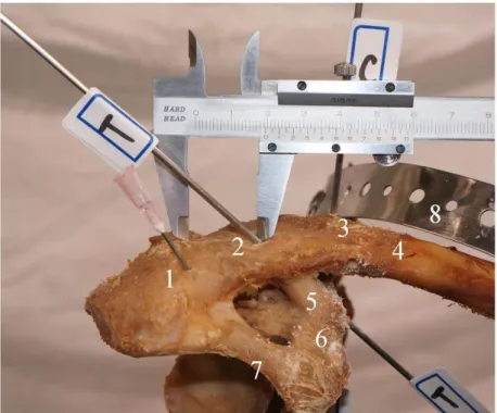
Fig. 1A
314
Distance from the lateral edge of the clavicle to the K-wire insertion in the trapezoid
315
ligament. An 18-G needle is placed within the acromioclavicular joint to mark the lateral
316
edge of the clavicle. 1: lateral edge of the clavicle; 2: insertion point of the K-wire in the
317
trapezoid ligament; 3. insertion point of the K-wire in the conoid ligament; 4. clavicle; 5.
318
trapezoid ligament; 6. coracoid process; 7. coracoacromial ligament; 8. metal plate fixing
319
the proximal clavicle and the scapular spine; T: K-wire drilled through the trapezoid
320
ligament; C. K-wire drilled through the conoid ligament.
321
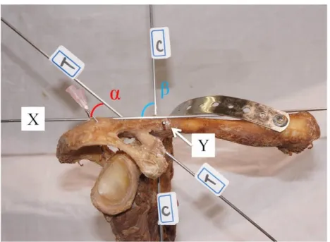
Fig. 1B
322
Coronal view of the right shoulder
323
X: horizontal baseline; reference K-wire set along the longitudinal axis of the clavicle
324
passing the anteroposterior midpoint of the distal and proximal clavicle; Y: reference K-
325
wire drilled from the anterior aspect of the clavicle to the posterior aspect, perpendicular
326
to wire-X; α: abduction angle between wire-X and -T; β: abduction angle between wire-
327
X and -C.
328
22
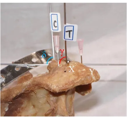
Fig. 1C
329
Lateral view of the right shoulder
330
γ: retroversion angle between wire-Y and -T; δ: retroversion angle between wire-Y and -
331
C.
332
Fig. 1D
333
C2: second conoid K-wire inserted at the midpoint of the anteroposterior diameter of the
334
clavicle on a line perpendicular to reference wire-X and parallel to wire-C; 9: insertion
335
point of the second conoid K-wire.
336
Fig. 2A
337
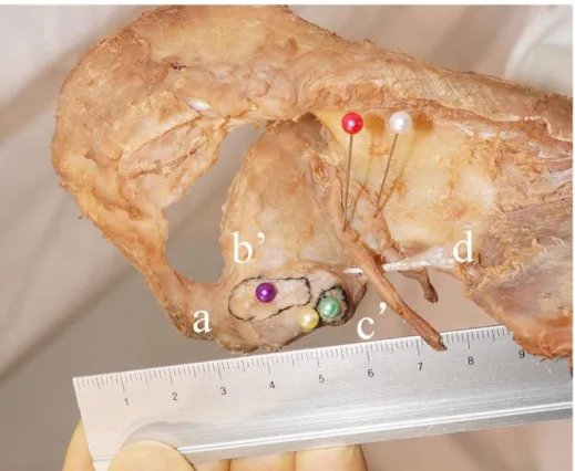
Anteroinferior view of the right shoulder
338
a: coracoid process; b: trapezoid ligament; c: conoid ligament; d: superior transverse
339
ligament of the scapula; white mark: suprascapular nerve; red mark: suprascapular artery;
340
T: K-wire drilled through the trapezoid ligament; C: K-wire drilled through the conoid
341
ligament; C2: K-wire drilled parallel to wire-C.
342
23
Fig. 2B
343
Superior view of the right shoulder. b′: attachment of the trapezoid ligament; c′:
344
attachment of the conoid ligament; purple mark: insertion point of the K-wire passing
345
through the trapezoid ligament; green mark: insertion point of the K-wire passing through
346
the conoid ligament; yellow mark: insertion point of the second conoid K-wire.
347
24
Tables
Table 1. Cadaver demographic characteristics and measurements of the insertion points and oblique angles of each K-wire at the clavicular surface.
Wire-T: K-wire drilled through the trapezoid ligament; Wire-C: K-wire drilled through the conoid ligament; Wire-C2: K-wire drilled parallel to wire-C; SD: standard deviation;
¶: paired t-test; †: Mann–Whitney U test; °: degree.
Characteristic, mean±SD (range) Male (n=17) Female (n=8) p-value
Age¶, years 73.9±13.2 (52–99) 79.5±9.6 (63–91) .30
Height¶, cm 163.5±8.2 (150–174) 154.3±6.0 (146–164) .009
Clavicular length¶, mm 164.8±10.6 (146–182) 147.8±11.1 (137–165) .001 Lateral edge to wire-T insertion¶, mm 21.6±4.4 (15.7–32.3) 18.2±4.5 (10.6–24.4) .08 Anterior border to wire-T insertion¶, mm 9.0±2.1 (5.0–12.9) 8.8±1.7 (6.4–11.0) .86 Lateral edge to wire-C insertion¶, mm 39.9±3.7 (32.7–47.3) 34.8±5.8 (26.8–40.5) .044 Posterior border to wire-C insertion¶, mm 6.3±1.8 (3.6–9.0) 5.4±0.78 (4.6–6.8) .12 Lateral edge to trapezoid/clavicle length¶ 0.132±0.027 (0.090–0.19) 0.122±0.027 (0.077–0.16) .41 Lateral edge to conoid/clavicle length¶ 0.243±0.025 (0.19–0.28) 0.234±0.026 (0.19–0.27) .43 Lateral edge to wire-C2 insertion¶, mm 37.7±4.1 (30.2–46.2) 33.9±5.7 (25.6–40.5) .066 Posterior border to wire-C2 insertion†, mm 10.3±1.7 (6.6–12.3) 9.6±1.7 (7.3–12.8) .26 Oblique angle of each K-wire
(α) Abduction angle, trapezoid¶ (°) 51.3±6.7 (41.2–64.8) 47.7±5.7 (40.0–57.8) .20 (β) Abduction angle, conoid¶(°) 88.2±3.0 (83.8–96.3) 88.2±7.6 (79.2–103.1) .99 (γ) Retroversion angle, trapezoid†(°) 88.6±10.0 (76.8–116.2) 92.5±6.1 (84.1–102.3) .14 (δ) Retroversion angle, conoid†(°) 82.1±7.3 (72.1–103.1) 81.7±1.7 (78.4–83.4) .67
25
Table 2. Correlation coefficients of the corresponding variables.
§: Pearson's correlation.
Correlation
coefficient p -value
Height and clavicular length § 0.52 .008
Clavicular length and lateral edge to wire-T insertion § 0.43 .034
Clavicular length and lateral edge to wire-C insertion § 0.60 .002
26
Table 3. Locations of each K-wire penetrating the medial cortex of the coracoid process and their relationships to the suprascapular nerve and artery.
Wire-T: K-wire drilled through the trapezoid ligament; Wire-C: K-wire drilled through the conoid ligament; Wire-C2: K-wire drilled parallel to wire-C; SD: standard deviation;
¶: paired t-test; †: Mann–Whitney U test.
Characteristic, mean±SD (range) Male (n=17) Female (n=8) p-value
Coracoid length¶, mm 43.7±4.0 (32.8–49.8) 39.0±2.6 (35.8–43.0) .006
Coracoid tip to wire-T exit point¶, mm 28.0±3.6 (22.5–33.2) 24.5±2.3 (20.3–27.8) .021 Coracoid surface to wire-T exit point¶, mm 9.8±2.6 (5.0–14.1) 8.5±2.6 (5.3–14.0) .27 Coracoid tip to wire-C exit point¶, mm 36.7±3.7 (29.0–43.8) 32.9±3.3 (27.1–37.1) .019 Coracoid surface to wire-C exit point¶, mm 14.7±4.2 (5.3–21.3) 14.2±4.9 (6.1–21.4) .79 Coracoid tip to wire-C2 exit point¶, mm 31.6±4.6 (22.6–40.1) 27.2±4.0 (22.8–33.8) .030 Coracoid surface to wire-C2 exit point¶, mm 14.3±3.4 (5.3–20.9) 13.1±4.7 (5.4–19.2) .47 Wire-T exit point to wire-C exit point¶, mm 11.1±2.9 (4.5–15.0) 11.2±4.2 (5.6–19.6) .95 Wire-T exit point to wire-C2 exit point¶, mm 7.5±2.8 (2.4–11.9) 7.9±3.6 (4.0–14.4) .79 Each K-wire exit point distance to neurovascular structures
Wire-T exit point to suprascapular nerve†, mm 21.5±6.5 (7.0–28.6) 20.8±4.6 (11.8–26.7) .51 Wire-T exit point to suprascapular artery†, mm 24.0±5.6 (10.0–30.8) 22.6±3.2 (15.8–24.9) .26 Wire-C exit point to suprascapular nerve¶, mm 12.4±3.1 (8.0–18.3) 11.2±4.5 (6.2–17.1) .43 Wire-C exit point to suprascapular artery¶, mm 15.7±2.6 (11.3–21.3) 14.8±4.0 (8.1–21.0) .52 Wire-C2 exit point to suprascapular nerve¶, mm 17.4±4.0 (11.2–25.0) 16.5±3.6 (9.6–22.2) .59 Wire-C2 exit point to suprascapular artery¶, mm 20.1±4.1 (13.8–29.7) 19.0±3.7 (12.0–24.8) .52
27
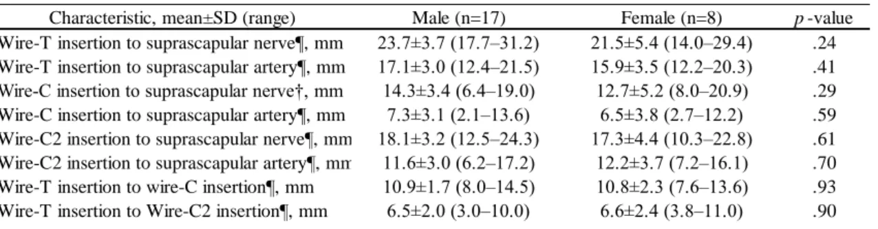
Table 4. Location of respective K-wire insertion points and the shortest distance from each K-wire to the neurovascular structures on the coracoid surface.
Wire-T: K-wire drilled through the trapezoid ligament; Wire-C: K-wire drilled through the conoid ligament; Wire-C2: K-wire drilled parallel to wire-C; SD: standard deviation;
¶: paired t-test. †: Mann–Whitney U test.
Characteristic, mean±SD (range) Male (n=17) Female (n=8) p-value
Wire-T insertion to suprascapular nerve¶, mm 23.7±3.7 (17.7–31.2) 21.5±5.4 (14.0–29.4) .24 Wire-T insertion to suprascapular artery¶, mm 17.1±3.0 (12.4–21.5) 15.9±3.5 (12.2–20.3) .41 Wire-C insertion to suprascapular nerve†, mm 14.3±3.4 (6.4–19.0) 12.7±5.2 (8.0–20.9) .29 Wire-C insertion to suprascapular artery¶, mm 7.3±3.1 (2.1–13.6) 6.5±3.8 (2.7–12.2) .59 Wire-C2 insertion to suprascapular nerve¶, mm 18.1±3.2 (12.5–24.3) 17.3±4.4 (10.3–22.8) .61 Wire-C2 insertion to suprascapular artery¶, mm 11.6±3.0 (6.2–17.2) 12.2±3.7 (7.2–16.1) .70 Wire-T insertion to wire-C insertion¶, mm 10.9±1.7 (8.0–14.5) 10.8±2.3 (7.6–13.6) .93 Wire-T insertion to Wire-C2 insertion¶, mm 6.5±2.0 (3.0–10.0) 6.6±2.4 (3.8–11.0) .90