Acta med. nagasaki. 5 : 99 - 107 (1960)
Six Cases of Leukemia Discovered at Mass Surveys of Nagasaki Atomic Bomb Survivors *1
Yasuro KAMOCHI, Bunro WATANABE , Noboru OZONO AND
Masanobu TOMONAGA *2
Second Department of the Institute for Nuclear Radiation Research , Hiroshima University School of Medicine, Hiroshima, Japan
Michito ICHIMARU, Shigeki TOYODA , Masami HAMASHIMA, AND Yoshitane KIKITSU *3
Second Department of Internal Medicine, Nagasaki University School of Medicine, Nagasaki, Japan
Received for publication August 31, 1960
1) Mass screening examinations were conducted on atomic bomb survi- vors in Nagasaki from 1954 to 1957. We found six cases of leukemia . 2) Five cases out of these six were chronic granulocytic leukemia and one was acute myeloblastic leukemia. 3) Three cases out of five chronic granulocytic leukemia are considered to have been in an early stage of this disease. Each of these cases showed leukocytosis, increase of basophils and a small number of immature cells in the peripheral blood. Decreased leueocyte alkaline phospatase and increased leukocyte catalase activity and increased basophils are important findings to be emphasized.
Ionizing radiations have been assumed as leukemogenic agents in man as well as experimental animals based upon : (a) an increased incidence of leukemia in the population exposed t o A-bombs in Hiroshima and Nagasaki 4)'011)")"), (b) observations by Court Brown and Doll upon. leukemia in cases of ankylosing spondylitis with therapeutic doses of x-rays'), (c) Simpso....'s research on. children, with therapeutic doses of x-rays for enlargement of the thymus gland 7) anal (d) various repcrts on the incidence of leukemia in x-ray labDratory workers.
At the present time, individual cases of radiation-induced leukemia can not be differen.tiated clinically or histo-pathologically from cases of "spontaneous" leukemia . However, in order to exaplain. the radiation. leukemogen.esis in. man it is important to analyse and report the leukemic cases occuring in. the irradiated population..
In. 1954 we started a mass screening examination ., with cooperation of the Ato- mic Bomb Casulty Commission in Nagasaki City and found one case of early chro- nic leukemia. As the mass survey continued over the years we have found a total of six cases of leukemia by the end of 1957, which are reported here in detail .
*1 Reported at the 20 th General Meeting of Japan Hematology Society on March 30. 1958
*2蒲 地 康 郎 ・渡 辺 紋 朗 ・小 園 昇 ・.朝 長 正 允 綿 市 丸 道 人 ・豊 田 成 樹 。浜 島 正 瑞 ・喜 々 津 良 胤
CASE REPORT
Case I. T.I. A 12-years-old junior high school boy.
He was exposed to the A-bomb in a one-storied wooden house located at 1406 meters from the hypocenter at the age of 3 years. He showed no acute radiation symptoms although he hed a laceration of his head. His developement' and growth
TABLE 1.
General and Hematologic Data on the First Examination
Patient, Sex T. I. M H.N. M K.T. M S. 1. M M.H. M T. M. F
Age at Time of Bomb 3 3 14 10 20 17
at Diagnosis 12 13 25 23 32 30
Exposed Distance (meters) 1406 1099 1924 2727 6220 2585
Acute Radiation Symptom 0 0 0 0 0 0
Type of Leukemia myelochr. . chr. myelo, chr. myelo. chr. myelo, chr. myelo. acute myelo.
Date 9/54 2156 12/56 12/57 11/57 12/57
Spleen (cm) 0.5 13 1.5 0 5.5 2
Liver (cm) 0 3 3 0 10 12
Lymph Nodes 0 0 0 0 0 -{-{-
Hb (%) 79 49 61 81 66 38
RBC (x 104) 426 301 378 432 342 137
~, Reticulocyte (%o) 17 23 43
Platelets (x 104) 94.5 12 9.8 154 13
w WBC 22.600 388.000 38.500 48.200 72.000 3.600
Blasts 0 1.5 0 0 1.0 0
C
Baso. 2.0 0.5 6.0 4.5 3.5 0.5
Eosin. 4.5 2.0 1.0 4.0 1.5 0 U
Promyelo. 0 39.5 1.5 1.0 27.0 0
d Myelo. 1.0 4.0 2.0 7.5 8.5 4.5
Metamyelo. 11.0 12.5 2.0 5.0 10.0 7.0
Q Z Band. 15.0 12.5 5.5 12.5 12.5 48.0
j Seg. 47.5 26.5 61.0 54.5 23.5 26.0
Lymph. 16.5 0 18.5 7.5 3.0 14.0
j Mono. 2.5 0 2.5 3.5 9.5 0.5
were normal and he did not suffer from any other specific diseases. When seen on September 8, 1954 during the mass survey (the number of the examined survi- vors was 3.333), it was found leukocytosis (22,600 per cu.mm.). He had no com- plaints but a firm spleen tip was palpated just under the left costal margine.
There was found mild anemia and leukocyte differential showed an increase of neutrophils with nuclear shift to the left and increased number of basophils. Nei-
ther myeloblasts nor promyelocytes were found. An obvious increase of promyelo- cytes were found in the bone marrow and the patient was considered to be in the
TABLE 2.
Myelogram on the First Examination
Patient T.I. H.N. K.T. S. 1. M. H. T.M.
Date 9/54 2/56 3/57 12/57 12/57 1/58
Nucleated Cell Counts 35 x 104 36 x 104 84 x 104 21 X 101
M y eloblasts 1 1.0 2.2 1.0 1.0 6.0 86.6
I P romyelo . 40.8 34.8 20.6 29.0 42.0 0.8
d M yelo. 8.0 3.0 13.2 13.6 7.0 0.4
Metamyelo. 21.0 16.8 10.2 8.4 11.4 0.4
.2 Band. 12.2 15.0 23.0 13.2 2.0 0.4
Seg. 6.6 21.2 12.6 22.6 26.6 0.4
Promyelo. 1.2 1.6 0 1.0 0 0.4
v Mvelo . 2.2 0.6 0.6 1.6 0 0
j 'o Metamyelo. 1.0 0.4 0 0.2 0 0
W Band . 1.2 0.2 1.0 1.0 0 0
Seg. 0.8 1,6 2.0 2.4 0 0
Baso. 1.0 1.0 1.2 0.6 0.8 0
Lymph. 0.4 0.6 1.4 4.2 0.8 0.2
Mono. 0.4 0 0.8 0 0 0
Proerythroblasts 0 0 0 0 0 0
Q) o Baso. 0.6 0 3.2 0.2 0 0
Poly. 0 0 2.0 0.4 1.4 0
v1 ~
Ortho. 0 0 0 0 0 0
c~ o Baso. 0 0 1.0 0 0 0
(U Cz Poly. 1.0 0 5.4 0.2 1.2 10.2
Z I Ortho. 0.6 0 0.8 0.2 0.8 0
Mitosis 0 0 0 0 0 0
Macrophages 0 0 0 0 0 0
v Lymphoides 0 0 0 0 0 0
v Plasma Cells 0 0 0 0 0 0
Megakaryocytes 0 0 0 0.2 0 0.2
Unidentified 0 1.0 0 0 0 0
early stage of ehronic granulocytic leukemia. He was treated with Nitromin, 6MP and Myleran with favorable results and at present he is doing well in complete remission. As shown in Fig. 1. leukocyte catalase was high and alkaline phospha- tase was low.
Case II. A 13-years-old junior high school boy.
At the age of 3 years he was exposed in a wooden house located 1.099 meters from the hypocenter. Although he had mechanical injuries nearly all over the body, he showed no acute radiation symptoms such as epilation, bleeding and stomatitis.
The other members of the family who were in the same house died. He felt exhausted after exertion sometimes since the summer of 1955. Leukocytosis (380,000 per cu.mm.) and splenomegaly were discovered at mass survey on Februa- ry 10, 1956 (the number of the examined survivors was 4049). The peripheral white cell differetial showed a typical picture of chronic granulocytic leukemia.
Fie. 1. Clinical course of case 1 (T.I. ).
Remarks: normal values of neutrophil alkaline phosphatase activity (N-APA) 14.2^-40.8 (Mean 28.0±1.2)12)
Remarks: normal values of leukocyte catalase activity (CAA) 0.74^-1.66 (Mean 1.23±0.04)13)
F] G. 2. Clinical course of case 2 (H,N,).
The patient required a higher dosage for longer period than case 1, although he responded well to the treatment with 6 MP and Myleran .. The duration of remi- ssion was also shorter as compared with case 1. He is now in complete remission and the spleen is not palpable .Values of leukocyte catalase and alkaline phosphatase are similar to those of case 1.
Case III. K.T. A 15-year-old male factory worker.
At the age of 14 years, he was exposed in open area 1924 meters from the
Fra, 3. Clinical course of case 3 (K. T. ).
hypocenter. His back was burned and he had fever but showed no epilation, bleeding or stomatitis. Leukocytosis (38,500 per cu.mm.) was discovered at the examination on December 20, 1956 (the number of the examined was 4049). At the same time very slight splenomegaly was also found. The peripheral blood smear exhibited a small number of immature leukocytes and an increase of basophils and myeloid proliferation was seen on myelogram and the patient was diagnosed as an early stage of chronic granulocytic leukemia. He responded well to 6 MP. We have had no subsequent direct contact with him but it was told that he had been working on a job without any difficulty. On August 2, 1957 he died in a sudden due to the perforation of peptic ulcer. The diagnosis was confirmed by autopsy.
WBC was about 50,000 per cu.mm. just before the death but the differential was unknown. Leukocyte catalase was in the normal range and alkaline phosphatase was low.
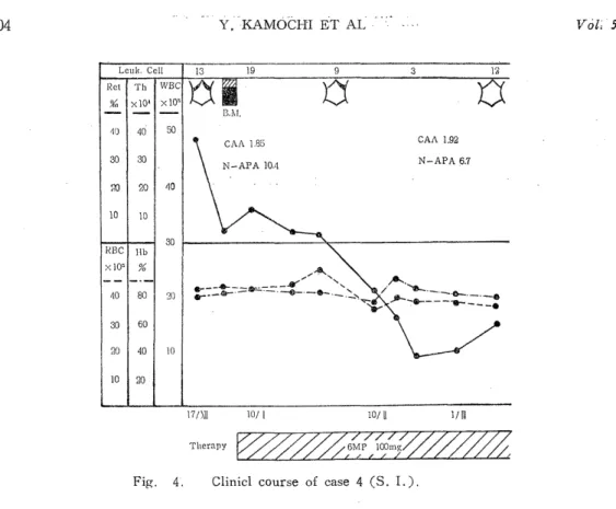
Case IV. S.I. A 23-year-old male factory worker.
At the age of 10 years, he was exposed at a distance of 2727 meters from the hypocenter. He had no trauma, burns or any acute radiation symptoms. Leukocy- tosis was found at the mass survey on December 17, 1957 (the number of the exa- mined was 2870). He had no subjective complaints. Leukocyte count was 48,200 per cu.mm. and the leukocyte differential showed a small number of immature
Fig. 4. Clinicl course of case 4 (S. I.).
leukocytes and an increase of basophils. The bane marrow picture also showed a proliferation of immature myeloid cells. The effect of 6 MP and Myleran was satisfactory and he is now working as a laborer. Leukocyte catalase was increased slightly and alkaline phosphatase was in the normal range. There were no splenome- galy and hepatomeagaly.
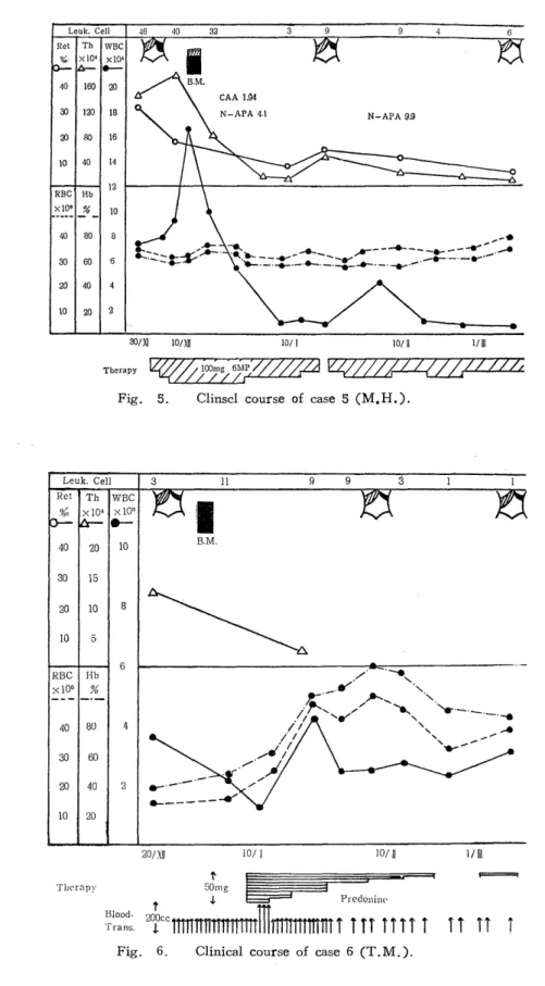
Case V. M.H. A 32-year-old male company employee.
At the age of 20 years,' he was exposed in a two-storied wooden building, 6.220 meters from the hypocenter and showed no acute radiation symptoms. He suffered from pulmonary tuberculosis at the age of 25 years and a thoraco-plasty was perfo- rmed at the age of 29 years. Leukocytosis (56,000 per cu.mm.) and moderate splenomegaly were discovered at the mass survey in October 1957 (simultaneously with Case IV). The leukocyte differential of the peripheral blood and bane marrow showed a typical picture of chronic gran.ulocytic leukemia. He responded well to 6 MP and Myleran treatment and lives now in good condition.. Leukocyte catalase was high and alkaline phosphatase was low.
Case VI. T.M. A 30-year-old married female.
At the age of 17 years, she was exposed in open area 2585 meters from the hypocenter. Her face and extremities were burned but she showed no acute radiation symptoms. At the age of 25 years she was hospitalized with pulmonary tuberculosis and a pleural decortication was performed. Severe anemia was found at the mass survey in Octover 1957 (simultaneously with Case IV and V). Leukopenia and thromb.)cytopenia and moderate splenomegaly were noticed at the same time.
Although the percentage of immature leukocytes was very small in the peripheral blood, bone marrow showed a distinct myeloblastic proliferation. Blood transfusion
Fig. 5. Clinscl course of case 5 (M.H.).
Fig. 6. Clinical course of case 6 (T. M. ).
and prednisolone `dd not irr:prave her ccn.diticn. an.d she expired on May 15, 1958.
The diagnosis of acute granulocytic leukemia was confirmed at the autopsy.
DISCUSSION
There has beenn consistent areerrent by rr.any werl=.ers on the following points : (1) The incidence of leukemia among the atomic b.)mb survivors in Nagasaki and Hiroshima is significantly high. (2) The incidence is higher in the more clo- sely exposed group. (3) The peak of incidence was in 1951 and 1952 and thereafter it seems to be decreasing. However , no one can dare to predict how long the leukemia incidence will continue to be high.
In 1954 Moloney and Lange ",I) reported two cases of subclinical leukemia found among the Hiroshima atomic bomb survivors. They pointed out (1) an increase of basophils, (2) leukocytosis associated with a small number of immature cells, (3) disturbance of the red cell and platelet series and (4) decreased values for leukocyte alkaline phosphatase.
Case' I, III and IV in our report may be considered to have been in the early stage of chronic granulocytic leukemia. Each showed a slight degree of leukocytosis, increase of basophils and a small number of immature cells in the peripheral blood.
Furthermore, the values of leukocyte alkaline phosphatase were decreased and leu- kocyte catalase were increased. These findings are consistent with Moloney's observation and seems to be very important for the diagnosis of early chronic granu- locytic leukemia.
Five case out of six were chronic granulocytic leukemia and only one case was acute granulocytic leukemia. This may be explained by the fact that the patients of acute leukemia usually visit doctors soon after its onset because of severity of their symptoms.
At the present time, it is impossible to determine whether these six cases were due to atmoic bomb radiation or not. Of the six cases, Case V was exposed at a long distance (6,220 meters) from the hypocenter, so that the dose of radia- tion may be almost zero. However, there is no question that Case I, II and III who were exposed within 2,000 meters, received a significant dose and also Case IV and VI who were exposed at the distance of about 2,500 meters, presu- mably received a considerable dose even under 29 rads. Although it is impossible to compare the detection rate of leukemia among the exposed with that among the non-exposed in the mass survey, the detection rate of leukemia in our mass survey is very high as compared with the incidence of leukemia in Japan (20-30 per million population per year). Lewis 5) reported a proportional relationship between radiation dose and the incidence of leukemia and recently Heyssel 2) and `lomonaga'-"),") described a linear relationship between the radiation air dose of the atomic bomb and the incidence of leukemia among survivors, who had been lightly shielded.
It is said that delayed effect of radiation may last for a lifetime. Therefore, it is necessary to examine the atomic bomb survivors with a long-term plan. The pericdic mass survey is of importance to study the late effects of radiation as well as for better management of survivors.
REFERENCES
1) COURTBROWN, W. M. AND DOLL, R. : Medical Research Council Special Series No. 295, Leukemia and Aplastic Anaemia in Patients Irradiated for Ankylosing
Spondylitis, London, Her Mejesty's Stationery Office, 1957.
2) HEYSSEL, R., BRILL, A. B., WOODBURY, L. A., NISHIMURA, E. T., GHOSE. T., HosHINO, T. AND YAMASAKI, M. : ABCC Technical Report, 1959.
3) ICHIMARU. M. : J. Kyu. Hem, Soc. 9 : 722 (1959) (Japanese).
4) LANGE, R. D., MOLONEY, W, C. AND YAMAWAKT, T. : Blood 9 : 574 (1954).
5) LEwis, E. B. : Science 125 : 9€5 (1957).
6) MOLONEY, W. C. AND LANGE, R. D. : Blood 9 : 663 (1954).
7) SIMPSON, C. L., HEYIPELMANN, L. H. AND FULLER, L. M. : Radiology 64 : 840 (1955).
8) TOMONAGA, M. Acta ffaem. Jap. 22 : 834 (1959) (Japanese).
9) TOMONAGA, M. Acta Haem, Jap. 20. Suppl. : 176 (1957) (Japanese).
10) TOMONAGA, M. AND BRILL, A. B. : ABCC Technical Report, 1959.
11) TOMONAGA, M., ITOGA, T., WATANABE, B. AND HAMASHIMA, M. : Acta Haem.
Jap. 22 Suppl. : 834 (1959) (Japanese).
12) TOYODA, S. : J. Kyu, Hem. Soc. in press (Japanese).
13) WATANABE, S., WAGA, M. AND ITO, T. : Acta Haem. Jap. 21 Suppl. : 301.
(1958) (Japanese).