Acta med. Nagasaki. 13: 85-93
Observation of Microcirculatory Change in the Great Occipital Nerve
Shobu SHIBATA and Masashichi KAWANO
Second Department of Surgery
Nagasaki University School of Medicine, Nagasaki, Japan
Received for publication , August 3, 1968
One-third of the patients with chronic post-traumatic headache have tenderness of the occipital nerves and, in lesser degree, of the trigeminal nerve. Various factors may contribute to this neuralgic reaction of these nerves which produce headach of extracranial pathology which we have been studying clinically and experimentally.
In this paper we report of the observation of changes in microcircula- tion of the great occipital nerve following occipital cooling and compression.
To reverse the impaired microcirculation we adovocate early ambulation and active exercise. Intramuscular injection of adrenaline is expected to produce similar effect on microcirculation which was observed under the perspective
microscope.
MATERIALS AND METHOD
To observe the effect of occipital compression the great occipital nerve of the rabbit was ligated in two places 3cm apart. The ligature was released 40 minutes later and microcirculation was observed during and after ligation. In some cases division of the nerve was added.
In the second series of experiment the occiput of the rabbit was cooled with ice bag at 5 degree C. and microcirculation of the nerve was studied for 6 hours. In the third series of experiment 0.013mg/kg of adrenaline was injected intramuscularly and observed to see the behavior of microcirculation during physical activity represented by
increased pulse rate, blood flow and blood pressure.
RESULTS
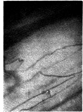
1. Effect of Compression: In the normal control the erythrocytes fill all lumens of the arteriole and the capillaries along the great occipital
*柴 田 尚武 ,川 野正七
nerve and flow as a homogeneous blood stream to one direction (Fig.
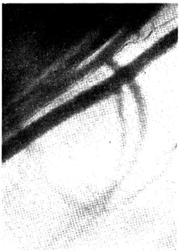
1). After 30 minutes of compression stasis in the venule appears show- ing nearly suspended blood flow with partial sludge phenomenon of the blood cells (Fig. 2). Due to marked stasis the capillaries are dis- tinctly visible in contrast to the control presenting sludge phenmenon and extravasation of the erythrocytes. Figure 3 shows the control, and Figure 4 shows microcirculation after 30 minutes of nerve compression.
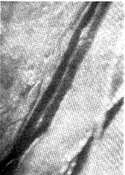
Stasis and sludge phenomenon are visible in the arteriole and the ve- nule. When the great occipital nerve was divided distally, the arteriole and the venule show strangulation associated with stasis and extrava- sation (Fig. 5) .
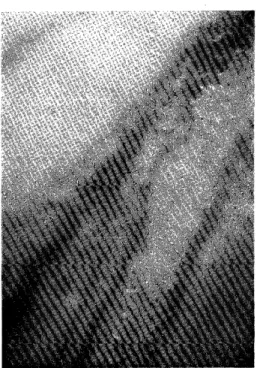
2. Effect of Cooling: The occipital region of the head was cooled with ice on 8 rabbits, and change in microcirculation of the nerve was stu- died one, three and six hours after initiation. Subdermal temperature drop to 5° to 10°C. in 5 or 10 minutes and was maintained around 5°C (Fig. 6). In contrast to the control (Fig. 7) movement of the vasucular wall becomes active after one hour of cooling (Fig. 8), showing irre- gularity of the diameter. Stasis and partial sludge phenomenon are reversed to the normal state, if cooling be stopped at this stage. After 3 hours of cooling the vessels are dilated and sludge phenomenon are common accompanied by interruption of capillary blood flow (Fig. 9).
Return to the normal blood flow of the capillary is not always possible at this stage, even if cooling was stopped. After 6 hours or longer cooling sludge phenomenon appear everywhere and rupture of the ve- nules accompanied by extravasation is found (Fig.10). Edema of the nerve is diffuse in this stage and all changes are irreversible.
3. Effect of Adrenaline: Adrenaline was injected intramuscularly, 0.013 mg per kilogram body weight, on rabbits to create the physical conditions similar to those in active exercise. Cardiac contraction mar- kedly increased in one minutes accompanied by increased pulse rate (plus 20) and blood pressure (plus 60mmHg.) In contrast to the control (Fig.11) marked increase of blood flow is found one minute after inje- ction of adrenaline (Fig.12).
DISCUSSION
These findings in the first and second series of experiments suggest
that continuous compression of the occipital region with pillow and
cooling with ice pillow customarily ill-advised and practised after head
iujury regardless of the grade of trauma, may give untoward effect on
microcirculation of the vasa nervosum of the great occipital nerve lea-
ding to prolonged hypoxia, increased irritability of the nerve and low-
ered pain threshold of the nerve manifested as neuralgic occipital hea-
dache. And ambulation and active exercise will improve microcircula-
tion of the nerve and contribute to healing of headache,
We are going to study the effect of therapeutic agents on micro- circulation of the nerve as a part of investigation of pathogenesis and treatment of chronic post-traumatic headache.
REFERENCES
1) FORBES, H. S.: The cerebral circulation I. Observation and measurment of pial vessels. Archiv. Nearol. Psychi., 19(5): %52-761, 1928.
2) KARJ_sIIIMA, K.: Experimental studies on microcirulation in the small blood vessels of the brain, especially on effects of artificial interception in arterioles
and venules. Nagaki Igakkai Zasshai, 39(10):838-853, 1964. (in Japanese with
English summary.)
3) M GGIO, E.: Microhemocirculation, observable variables and their biologic control. C.C. Thomas, Springfield, 1967.
4) TUCHI`IA, M. et al: Microcirculation. Chugaiigakushya, Tokyo, 1965.
5) ZWEIF cli, B. W.: Functional behavior of the microcirculation. C. C. Thomas,
Springfield, 1961.
Fig. 1. Microcirculation of the great occipital nerve in rabbit (control).
Fig. 2. 30 minutes after compression of the great occipital nerve.
Fig. 3:Control,
Fig. 4. 30 minutes after compression of the great occipital nerve.
Fig. 5. Amputation .
Fig. 6. Subdermal temperature of the occiput following cooling with ice.
Fig. 7. Control.
Fig. 8. After one hour of cooling.
Fig. 9. After three hours of cooling.
Fig. 10. After six hours of cooling.
Fig. 11. Control.