REPORTS OF THE FACULTY OF
No.10, 83‑90 (1977) ENGINEERING, NAGASAKI UNIVERSITY 83
Syntheses and Characterization
Containing Nucleic Acid
of Polymers Bases
Kyoko
by
HIRAOKA* and Tetsuo YOKOYAMA*
SYNOPSIS
Two different types of the polymers containing nucleic acid bases in backbone or in pendant groups were prepared. The polymers of the first type were polyureas obtained by the polyaddition reaction of uracil and adenine with hexamethylene diisocyanate (HMDI). The second type, that is .cationic polyurethanes containing nucleic acid bases in pendant groups, were obtained by Menschutkin reaction of halogenated derivatives of u;acil and adenine with a linear polyurethane containing, tertiary nitrogen atoms which was based on HMDI and N‑methyldiethanolamine. Base‑base interactions were studied for the polymers by UV and NMR spectra. A relatively high value of hypochromicity, ca. 19%, was observed for the mixture of the ionic polyurethane with uracil pendant and herring'sperm DNA. Complementary hydrogen bonding interaction was detected for the mixture of the ionic polyurethane with adenine pendant and that with uracil pendant. Nonthrombogenic character of the polymers was examined according to the modified Lee‑White method. The ionic polyurethanes with adenine and uracil pendant exhibited fairly good anti‑clotting property.
INTRODUCTION
It is expected that the synthetic polymers containing nucleic acid bases in the structure are highly functional materials because of their ability for complementary base pairing. They may exhibit template characteristics through this specific interaction which governs the process of replication and the genetic code transcription in vivo. Specific catalytic activity is also expected in these polymers. Further‑
more, if they obtain helical and double strand structure similar to DNA through their se‑
lective hydrogen bonding and specific inter‑
action between bases, the possible conforma‑
.Department of Materials Science and Engineering
tional response caused by the chemical and physical excitation may offer a fuhdamental mechanism for the design of new mechanoche‑
mical system.Thus the polymers will work as a novel functional material and will serve as a useful model for the elucidation of delicate, mechanism realized in biopolymers. From this viewpoint a number of studies have been carried out on the syntheses and characteri‑
zation of the polymers containing nucleic acid
bases. .
Jones et al.') synthesized 5'‑O‑acryloyluri‑
dine and 5'‑O‑acryloylthymidine and found that
the polymers of these compounds have inter‑
84
actions with denatured DNA.
prepared a variety of similar compounds, which were mainly vinyl compounds, and observed hypochromic effect in UV spectra owing to selective interactions between the polymers.
Kaye3} and Pitha et al.̀) studied the interactions between poly(9‑vinyladenine) and poly U by CD and concluded the two‑dimensional network structure for the complex of the two polymers.
Seita et al.5)synthesized the polymers contain‑
ing nucleic acid bases and acid esters, and found strong interactions between their poly‑
mers and DNA. Okubo et al.6) prepared and studied the cationic polymers from poly (4‑pyri‑
dylethylene) by quaternization with chloroethyl derivatives of nucleic acid bases.
In this paper the authors attempted to prepare the novel polymers in which nucleic acid bases are introduced in backbone or side chain, and determined hydrogen bonding and stacking interaction between bases. The poten‑
'
tiality as biomaterials was also examined for '
the prepared polymers. . '
'
EXPERIMENTAL
1. Reagents
Uracil, 5‑bromouracil, adenine, and herring sperm DNA were commercial grade and used without further purification. Other starting materials and solvents were purified by con‑
ventional methods.
2. Synthesis of poly [2,4‑pyrimidinedione‑
1,3‑diyl) icarbonyliminohexamethyleneimino‑
carbonyl] (Ura‑HMDI) .
A mixture of uracil (1.12 g; O.Ol mol) and HMDI (1.68 g; O.Ol mol) in 30 ml of N, N‑
dimethylformamide (DMF) was stirred at 800C for 1 hr. Then the reaction mixture was poured into a large amount of water. A white powder precipitated was filtered off, washed with water, and dried at 800C in vacuo until a constant weight was reached. This compound wi!1 be abbreviated as Ura‑HMDI. Yield 50%.
K. HIRAOKA AND T.
Takemoto et al.2)
YOKOYAMA
Found Ct 50.19 H: 6.84 N: 19.68 Calcd C: 51.41 H: 5.76 N: 22.83
[v] == O.1 (m‑cresol, 300C)
3. Synthesis of poly[imino(6,9‑purindiyl) carbonyliminohexamethyleneiminocarbonyl]
(Ade‑HMDI)
A mixture of adenine (1.35 g; O.Ol mol) and HMDI (1.68 g; O.Ol mol) in 40 ml of N, N‑
dimethylacetamide (DMA) was stirred at 600C.
After 30 min the reaction mixture that was inhomogeneous at the beginning of the reaction became transparent, and then a white powdery precipitate started to separate out. During the course of the reaction the precipitate increased in its amount. The extent of reaction was followed by titrating unreacted NCO group according to amine equivalent method. After 72 hr no residual NCO group was detected. The product was filtered off, washed with DMA, and dried at 600C in vacuo. This compound will be abbreviated as Ade‑HMDI. Yield 40%.
Found C:50.21 H:5.61 N:33.96 Calcd C:51.46 H:5.66 N:32.33
4. Synthesis of poly[oxyethylene(methyl‑
imino)ethyleneoxycarbonyliminohexame‑
thyleneiminocarbonyl] (N‑MePU) A mixture of N‑methyldiethanolamine
(17.90 g; O.15 mol) and HMDI (25.22 g; O.15 mol) in 200 ml of DMA was stirred at 800C for 3hr. The reaction mixture was poured into a large amount of water. A white powder pre‑
cipitated was filtered off, washed with water, and dried at 800C in vacuo. This compound will be abbreviated as N‑MePU. Yield 80%.
Found C: 54.36 H: 8.77 N: 14.98 Calcd' C: 54.44 H: 8.71 N: 14.63
5. Synthesis of poly[oxyethylene[methyl‑5‑
(2,4 ( IH ,3H ) ‑pyrimidinedionyl) imino ] ethyl e ‑
neoxycarbonyliminohexamethyleneiminocar‑
bonyl bromide] (PU‑Ura)
A mixture of N‑MePU (2.51 g; 8.7 mmol)
and 5‑bromouracil (2.0 g; 10.4 mmol) in DMA
POLYMERS CONTAINING NUCLEIC ACID
was stirred at 800C for 160 hr. By pouring the reaction mixture into a large amount of water, a white powder was precipitated. It was filtered off, washed with water, and dried at 800C in vacuo. This compound will be abbreviated as PU‑Ura. Yield 30%.
Found C: 41.38 H: 5.78 N: 14.98 Calcd C:42.68 H:5.85 N:14.63 6. Synthesis of poly[oxyethylene[methyl‑
2‑hydroxy‑2‑(IH‑purin‑6‑amino‑9‑yl) ethyl‑
imino]ethyleneoxycarbonyliminohexamethyl‑
eneiminocarbonylbromide] (PU‑AdeEBH)
A mixture of adenine (2.00 g; 14.7 mmol), epibromohydrin (2.08 g; 15.2 mmol) , and a trace amount of anhydrous potassium carbonate in 40 ml of DMF was stirred at 600C. After 24 hr N‑MePU (O.96 g; 3.3 mmol) was added to the reaction mixture. After 100 hr, the small amount of the unreacted adenine was discarded by filtration. The filtrate was poured into a large amount of acetone, and then a brown solid was precipitated. It was filtered off, washed with acetone, and dried at 600C in vacuo. This will be abbreviated as PU‑AdeEBH. Yield 30%.
' Found ' C: 41.41 H: 6.42 N: 17.79 Calcd C:45.07 H:6.32 N:20.03
7. Synthesis of 9‑(2'‑bromeethyl)adenine (AdeEtBr)
A mixture of adenine (2.70 g; O.02 mol), ethylene carbonate (1.76 g; O.02 mol), and a trace amount of sodium hydroxide in 40 ml of DMF was stirred at llOOC for 1 hr7). After cooling, the solvent was removed to complete dryness under reduced pressure. Recrystalli‑
zation of the residue from ethanol gave 9‑
(2'‑hydroxyethyl) adenine (AdeEtOH) as color‑
less plates.
In a three‑necked round‑bottomed flask equipped with a stirrer and a dropping funnel was placed 25 ml of pyridine contain'ing AdeEtOH (O.Ol mol; 1.8 g). The flask was cooled externally with crushed ice and salt to
‑10eC. PBr(1.08 g;4mmol)was added slowly to
BASES 85
the flask during agitation. After 30 min the temperature of the reaction mixture was raised to room temperature and was allowed to stand overnight. Then the solvent was removed under reduced pressure. Recrystallization of the residue from ethanol gave 9‑(2'‑bromoethyl) adenine (AdeEtBr) as white needles.' It was filtered off and dried at 600C in vacuo. Yield 20%.
Found C:32.19 H:3.90 N:26.78 Calcd C:34.73 H:3.34 N:28.93
8. Synthesis of poly[oxyetylene[methyl‑2‑
(purin‑6‑amino‑9‑yl)ethylimino] ethyleneoxy‑
carbonyliminohexamethyleneiminocarbonyl
bromide] (PU‑AdeEtBr)
A mixture of N‑MePU (O.38 g; 1.3 mmol) and AdeEtBr (O.32 g; 1.3 mmol) in 40 ml of DNA was stirred at 60eC for 100 hr. By pouring the reaction mixture into acetone, a brown precipitate appeared. It was filtered off, washed with acetone, and dried at 600C in vacuo. This compound will be abbreviated as PU‑AdeEtBr.
9. Ultraviolet absorption spectra
Ultraviolet absorption spectra of the obtained polymers, DNA, and the mixtures of the polymers and DNA were measured in dilute
solutions of Walpole buffer solution (CH3COOH‑CH3COONa pH 4.5) by using a Hitachi UV‑VS spectrometer model 323 and fused quartz cells of approximately 1.0 cm path.
All spectra were recorded at room temperature.
In the case of the mixtures of the prepared polymers and DNA, the separate solutions dis‑
solving each component were mixed and
allowed to stand 2 days before spectroscopic measurement in order to make sure inter‑
polymer interactions. An apparent hypochro‑
micity was calculated according to the follow‑
ing equation,8) where m and n are the volume fractions of the solution of polymer a and that of polymer b, and Ia,Ib, and Ia+b the absor‑
bances of the solutions of polymer a, polymer b,
and the mixture of the two solutions, respective‑
IY.
'
% Hypochromicity=100 (1L m II.atb. I, )
' 10. Nuclear magnetic resonance s.pectra Nuclear. magnetic resonance spectra were
recorded on a Nihon Denshi NMR spectrome‑
ter model JNM‑MH‑100 operating at 100Mc.
TMS and DSS internal standards were used for DMSO‑d6‑benzene‑d6 mixed solution and for D20‑D2S04 solution, respectively, The measure‑
ments were made on O.05, O.1, and O.15 mol solution at room temperature, 500, and 750C.
11.' Examination of anti‑clotting property of ' the polymers '
Modified Lee‑White method9) was used for the examination of nonthrombogenic character of the polymers. Two test tubes of 10 mm diameter were prepared for each polymer. The inside walls of test tubes were coated with the obtained polymers. For the control, two tubes were not treated by polymer. An appropriate amount of whole human blood' was withdrawn
HIRAOKA AND T. YQKOYAMA
from an arm vein milliliter of blood
using a small syringe. One just withdrawn was trans‑
fered into cach tube. The one of the two tubes for each polymer was rotated endwise every thirtyseconds. The other tube was left without any disturbance. At the point at which the blood no longer flows from its position but maintains its surface contour when inverted, the other tube was started to rotate. The point at which the blood in the second tube no longer flowed as described above was taken as the end point. The time from withdrawing of the blood from vein to final clotting was recorded as clotting time.
RESULTS AND DISCUSSION
1. Preparation
The preparation of the polymers containing nucleic acid bases in backbone is illustrated in Fig. 1. The reactions proceeded smoothly. The obtained polymers, Ura‑HMDI and Ade‑HMDI, were identified with those expected from the reaction scheme. The polymers were white
Fig.
HN‑ Arfo
NH o' Uracit
OCN(CH2)6NCO
HMDI
NH2
Na",}ii, H Adenine
t‑̀‑ NH
,‑vt*,‑OcoN"‑(cH2)6‑""Oc'hv Xlhr<" Il).(cH2)6xs"‑Nv'
Ura‑HMDI Ade‑HMDI
' 1 Synthetic route of the polymers containing nucleic acid bases '
in
powder.
Since the polymerization mechanism is essentially polyaddition of isocyanate with amine, pyrimidine and purin bases were polymerized by the formation of substituted urea linkage. As a result of it, N‑H groups of nucleic acid bases are used up after polymeri‑
zation. This consumption of N‑H groups will cause the decrease in basic character of the nucleic acid bases and the lack of specific interactions between bases. Though the poly‑
mers obtained consist of nucleic acid base structure, it seems appropriate to regard them as novel polyureas with specific heterocyclic ring structure in backbone.
In order to keep the character of nucleic acid bases, it is better to incorporate bases as pendant groups to some polymeric stems. This is in line with the structure of polynucleotides.
CH3 OCN(CH2tsNCO HO(CH2)2‑rk‑(CH2)20H HMD! N‑MethytdiethanoE amine
‑‑T‑
9H3 oH Ho
,‑v O(C H2 )2‑N‑(C H2)2 0CN ‑‑ (C H2 )6‑ NC tv
N‑MePU
Br' rvo HNN.NH o 5‑Bromouracit
POLYMERS CONTAINING NUCLEIC ACID BASES
In the case of uracil and
87
adenine, the base is connected to the ribose at Ni and Ng position, respectively. In Fig. 2 are shown the synthetic route of the polymers containing nucleic acid bases as pendant groups studied in this work.
The stem polymer is a linear polyurethane containing tertiary nitrogen atom to which halogenated derivatives of nucleic acid bases add by Menschutkin reaction, resulting in the quaternizationof nitrogen atom. Thus, the resultant polymer obtain cationic character in addition to the heterocyclic ring structure of nucleic ・acid base. In the case of uracil, Cs position was used for the connecting site because 5‑bromuracil was obtained commer‑
cially. As for adenine, it was necessary to prepare halogenated intermediates. The Ng position was available for this purpose in most cases except for AdeEBH. In the latter case the
. NH2
Nitl{NIIiit(>wtt2cH2oco
H
Adenine
ji CNHo2,C Hc H2B,・
NH2
N,?i,il)I(.>
CH2CHCH2Br 6H AdeEBH
NH2
"i,,hr,,>
t'
CH2CH20H lPlk, ., ,
NH2 Nk7i,il)ll(>
t CH2CH2lk
AdeEtBr
Fig. 2
r‑NcOo.vRpaA.o2,".v' 'N'neO'‑v!'H,Ii'E.lo8N"‑‑
Hte,,O f",(ickYO,"
NH2 PU‑Ura PU‑AdeEBH
gSrY on
utphse.ti,CrOUte of the polymers containing nucleic acid bag
Lme,,A.g・M.v,2H,A.
CH2, Bti‑
,
N kH2 ‑, Nf'or'N)
NH2 PU‑AdeEttr
es in pendant
88 KHIRAOKA AND T.
selectivity of the reaction of epibromohydrin with adenine was low, giving a mixture of the two isomers, 9‑(2‑hydroxy‑3‑bromopropyl,) adenine and 3‑(2‑hydroxy‑3‑bromopropyl) adenine. Because of the similar properties of the two isomers, the further elaborative separa‑
tion was avoided and the mixture was used for the next step. The use of ethylene carbonate instead of epibromohydrin, yielded the highly selective addition and therefore led the single' halogenated derivative (AdeEtBr) .The reaction of halogenated intermediates with N‑MePU
proceeded almost quantitatively by sufficiently
prolonged reaction. .
Thus the five different polymers, Ura‑
HMDI, Ade‑HMDI, PU‑Ura, PU‑AdeEBH, and PU‑AdeEtBr were obtained.
2. Base‑base interaction
It is a well known phenomenon that
polynucleotides show hypochromism, which is the decrease in absorbance per chromophore in the polymer compared to that of the monomer.
The origin of hypochromism has been ex‑
plained in different ways. Tinoco and RhodesiO・'i)
attributed hypochromism to dipole‑dipole interaction between transition moments in neighboring oscillators. This has been termed an off‑resonance interaction because the interacting dipoles originate from different transitions. When the bases are arranged e in verticai stacking arrangement, that is, E
base planes are arranged parallel and one above another, this model predicts hypochro‑ 2si O'2
mism. Bolton and Weiss'2) have attributed hypochromism to a local field effect, which originates from the induced electric dipole moments in neighboring oscillators. Since self‑
interaction of single transition of the monomer Oo
is considered, this has been termed a resonance interaction. This resonance effect can lead to
Fig. 3 hypochromism even in the absence of secondary
structure or vertical stacking of the bases.
Later, Thomas and Kyogokui3) showed that the
YOKOYAMA
dimerichydrogen‑bonded complexes between bases (complementary base pairing) lead to hypochromism, and theretore concluded that parallel stacking of the bases is not the only condition for hypochromism.
By consulting these studies, it seems safe to consider that the existence of hypochromism suggests base‑base interactions which may be hydrogen bonding or vertical stacking inter‑
actlon.
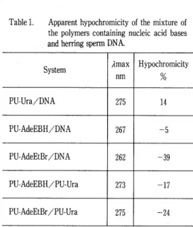
Fig. 3 shows example of hypochromic effect of PU‑Ura mixed with DNA in Walpole buffer at room temperature. The absorbance at 26e nm, when plotted against the volume fraction of PU‑Ura, show a negative deviation from Beer's low, reaching the relatively high value of 14%
hypochromicity at volume fraction of O.9. Table 1 summarizes the results of hypochromicity between the prepared polymers and DNA.
Though the accurate molar relationships are not known because of uncertainty of the struc‑
ture of DNA, it is concluded that the synthetic polymers in this study can interact with DNA,
O.4
O.5 1.0
Vot.fraction ot PU・‑Ura
Comparison of observed and Beer's law absorbances at 275 nm for the
mixture of PU‑Ura and DNA in
Walpole buffer.
Table 1.
POLYMERS CONTAINING NUCLEIC ACID BASES
Apparent hypochromicity of the mixture of the polymers containing nucleic acid bases and herring sperm DNA.
System Amax
nm
HYpochromicity
o/o
PU‑Ura/DNA 275 14
PU‑AdeEBH/DNA 267 ‑5
PU‑AdeEtBr/DNA 262 ‑39
PU‑AdeEBH/PU‑Ura 273 ‑17
PU‑AdeEtBr/PU‑Ura 275 ‑24
In Walpole buffer solution at room temperature,
' '
which may be vertical stacking interactions.
Alternatively, interaction between bases‑ can be studied by NMR spectraì). Table 2 shows the results of NMR spectra in D20‑D2S04 solution. The addition of D2S04 was necessary for solubility problem. It is seen that the dilution shift and temperature shift of C6‑H proton of PU‑Ura are trivial to draw any conclusion on base‑base interactions. Also, the trivial downfield shift observed for the mixture of PU‑Ura and PU‑AdeEtBr does not give any evidence for the complex formation.
Table2 Shifts in the resonant frequency of the C6 proton of PU‑Ura upon the addition of PU‑AdeEtBr.
Conc. Temp. 6(C6‑H)
M/1 OC ppm ppm A6
89
In nonaqueous solvents (DMSO and DMF) , the proton resonances are essentially inde‑
pendent of concentration. DMSO and DMF are hydrogen bonding acceptors and interact with bases with the result that solute molecules are interrupted to associate with each other.
However, it has been observed that the use an equal volume mixture of benzene and DMSO is effective to detect a downfield hydrogen
bonding shift of N‑H proton in the base pairing interactions.'5) According to their observation an equal volume mixture of benzene and DMSO was used in this study. The results are shown in Table 3. As seen from the table, the resonance peak at around 7.3‑7.7 ppm, which is assigned to N3‑H proton signal of pyrimidine ring of PU‑Ura, showed a downfield shift (‑O.1 ppm) upon mixing with PU‑AdeEtBr. By elevating temperature from 25 to 50 to 750C a marked upfield shifts are observed. Thus it is concluded that considerable self‑association occurs be‑
tween uracil and adenine moiety due to hydrogen bonding interaction.
Table3. Chemical shifts of the hydrogen bonding protons of PU‑Ura.
System PU‑Ura PU‑Ura
PU‑Ura PU‑Ura/
PU‑AdeEtBr O.05
O.05
O.10
O.05‑
O.05 50
75
75 75
8.55
8.47
8.48 8.48
System Temp. 6(N3‑H) A6
oc ppm ppm
PU‑Ura
PU‑Ura/PU‑AdeEtBr
PU‑Ura/PU‑AdeEtBr
PU‑Ura/PU‑AdeEtBr
25
25
se
75
7‑, 59