Ocular Diseases on Naru Island, Japan: a Seven-year Survey
Hirohiko HAYASHIDA, Takashi KITAOKA
Department of Ophthalmology and Visual Sciences, Graduate School of Biomedical Sciences, Nagasaki University
Purpose: To examine the characteristics over seven years of ocular diseases in a closed society on Naru Island in Nagasaki Prefecture.
Subjects: Annual eye examinations were performed dur- ing routine physical examinations on Naru Island, Japan from 1995 to 2002. We examined 639 (213 males and 426 females) in1995, 579 (196 males and 383 females) in1998 and 421 (159 males and 262 females) in 2002.
Results and Discussion: Pterygium appeared to increase with age. The prevalence of cataract was higher in woman than in man. Cataract was more prevalent in those with pterygium than in those without one. The prevalence of pseudoexfoliation increased with age, and it was greater in those with cataract than in those without one.
Conclusions: The incidence of degenerative and age- related diseases in this population followed for seven years showed a tendency to increase with age.
ACTA MEDICA NAGASAKIENSIA 48: 15-21, 2003
Key Words: population-based study, Naru island, incidence of ocular diseases, prevalence of ocular diseases
Island annually from 1995 to 2002. Naru Island is an isolated off-shore island in Nagasaki Prefecture in western Kyushu, Japan. It is one of many isolated is- lands where the residents form a socially, historically, genetically and culturally specific society. Naru Island has a population of about 4,000 with no ophthalmolo- gists.
The Naru Island Eye Study is a population-based study of ocular diseases, conducted by the Naru Town Office and supported by the Department of Ophthalmology of Nagasaki University Hospital. We examined for 7 years from 1995 to 2002 and recorded the prevalence, characteristics, and natural course of ocular diseases.
We can study not only the prevalence but also the in- cidence of eye diseases. While association with risk factors can be estimated by cross-sectional data, inci- dence data are necessary to develop insights regarding potential causative factors. We studied the cumulative incidence of ocular diseases observed in the popula- tion from 1995 to 2002. Such a study is rare even in the above-mentioned population-based studies.
Introduction Subjects and Methods
There are several eye studies of large populations:
Framingham Eye Study, Massachusetts, US", Beaver Dam Eye Study, Wisconsin, US2', Blue Mountains Eye Study, Australia", Barbados Eye Study, West Indies"', National Health and Nutrition Examination Survey, US') and Hisayama Eye Study, Japan." The diagnos- tic method, criteria and age range vary from study to study. These reports have greatly contributed to the analysis of factors related to prevalence, cause, pro- gression, clinical features and relationship among dis- eases.
We examined the eyes of 10% - 14% residents of Naru
Address Correspondence: Hirohiko Hayashida, M.D.
Department of Ophthalmology and Visual Sciences, Graduate School of Biomedical Sciences, Nagasaki University, 1-7-1 Sakamoto, Nagasaki 852-8501, Japan
TEL: +81-95-849-7345, FAX: +81-95-849-7347 E-mail: [email protected]
Study population
In 1995, we examined 639 (213 males and 426 fe- males) residents out of the total population of 4,674 (2,275 males and 2,399 females). In 1998, we examined 579 (196 males and 383 females) residents out of 4,396 (2,104 males and 2,292 females), and in 2002, we examined 421 (159 males and 262 females) residents out of 3,949 (1,895 males and 2,054 females).
In the analysis, we excluded those who were aged under 40 years or over 80 years at the time of exami- nation. Thus, 612 (204 males and 408 females), 544 (182 males and 362 females) and 402 (147 males and 255 females) were remained for analysis among resi- dents examined in 1995, 1998 and 2002, respectively.
Ethical approval for the study was obtained from the Naru Town Office and written informed consent was obtained from each resident we examined.
Ophthalmic examination
The examination included visual acuity, autorefractometry, applanation tonometry, slit lamp biomicroscopy, indirect ophthalmoscopy and non-mydriatic fundus photography, and was conducted by seven ophthalmologists. Applanation tonometry was performed on all individuals. A detailed high-magnification slit lamp biomicroscopy assessment of the anterior segment was performed. Pterygium was defined as the presence of a raised fleshy growth that crosses the limbus and encroaches onto the clear cornea. Individuals who had a history of pterygium surgery were included in the pterygium group. Slit lamp biomicroscopy was used to identify cataract. We esti- mated the angle width by van Herick's method. Narrow angle was diagnosed when the ratio of anterior cham- ber to cornea was less than 1/3. Pseudoexfoliation was diagnosed by the presence of typical layered white deposits on the anterior lens surface. Increased cupping was diagnosed when the cup to disc ratio was larger than 0.5. Glaucoma was diagnosed by fur- ther examinations in the ophthalmology clinic. Ocular hypertension was diagnosed when tension exceeded 21 mmHg.
Statistical analysis
The prevalence and the cumulative incidence were analyzed for respective ocular diseases on the basis of logistic regression model with sex and age as covariates. Starting from the full model including in- teraction term of sex and age as well, we selected the most appropriate model by means of AIC. Once the most appropriate model was selected significance tests of parameters in the model were conducted using like- lihood ratio statistics. Association between cataract and pterygium (or pseudoexfoliation) was analyzed in a similar way. FREQ, LOGISTIC and UNIVARIATE in the SAS- system') were used for the calculations.
Results
The prevalence of ocular diseases by age is shown in Tables IA, 1 B and IC, and. the incidence of ocular diseases by age is shown in Table 2.
Pterygium
The prevalence of pterygium was 8.7%, 10.7% and 13.2% in 1995, 1998 and 2002, respectively (Tables 1A- 1Q, and showed a significant increase with age: an in- crease in the odds of the prevalence by 1 year
Hirohiko Hayashida et al : Ocular Diseases on Naru Island
Table IA. Prevalence of ocular diseases by age in residents of Naru Island, Nagasaki, Japan, examined in 1995
Ocular disease Age at examination (years) Total
40-49 50-59 60-69 70-79
Pterygium 5/99 (5.1)a 13/136(9.6) 32/276(11.6) 13/101 (12.9) 53/612(8.7)
Trichiasis 1/99(1.0) 5/136(3.7) 4/276(l.4) 1/101 (1.0) 11/612 (1.3)
Cataract 1/99(1.0) 10/136(7.4) 108/276 (39.1) 68/101 (67.3) 187/612 (30.6)
Pseudophakia 0/99 0/136 0/276 6/101 (5.9) 4/612(0.7)
Pseudoexfoliation 0/99 3/136(2.2) 10/276 (3.6) 7/101 (6.9) 20/612 (3.3)
Narrow angle 0/99 5/136(3.7) 14/276 (5.1) 5/101 (5.0) 24/612 (3.9)
Glaucoma and ocular hypertension 1/99(1.0) 4/136(2.9) 4/276(l.4) 0/101 9/612(l.5) Enlargement of cupping of optic disc 4/99(4.0) 6/136(4.4) 23/276 (8.3) 6/101 (5.9) 39/612 (6.4) Chorioretinal atrophy 0/99 3/136(2.2) 4/276(1.4) 4/101 (4.0) 11/612 (1.8) Age-related macula degeneration 0/99 0/136 1/276(0.4) 1/101 (1.0) 2/612(0.3) a) Number of cases/number of residents examined (%).
Table 113. Prevalence of ocular diseases by age in residents of Naru Island, Nagasaki, Japan, examined in 1998
Ocular disease Age at examination (years) Total
40-49 50-59 60-69 70-79
Pterygium 4/90(4.4)' 10/106 (9.4) 28/223 (12.6) 16/125 (12.8) 58/544 (10.7)
Trichiasis 0/90 2/106(l.9) 5/223(2.2) 2/125(l.6) 9/544(l.7)
Cataract 6/90(6,7) 9/106(8.5) 121/223 (54.3)) 98/125 (78.4) 234/544 (43.0)
Pseudophakia 0/90 0/106 6/223(2.7) 8/125(6.4) 14/544 (2.6)
Pseudoexfoliation 1/90(1.1) 1/106(0.9) 14/223 (6.3) 10/125 (8.0) 26/544 (4.8)
Narrow angle 2/90(2.2) 3/106(2.8) 8/223 (3.6) 8/125(6.4) 21/544 (3.9)
Glaucoma and ocular hypertension 0/90 3/106(2.8) 7/223(3.1) 3/125(2.4) 13/544 (2.4) Enlargement of cupping of optic disc 3/90(3.3) 4/106 (3.8) 17/223 (7.6) 11/125 (8.8) 35/544 (6.4) Chorioretinal atrophy 1/90(1.1) 1/106(0.9) 2/223(0.9) 2/125(l.6) 6/544(l.1) Age-related macula degeneration 1/90(1.1) 0/106 3/223 (1.3) 2/125(l.6) 6/544(l.1) a) Number of cases/number of residents exzmined (%).
Table IC. Prevalence of ocular diseases by age in residents of Naru Island, Nagasaki, Japan, examined in 2002
Ocular disease Age at examination (years) Total
40-49 50-59 60-69 70-79
Pterygium 2/45 (4.4)' 7/90(7.8) 20/143 (14.0) 24/124 (19.4) 53/402 (13.2)
Trichiasis 0/45 2/90(2.2) 4/143 (2.8) 4/124(3.2) 10/402 (2.5)
Cataract 0/45 9/90(1.0) 47/143 (32.9) 96/124 (77.4) 152/402 (37.8)
Pseudophakia 0/45 0/90 4/143(2.8) 9/124(7.3) 13/402 (3.2)
Pseudoexfoliation 1/45(2.2) 2/90(2.2) 3/143(2.1) 9/124(7.3) 15/402 (3.7)
Narrow angle 0/45 2/90(2.2) 5/143 (3.5) 4/124(3.2) 11/402 (2.7)
Glaucoma and ocular hypertension 0/45 3/90(3.3) 7/143 (4.9) 4/124(3.2) 14/402 (3.5) Enlargement of cupping of optic disc 2/45(4.4) 4/90(4.4) 9/143(6.3) 8/124(6.5) 23/402 (5.7)
Chorioretinal atrophy 1/45(2.2) 1/90(1.1) 0/143 1/124(0.8) 3/402(0.7)
Age-related macula degeneration 0/45 0/90 1/143(0.7) 3/124(2.4) 4/402(l.0) a) Number of cases/number of residents examined (%).
increase in age was 1.03-fold (95% confidence interval, CI: 1.0-1.06) in 1995, 1.04-fold (95% CI: 1.01-1.08) in 1998 and 1.05-fold (95% CI: 1.02-1.09) in 2002, respec- tively.
The cumulative incidence of pterygium was 5.9%
(Table 2). An increase in the odds of the cumulative
Hirohiko Hayashida et al : Ocular Diseases on Naru Island
Table 2. Cumulative incidence of ocular diseases by age in residents of Naru Island, Nagasaki, Japan (1995-2002)
Ocular disease Age at examination 1995 (years) 'fatal
40-49 50-59 60-69 70-79
Pterygium 1/64 (1.6)° 6/93(6.5) 13/170 (7.6) 2/43(4.7) 22/370 (5.9)
Trichiasis 3/68(4.4) 3/99(3.0) 3/187(l.6) 0/48 9/402(2.2)
Cataract 4/68(5.9) 22/96 (22.9) 70/116 (60.3) 16/20 (80.0) 112/300 (37.3'
Pseudophakia 0/69 0/102 11/190 (5.8) 5/47(10.6) 16/408 (3.9)
Pseudoexfoliation 0/69 3/99(3.0) 11/183 (6.0) 0/45 14/396 (3.5)
Narrow angle 4/69(5.8) 5/100 (5.0) 9/179(5.0) 0/47 18/395 (4.6)
Glaucoma and ocular hypertension 1/69(l.4) 0/100 6/187(3.2) 1/49(2.0) 8/405(2.0) Enlargement of cupping of optic disc 4/68(5.9) 3/99(3.0) 5/177(2.8) 3/46(6.5) 15/390 (3.8)
Chorioretinal atrophy 1/69(l.4) 0/99 0/188 0/46 1/402(0.2)
Age-related macula degeneration 0/69 1/102(l.0) 5/190(2.6) 1/49(2.0) 7/410(l.7)
a) Number of new cases of the disease/number of residents without the disease in 1995 (%).
incidence by 1 year increase in age was 1.03-fold (95%CI: 0.98-1.08) and was not statistically significant.
Trichiasis
The prevalence of trichiasis was 1.8%, 1.7% and 2.5%
in 1995, 1998 and 2002, respectively (Tables 1A-1Q. A decrease in the cumulative incidence of trichiasis with age was observed (Table 2), but it was not statistically significant (odds ratio, OR=0.96, 95% CI: 0.89-1.03).
Cataract
The prevalence of cataract was 30.6%, 43.0% and 37.8% in 1995, 1998 and 2002, respectively, and showed a significant increase with age: an increase in the odds of the prevalence by 1 year increase in age was 1.20-fold (95% CI: 1.16-1.25) in 1995, 1.20-fold (95%
CI: 1.17-1.25) in 1998 and 1.23-fold (95% CI: 1.18-1.29) in 2002, respectively (Tables IA-1C).
The cumulative incidence of cataract was 37.3% and a significant increase in the odds of the cumulative in- cidence by 1 year increase in age was demonstrated (OR=1.20, 95%CI: 1.15-1.26).
Table 3 presents the prevalence of cataract by sex and age in residents examined in 1995, 1998 and 2002. The prevalence of cataract was higher in woman than in man for three years but the difference between two sexes was not statistically significant:
the odds of the prevalence in woman as compared with that in man was 1.52 (95% CI: 0.99-2.63) in 1995,
1.43 (95% CI: 0.91-2.27) in1998 and 0.87 (95% CI: 0.51- 1.49) in 2002.
No significant difference was observed between woman and man in the incidence of cataract as seen in Table 4.
Table 3. Prevalence of cataract by sex and age in residents of Naru Island, Nagasaki, Japan
Year of examination Sex' Age at examination (years) Total
40-49 50-59 60-69 70-79
1995 M 0/20 2/24 (8.3)6 38/107 (35.5) 32/53 (60.4) 72/204 (35.3)
F 1/79(l.3) 8/112 (7.1) 70/169 (41.4) 36/48 (75.0) 115/408 (28.2)
1998 M 1/17(5.9) 1/18(5.6) 41/82 (50.0) 50/65 (76.9) 93/182 (51.1)
F 1/17(5.9) 8/88 (9.1) 80/141 (56.7) 48/60 (80.0) 141/362 (39.0)
2002 M 0/16 2/23(8.7) 18/43 (41.9) 50/65 (76.9) 70/147 (47.6)
F 0/29 7/67(10.4) 29/100 (29.0) 46/59 (78.0) 82/255 (32.2)
a) M=male, F=female.
b) Number of cases/number of residents examined (%).
Table 4. Cumulative incidence of cataract by sex and age in residents of Naru Island, Nagasaki, Japan (1995-2002)
Sexa Age at examination 1995 (years) Total
40-49 50-59 60-69 70-79
M 1/11 (9.1)6 2/12(16.7) 33/52 (63.5) 10/13 (76.9) 46/88 (52.3) F 3/57(5.3) 20/84 (23.8) 37/64 (57.8) 6/7(85.7) 66/212 (31.1)
a) M=male, F=female.
b) Number of new cases of cataract/number of residents without cataract in 1995 (%).
Table 5 presents the prevalence of cataract by pterygium. No significant difference was observed in the prevalence of cataract between residents with pterygium and those without pterygium in 1995 (p=0.94, chi-square test), while the prevalence of cata- ract was significantly higher in residents with pterygium than in those without pterygium in 1998 (p=0.005, chi-square test) and 2002 (p=0.001, chi-square test). However, the difference became insignificant ex- cept for 2002 after adjustment for sex and age as seen in Table 5.
Table 5. Prevalence of cataract by pterygium
Year of examination Pterygium Odds ratio` (95% confidence interval)
Yes No
1995 19/63 (30.2)' 168/549 (30.6)6 0.73 (0.38-1.37)
1998 35/58 (60
.3) 199/486 (41.0) 1.73 (0.90-3.44)
2002 32/53 (60
.3) 120/349 (34.4) 2.44 (1.19-5.17)
a) Number of cases of cataract/number of residents examined with pterygium (%).
b) Number of cases of cataract/number of residents examined without pterygium (%).
c) Odds ratio after adjustment for sex and age.
Pseudoexfoliation (PE)
The prevalence of pseudoexfoliation was 3.3%, 4.8%
and 3.7% in 1995, 1998 and 2002, respectively (Tables lA-IC), and showed a significant increase with age: an increase in the odds of the prevalence by 1 year
increase in age was 1.09-fold (95% confidence interval, CI: 1.03-1.17) in 1995, 1.08-fold (95% CI: 1.03-1.14) in 1998 and 1.06-fold (95% CI: 1.0-1.13) in 2002, respec- tively.
The cumulative incidence of pseudoexfoliation was 3.5% and an increase with age was suggested (Table 2). However, the formal analysis indicated no signifi- cant increase with age (OR=1.03, 95% CI: 0.97-1.10).
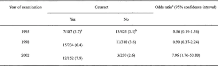
Table 6 presents the prevalence of pseudoexfoliation by cataract. No significant difference was observed in the prevalence of pseudoexfoliation between residents with cataract and those without cataract in 1995 (p=0.66, chi-square test) and 1998 (p=0.12, chi-square test), while the prevalence of pseudoexfoliation was significantly higher in residents with cataract than in those without cataract in 2002 (p=0.001, chi-square test). The difference remained significant in 2002 after adjustment for sex and age (Table 6).
Table 6. Prevalence of pseudoexfoliation by cataract
Year of examination Cataract Odds ratio` (95% confidence interval)
Yes No
1995 7/187 (3.7)° 13/425 (3.1)6 0.56 (0.19-1.56)
1998 15/234 (6
.4) 11/310 (3.6) 0.90 (0.37-2.24)
2002 12/152 (7.9) 3/250(2.6) 7.96 (1.76-50.80)
d) Number of cases of pseudoexfoliation/number of residents examined with cataract (%).
e) Number of cases of pseudoexfoliation/number of residents examined without cataract (%).
0 Odds ratio after adjustment for sex and age.
Narrow angle
The prevalence of narrow angle was 3.9%, 3.9% and 2.7%, in 1995, 1998 and 2002, respectively (Tables 1A- IQ and showed a trend to increase with age: an in- crease in the odds of the prevalence by 1 year in- crease in age was 1.07-fold (95% CI: 1.02-1.13) in 1995, 1.05-fold (95% CI: 1.00-1.11) in 1998 and 1.04-fold (95%
CI: 0.98-1.12) in 2002, respectively.
The cumulative incidence of narrow angle was 4.69% (Table 2) and no significant correlation with age was observed (OR=1.01, 95% CI: 0.96-1.07).
Glaucoma and ocular hypertension
The prevalence of glaucoma and ocular hyperten- sion was 1.5%, 2.4% and 3.5% in 1995, 1998 and 2002, respectively, and the cumulative incidence of glau- coma was 3.5%. Glaucoma with pseudoexfoliation was not found either in 1998 or 2002. However, one of the 612 residents (0.2%) examined in 1995 had the com- bined diagnosis.
Hirohiko Hayashida et al : Ocular Diseases on Naru Island
Enlargement of optic disc cupping
The prevalence of enlargement of the optic disc cup was 6.4%,..6.4% and 5.7% in 1995, 1998 and 2002. The incidence of enlargement of the optic disc cup was 3.8%.
Chorioretinal atrophy
The prevalence of chorioretinal atrophy was 1.8%, 1.1% and 0.7% in 1995, 1998 and 2002, respectively, and the cumulative incidence of chorioretinal atrophy was 0.2%.
Age-related macular degeneration (AMD)
The prevalence of AMD was 0.3%, 1.1% and 1.0% in 1995, 1998 and 2002, respectively, and the cumulative incidence of AMD was 1.7%.
Diabetic retinopathy
Diabetic retinopathy was noted in 1 person (0.2%) of the 612 residents examined in 1995, in 2 (0.4%) of the 544 residents examined in 1998 and in 2 (0.5%) of the 402 residents examined in 2002.
Genetic diseases
Rci111L15 p'ig111entuta was found In 3 (V.5%) of the 612 residents examined in 1995, 1 (new patient, 0.2%) of the 544 residents examined in 1998 and no new pa- tient among the 402 residents examined in 2002.
Discussion
Many population-based eye studies have reported age differences in the prevalence of ocular diseases.
However, we studied the prevalence and the incidence for 7 years on Naru Island. The characteristics of our
examined population are unique. Naru Island is com- pletely isolated and its population is decreasing, never increasing, and growing older year by year. The envi- ronment and occupations (fishing and farming) are un- changed. Thus this population is very suitable for a study of the natural course of certain age-related and degenerative diseases. Some diseases specific for the distinct are also discovered easily.
One of the prominent ocular diseases with a high prevalence on Naru Island was pterygium. Studies that included populations of all ages showed, the prevalence of pterygium to be 0.5% in South Africa,"
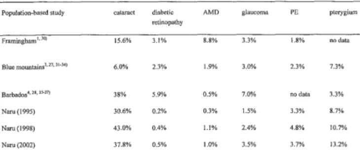
Table 7. Comparison of prevalence of ocular diseases in main population-based studies
Population-based study cataract diabetic AMD glaucoma PE pterygium
retinopathy
Framingham'' 10 15.6% 3.1% 8.8% 3.3% 1.8% no data
Blue mountains3.27' J1-34) 6.0% 2.3% 1.9% 3.0% 2.3% 7.3%
Barbados'. 21,11-37) 38% 5.9% 0.5% 7.0% no data 3.3%
Nam (1995) 30.6% 0.2% 0.3% 1.5% 3.3% 8.7%
Nara(1998) 43.0% 0.4% 1.1% 2.4% 4.8% 10.7%
Nam (2002) 37.8% 0.5% 1.0% 3.5% 3.7% 13.2%
AMD: age-related macular degeneration. PE: pseudoexfoliation.
0.3% in the Solomon Islands," 3.3% in The Barbados Eye Study, West Indies") (Table 7) and 0.7% in Copenhagen, Denmark.") The prevalence of pterygium in the State of Victoria, Australian)) was 2.8% in par- ticipants over 40 years of age. On Naru Island it was higher than in these studies, but a similar incidence has been reported in Okinawa, Japan (6.5%),12) in Eskimos in Greenland (8.6%)10) and in The Blue Mountains Eye Study, Australia (7.3%) 3) (Table 7). The Barbados Eye Study, West Indies') showed that the incidence of pterygium was related to race, so there might be a racial difference. However, it is well-known that pterygium is more common in very sunny, dusty, windy and dry environments."' These environmental conditions are prevalent in Naru.
The prevalence of pterygium on Naru Island in- creased apparently. 'LPILLe statistical significance was not shown in 1995 but was shown in 1998 and 2002.A similar tendency has been reported in several studies,'- 4 , 11, 12) although the incidence of pterygium did not increase with age. An increase in the odds of the cumulative incidence by 1 year increase in age was 1.32-fold (95%CI: 0.86-2.12) and was not statisti- cally significant. The age may not be a great risk fac- tor for pterygium.
Several studies 4 ,11,14.15) reported an increased risk of pterygium with exposure to ultra-violet radiation.
Such exposure has also been implicated as an in- creased risk of cataract. In our Naru Island Eye Study in 1998 and 2002, we found a statistically significant
difference in the incidence of cataract in eyes with and without pterygium. In The Blue Mountains Eye Study, Australia"), pterygium was associated with subcapsular cataract, but it was not associated with lens opacities in the Australian study by McCarty et al,' in the Barbados Eye Study, West India 4) or in the K Island Eye Study, Amami Islands, Japan.")
The prevalence of cataract significantly increased with age in residents examined in 1995, 1998 and 2002, while a significant difference in the prevalence
of cataract was not demonstrated between woman and man. A higher prevalence of age-related lens opacities in women than in men has been suggested in several studies."-"' The Beaver Dam Eye Study") reported that women were more severely affected with nuclear and cortical opacities than were men.
The Barbados Eye Study") reported that after adjust- ing for age, lens opacities were more frequent in women than in men. The possibility of hormonal ef- fects on cataractogenesis needs further study.
The present study indicated a significant increase in the prevalence of pseudoexfoliation with age. Several population-based studies"-") have reported that the prevalence of pseudoexfoliation increases with age.
However, these reports are cross-sectional studies. An increase with age was suggested in the incidence of pseudoexfoliation (Table 2) though the increase was not statistically significant (OR=1.43, 95% CI: 0.78-2.78).
The Framingham Eye Study, Massachusetts, US') re- ported an association of pseudoexfoliation with senile lens changes but the association was not statistically significant. In the Naru Island Eye Study, a significant difference in the prevalence of pseudoexfoliation was shown between the residents with and without cata- ract who were examined in 2002. Taylor 21) found a significant association between pseudoexfoliation and cataracts in Australian aborigines over 60 years of age. Since pseudoexfoliation and cataract are age- related, it would be probable that the observed asso- ciation was due to aging.
Glaucoma with pseudoexfoliation was not found in the present study. It is very interesting that pseudoexfoliation was not associated with glaucoma on Naru. The same phenomenon has been noted by Toda in the Kamigoto Islands near Naru Island.")
The prevalence of pseudophakia increased in 7 years of the period of the present study (Tables IA- 1Q. In 1995 when we started this examination, pseudophakia was 0.7%, but it was noted 3.2% in 2002.
We recommended cataract surgery for those who had low visual acuity caused by cataracts. We are confi- dent that the recommendation was valuable.
The prevalence of trichiasis showed no increase with age. A decrease in the cumulative incidence of trichiasis with age was observed but it was not the statistically significant. Trichiasis seems to have no association with age.
The prevalence of age-related macular degeneration (AMD) in white populations was 1.6% in the Beaver Dam Eye Study, Wisconsin, US 2, 1.7% in the Rotterdam Eye Study, The Netherlands26) and 1.9% in the Blue Mountains Eye Study, Australia,27) while in black populations the prevalence of AMD was 0.6% in the
Barbados Eye Study, West Indies"' and 0.22% in the Baltimore Eye Study, US."' The prevalence of AMD in Hisayama, Japan" was 0.87%. The prevalence of AMD on Naru Island was 0.3% in 1995, 1.1% in 1998 and 1.0% in 2002 and wass similar to that of white popula- tions.
The prevalence of diabetic retinopathy on Naru Island was 0.2-0.5% over 7 years, which was much lower than that observed in several other studies (Table 7). The diet of Naru Islanders includes more fish and vegetables than meat.
Naru Island is completely isolated and the popula- tion is very stable. Thus genetic diseases may proba- bly be present in some families. We found 4 patients with retinitis pigmentosa among those who received one examination during the 7 years, probably because of its long history and no possibility of treatment. In Japan the prevalence of retinitis pigmentosa is 1/4,000-1/6,000, so 4 patients in a population of about 4,000 population is very high, probably because of the geographically and historically closed society of Naru.
We examined the residents in Naru Island, Nagasaki Prefecture from 1995 to 2002 and found the changes in the prevalence of ocular diseases and recorded the natural course of several of them. We could obtain im- portant information on the changes of ocular diseases with age, and we believe our Naru Island Eye Study contributes greatly to ophthalmological knowledge.
Acknowledgement
We express sincere thanks to Dr. Tsugio Amemiya, Professor of Department of Ophthalmology and Visual Sciences, Graduate School of Biomedical Sciences, Nagasaki University, who designed, organized and commenced Naru Island Eye Study, for his encourage- ment, valuable suggestions and advices.
We also express sincere thanks to Dr. Yoshisada Shibata, Professor and Chairman of Department of Radiation Epidemiology, Atomic Bomb Disease Institute, Nagasaki University School of Medicine, for his statis- tical advice and his revision of this manuscript.
References
1 . Hiller R, Sperduto RD, Krueger DE: Pseudoexfoliation, intraocular pressure, and senile lens changes in a population-based survey.
Arch Ophthalmol 100: 1080-1082, 1982
2. Klein R, Klein BE, Linton KL: Prevalence of age-related maculopathy: The Beaver Dam Eye Study. Ophthalmology 99: 933-
943, 1992
3. Panchapakesan J, Mitchell P, Hourihan F: Prevalence of pterygium and pinguecula: the Blue Mountains Eye Study. Aust N Z J
Hirohiko Hayashida et al : Ocular Diseases on Naru Island
Ophthalmol 26 (supp l l ): S2-S5, 1998
4. Luthra R, Nemesure BB, Wu S-Y, et al: Frequency and risk factors for pterygium in the Barbados Eye Study. Arch Ophthalmol 119:
1827-1832, 2001
5. Klein R, Rowland ML, Harris MI: Racial/ethnic differences in age- related maculopathy: Third National Health and Nutrition
Examination Survey. Ophthalmology 102: 371-381, 1995
6. Oshima Y, Ishibashi T, Murata T, et al: Prevalence of age related maculopathy in a representative Japanese population: The
Hisayama study. Br J Ophthalmol 85: 1153-1157, 2001
7. SAS Institute. SAS/Stat User's Guide. Version 6. 4th edn. SAS Institute, Cary, NC, 1990
8. Hill JC, Maske R: Pathogenesis of pterygium. Eye 3: 218-226, 1989 9. Verlee DL: Ophthalmic survey in the Solomon Islands. Am I Ophthalmol 66: 304-319, 1968
10. Norn MS: Prevalence of pinguecula in Greenland and in Copenhagen and its relation to pterygium and spheroid degenera-
tion. Acta Ophthalmol 57: 96-105, 1979
11. McCarty CA, Fu CL, Taylor HR: Epidemiology of pterygium in Victoria, Australia. Br J Ophthalmol 84: 289-292, 2000
12. Yamakawa R, Nagamine Y, Koibuchi H, et al: Frequency of pterygium in Okinawa Prefecture. Folia Ophthalmol Jpn 47: 587- 591, 1996
13. Duke-Elder S. System of Ophthalmology Vol. VIII Diseases of the Outer Eye Part I. (Kimpton H.; C. V. Mosby Company, St. Louis)
pp.573-582, 1965
14. Perkins ES: The association between pinguecula, sunlight and cataract. Ophthalmic Res 17: 325-330, 1985
15. Taylor HR, West SK, Rosenthal FS, et al: Corneal changes associ- ated with chronic UV irradiation. Arch Ophthalmol 107: 1481-
1484, 1989
16. Lim R, Mitchell P, Cumming RG: Cataract associations with pinguecula and pterygium: the Blue Mountains Eye Study. Am J
Ophthalmol 126: 717-719, 1998
17. Sasaki H, Asano K, Kojima M, et al: Epidemiological survey of ocular diseases in K Island, Amami Islands: Prevalence of cataract
and pterigium. J Jpn Ophthalmol Soc 103: 556-563, 1999
18. Kahn HA, Leibowitz HM, Ganley JP, et al. The Framingham Eye Study, I: outline and major prevalence findings. Am J Epidemiol
106: 17-32, 1977
19. Sperduto RD, Seigel D. Senile lens and senile macular changes in a populationbased sample. Am J Ophthalmol 90: 86-91, 1980 20. Klein BEK, Klein R, Linton KLP. Prevalence of age-related lens
opacities in a population: the Beaver Dam Eye Study.
Ophthalmology 99: 546-552, 1992
21. Leske MC, Connell AM, Wu SY, et al: Prevalence of lens opacities in the Barbados Eye Study. Ach Ophthalmol 115: 105-111,1997 22. Asved H: The geographical distribution of fibrillopathia
epitheliocapsularis. Acta Ophthalmol 47: 792-810, 1969
23. Taylor HR, Hollows FC, Moran D: Pseudoexfoliation of the lens in Australian aborigines. Br J Ophthalmol 61: 473-475, 1977 24. Taylor HR: Pseudoexfoliation, an enviromental disease? Trans
Ophthalmol Soc UK 99: 302-307, 1979
25. Toda S: Retinitis pigmentosa and color vision deficiency in Kamigoto Island, Nagasaki Prefecture. J Jpn Ophthalmol Soc 101:
669-676, 1997
26. Vingerling JR, Dielemans I, Hofman A, et al: The prevalence of age-related maculopathy in the Rotterdam Study. Ophthalmology
102: 205-210, 1995
27. Mitchell P, Smith W, Attebo K, et al: Prevalence of age-related maculopathy in Australia: The Blue Mountains Eye Study.
Ophthalmology 102: 1450-1460, 1995
28. Schachat AP, Hyman L, Leske MC, et al: Features of age-related macular degeneration in a black population: The Barbados Eye
Study Group. Arch Ophthalmol. 113: 728-735, 1995
29. Friedman DS, Katz J, Bressler NM, et al: Racial differences in the prevalence of age-related macular degeneration: the Baltimore Eye
Survey. Ophthalmology 106: 1049-1055, 1999
30. Kini MM, Leibowitz HM, Colton T, et al: Prevalence of senile cata- ract, diabetic retinopathy, senile macular degeneration, and open
angle glaucoma in the Framingham Eye Study. Am J Ophthalmol
85: 28-34, 1978
31. Mitchell P, Cumming RG, Attebo K, et al: Prevalence of cataract
in Australia. Ophthalmology 104: 581-588, 1997
32. Mitchell P, Attebo K, Wang JJ, et al: Prevalence of diabetic retinopathy in an older community: the Blue Mountains Eye
Study. Ophthalmology 105: 406-411, 1998
33. Mitchell P, Smith W, Attebo K, et al: Prevalence of open-angle glaucoma in Australia: the Blue Mountains Eye Study.
Ophthalmology 103: 1661-1669, 1996
34. Mitchell P, Wang JJ, Smith W: Association of pseudoexfoliation
syndrome with increased vascular risk. Am J Ophthalmol 124: 685- 687, 1997
35. Leske MC, Schachat A, Hyman L, et al: Prevalence of lens opaci- ties in the Barbados Eye Study. Ach Ophthalmol 115: 105-111,
1997
36. Leske MC, Schachat AP, Connell AM, et al: Diabetic retinopathy in a black population: the Barbados Eye Study. Ophthalmology
106: 1893-1899, 1999
37. Leske MC, Connell AM, Wu SY, et al: Risk factors of open-angle glaucoma. Ach Ophthalmol 113: 918-924, 1995