Fukushima Medical University
福島県立医科大学 学術機関リポジトリ
This document is downloaded at: 2021-11-07T23:13:52Z
Title Anti-fibrotic effect of CCR2 antagonist on experimental murine scleroderma induced by bleomycin( 本文 )
Author(s) 石川, 真郷
Citation
Issue Date 2019-03-22
URL http://ir.fmu.ac.jp/dspace/handle/123456789/986
Rights
This is the pre-peer reviewed version of the following article:
[Exp Dermatol.], which has been published in final form at [https://doi.org/10.1111/exd.14088]. This article may be used for non-commercial purposes in accordance with Wiley Terms and Conditions for Use of Self-Archived Versions.
DOI
Text Version ETD
1
学位論文
Anti-fibrotic effect of CCR2 antagonist on experimental murine scleroderma induced by bleomycin
(ブレオマイシン誘導皮膚硬化モデルにおける
CCR2
拮抗薬の抗線維化効果)福島県立医科大学大学院 医学研究科 博士課程 皮膚・粘膜学
石川 真郷
2
概要
全身性強皮症(
Systemic sclerosis :SSc
)は、皮膚、肺を含む様々な臓器に線維化をきたす結合 織疾患である。線維化が生じる機序は明らかになっておらず、現在まで有効な治療法は極めて少 ない。Monocyte chemotactic protein-1 (MCP-1)
はマクロファージや血管内皮細胞から産生される、単球や
T
細胞の主要な遊走・活性化因子である。強皮症硬化皮膚の表皮・浸潤単核細胞・血管 内皮細胞にMCP-1
が強く発現していることや、MCP-1
の受容体CCR2
も活動性のある強皮症患 者皮膚では強く発現していることが報告され、SSc
においてMCP-1
とCCR2
のシグナル経路が強 皮症の皮膚硬化の機序で重要な位置を占めている可能性が示唆されてきた。本研究では、ブレオマイシン(
BLM
)誘導性強皮症モデルマウスを用いて、CCR2
受容体拮抗薬(
RS-504393
)を局所投与し、皮膚硬化および肺線維化に与える影響について検討した。C3H/HeJ
マウス(
6
週令、雌, n=18
)の背部に、RS-504393
を、濃度が4mg/kg (n=6)
、8mg/kg (n=6)
となるよ うに調節し、100 μl
皮内注射した。6
時間後に同部位に ブレオマイシン(125 μg/ml
)を100 μl
皮 内注射した。週3
回4
週間投与した後、6mm
デルマパンチで背部のブレオマイシン注射部位の皮 膚および肺を採取し、病理組織学的、生化学的な検討を行った。 コントロールとしてRS-504393
の代わりにPBS
を投与した群(n=6)
を用いた。PBS+BLM
群と比較して、RS-504393+BLM
群で は組織学的な皮膚硬化の抑制が認められ、高濃度のRS-504393
で真皮厚は有意に減少し、皮膚 組織中の可溶性コラーゲン量は減少傾向を示した。また、真皮内に浸潤する肥満細胞数は、RS-
504393
投与により有意に減少した。さらに、真皮内の筋線維芽細胞数は、高濃度のRS-504393
投与により有意に減少した。肺組織においては、
RS-504393
投与により肺胞壁の肥厚および炎症細 胞浸潤の減少を認め、肺線維スコア(Ashcroft score
)は減少傾向を示し、高濃度のRS-504393
で 肺組織中の可溶性コラーゲン量は有意に減少した。以上より、
RS-504393
はブレオマイシン誘導性強皮症モデルマウスにおいて、皮膚および肺の 線維化を抑制することが示唆された。この結果は、SS
cに対する抗線維化薬としてRS-504393
の可 能性を示唆する。3
Abstract
Background
The mechanisms of Systemic sclerosis (SSc) are complex and multiple fibrogenic mediators have been detected in SSc, including monocyte chemotactic protein-1 (MCP-1). Previous studies highlighted the role of the MCP-1/CCR2 (major receptor for MCP-1) signaling pathway in fibrotic disease and identified the blockade of the MCP-1/CCR2 signaling pathway as a therapeutic target for treating fibrosis.
Objective
The purpose of this study was to investigate the efficacy of CCR2 antagonist RS-504393 in a mouse model of scleroderma induced by bleomycin.
Methods
We divided all mice (n=18) into 3 groups. One hundred microliters of the RS-504393 solution, a dose of 4 mg/kg/day (n=6) or 8 mg/kg/day (n=6), were administered via intradermal injection into the shaved back of the mice 6 hours before one hundred microliters bleomycin injection (125 µg/ml) once daily for three days/week for four weeks. PBS was used as control for RS-504393 under same protocol (n=6).
Results
Histopathological examination showed RS-504393 + bleomycin mice showed suppressed induction of dermal sclerosis and decrease in dermal thickness. The amount of collagen in skin sites of RS- 504393 +bleomycin showed tendency to decrease. The number of mast cells and myofibroblasts in RS-504393 + bleomycin skin sites was significantly lower compared with PBS + bleomycin skin sites.
Semiquantitative histopathological scoring of lung showed the inhibition of fibrosis. The amount of collagen in the lung of RS-504393 + bleomycin mice was lower compared with PBS + bleomycin.
Conclusion
These data suggested that CCR2 antagonist RS-504393 may have a potential as a therapeutic agent
for SSc.
4
Abbreviation
SSc: Systemic sclerosis:
TGF- β: transforming growth factor-β1
CTGF: connective tissue growth factor
MCP-1: monocyte chemotactic protein-1
CCR2: C-C chemokine receptor type 2
CXCR1: CXC chemokine receptor 1
CCR1: C-C chemokine receptor type 1
CCR3: C-C chemokine receptor type 3
PBS: phosphate-buffered saline
H&E: hematoxylin and eosin
α-SMA: α-smooth muscle actin
IL-4: Interleukin-4
5
Introduction
Systemic sclerosis (SSc) is a chronic, multisystem disease with distinctive pathogenetic features, comprising vascular derangement, immune system activation, tissue fibrosis and a heterogeneous clinical profile (1). It is known that bleomycin, a chemotherapeutic agent used for the treatment of several types of malignancy, induced pulmonary fibrosis and dermal sclerosis as adverse effect, resembling symptoms of SSc (2). The mechanisms are complex and multiple fibrogenic mediators have been detected in SSc, including transforming growth factor-β (TGF-β), connective tissue growth factor (CTGF), and monocyte chemotactic protein-1 (MCP-1) (3–5).
MCP-1, belongs to the C-C chemokine superfamily, is an important cytokine for recruiting monocytes, T cells, and dendritic cells to the sites of inflammation (6). Previous studies have suggested that MCP-1 is strongly expressed in the epidermis, inflammatory mononuclear cells, and vascular endothelial cells in the sclerotic skin of SSc patients (7). In addition, up-regulation of MCP- 1 occurred in early murine sclerodermatous graft-versus-host disease, a model for human scleroderma, and preceded infiltration of immune cells and skin thickening (8). These studies suggested that the involvement of MCP-1 in the pathogenesis of SSc. The assumed mechanisms of MCP-1’s contribution to fibrotic diseases including SSc are regulating fibrogenic cytokine expression and fibroblast responsiveness to TGF-β, promoting mast cells infiltration (9–12) and myofibroblasts accumulation (13). Other studies demonstrated that an activation loop of tissue fibrosis centered on MCP-1 might be formed in SSc (14). MCP-1 is produced by vascular endothelial cells and fibroblasts, makes macrophages and T cells migrate around fibroblasts, and induces the production of TGF-β and PDGF from these activated infiltrating cells. As a result, these growth factors promote collagen production from fibroblasts (15). MCP-1 stimulates fibroblast collagen expression via specific receptors and endogenous upregulation of TGF-β expression, which then results in autocrine stimulation of collagen gene expression (16). In addition, MCP-1 induces differentiation into Th2 cells rather than Th1 cells (17). Th2 cells that migrate around the fibroblasts affect collagen production from fibroblasts via IL-4 production (18). Furthermore, activated fibroblasts produce MCP-1, which forms an activation loop of tissue fibrosis centered on MCP-1.
On the other hand, CCR2, major receptor for MCP-1, deficiency improved bleomycin-induced
pulmonary fibrosis (19), and administration of an anti-MCP-1 neutralizing antibody reduced
bleomycin-induced dermal sclerosis (4). In another study, Kitagawa K et al. demonstrated that,
administration of the CCR2-selective antagonist RS-504393 reduced kidney fibrosis (20). Their
6
study and other studies highlighted the role of the MCP-1/CCR2 signaling pathway in fibrotic disease and identified the blockade of the MCP-1/CCR2 signaling pathway as a therapeutic target for treating fibrosis.
The purpose of this study was to investigate the efficacy of CCR2 antagonist RS-504393 in a mouse model of scleroderma induced by bleomycin. RS-504393 is a specific small organic molecule antagonist of CCR2 and inhibits the receptor by occupation of a binding site (21); however, it is not a chemotaxis agonist and does not stimulate any postreceptor signaling. RS-504393 specifically inhibits MCP-1 and MCP-3 signaling through CCR2, but is not an antagonist of CXCR1, CCR1, or CCR3 (20). Bleomycin-induced scleroderma model is widely used because skin and lung fibrosis resembling scleroderma can be easily induced by repeated local injections of bleomycin (22). In addition, in this model, expression of MCP-1 and CCR2 was elevated at both protein and mRNA levels in the lesional skin (4). MCP-1 as well as CCR2 were detected on the infiltrating mononuclear cells at early stages following bleomycin treatment and on the fibroblasts at latter stages in the sclerotic skin (4). We focused on the fibrogenic function of MCP-1/CCR2 signaling in SSc by analyzing the efficacy of local CCR2 antagonist administration in the mouse model of scleroderma induced by bleomycin.
Materials and methods
Animals
C3H-HeJ female mice (specific pathogen-free, six weeks old, weighing about 20 g, n=18) were purchased from CLEA Japan, Inc. (Tokyo, Japan). Animals were housed in plastic cages at room temperature (RT; 21-24℃) on a 12-hour light/dark cycle. Food and water were given ad libitum.
All animal experiments in this study were approved by the Animal Care Committee of Fukushima Medical University on May 23, 2017 (No. 29060).
Development of dermal sclerosis by bleomycin
The scleroderma mouse model was established according to the previous method (21). Bleomycin
(Nippon Kayaku Co., Ltd., Tokyo) was dissolved in phosphate-buffered saline (PBS) at a
concentration of 125 μg/ml. One hundred microliters of bleomycin were injected into the shaved
back of the mice once daily for three days/week for four weeks.
7 Administration of RS-504393
RS-504393 (Cayman chemical, Co., Michigan, USA) powder was diluted in PBS. We divided all mice (n=18) into 3 groups. One hundred microliters of the RS-504393 solution, a dose of 4 mg/kg/day (n=6) or 8 mg/kg/day (n=6), were administered via intradermal injection into the shaved back of the mice 6 hours before bleomycin injection once daily for three days/week for four weeks.
All injections were performed at the same site with a 27-gauge needle. PBS was used as control for RS-504393 under same protocol (n=6).
Histopathological analysis of dermal fibrosis and dermal thickness measurement
Six-millimeter punch biopsy was performed on injected skin sites on the following day of the final treatment. Each skin piece was cut into two; one was used for paraffin-embedded sectioning (5 μm sections), which was deparaffinized, rehydrated, and stained with hematoxylin and eosin (H&E) or Elastica-Masson, and the other was immediately stored in liquid nitrogen for cryosectioning, that was used for measurement of amount of collagen. For evaluation of skin sclerosis, dermal thickness was measured on photomicrographs of the H&E-stained sections, measuring the distance between the epidermal-dermal junction and the dermal-fat junction at ten randomly-selected sites, that were between rete ridges, per section under a magnification of × 100. Infiltrating mast cells in the dermis were stained with toluidine blue, and stain-positive cells were counted in ten randomly-selected sites per section under a light microscopic field at a magnification of × 400.
Histopathological assessment of lung fibrosis
Lungs were excised from each mouse at the same time of the skin biopsy. Lung tissue was stained using H&E staining. The severity of lung fibrosis was determined by semiquantitative histopathological scoring using light microscopy at a magnification of × 100 (Ashcroft score) (23).
Lung fibrosis was scored on a scale of 0–8 by examining 45 randomly chosen fields (5 lobes/mouse × 3 sections/lobe × 3 fields/section) for each mouse (0: Normal lung, 1: Minimal fibrous thickening of alveolar or bronchiolar walls, 3: Moderate thickening of walls without obvious damage to lung architecture, 5: Increased fibrosis with definite damage to lung structure and formation of fibrous bands or small fibrous masses, 7: Severe distortion of structure and large fibrous areas; "honeycomb lung" is placed in this category, 8: Total fibrous obliteration of the field. If there was any difficulty in deciding between two odd numbered categories, the field would be given the intervening even numbered score).
Immunohistochemical staining
Alpha-Smooth muscle actin (α-SMA) is used as a marker for a subset of myofibroblasts which are
regarded as important effector cells of tissue fibrogenesis (24). Multiple 5 μm sections of paraffin-
8
embedded skin samples were deparaffinized and rehydrated. Immunolabeling of α-SMA was performed, followed by detection with an avidin-biotin-peroxidase complex with primary rabbit anti–
mouse α-SMA antibody diluted 1:50 in PBS (Abcam, Tokyo, Japan). Appropriate biotinylated secondary antibodies and color development with DAB were used (Dako, Tokyo, Japan). Stain- positive cells were counted in ten randomly-selected dermal sections under a light microscopic field at a magnification of × 400.
Measurement of amount of collagen
Collagen deposition was estimated by determining the total collagen content of the cryopreserved skin and lung using Sircol Soluble Collagen Assay Kit (Biocolor Ltd., UK). After adding 500 μl of dye reagent to the ground skin and lung, tubes were placed in a gentle mechanical shaker for 30 min.
Then, the mixture was centrifuged, followed by discarding of the supernatant. After adding 500 μl of alkali reagent, the final product was measured against soluble collagen standards at 540 and 555 nm.
Statistical analysis
Results were expressed as mean ± SD and analyzed using the Bell Curve for Excel software (version 2.15; Social Survey Research Information Co., Ltd., Tokyo). Data was analyzed by Kruskal-Wallis test and Steel-Dwass test. P < 0.05 was considered significant.
Results
Local injection of CCR2 antagonist inhibits dermal sclerosis induced by bleomycin
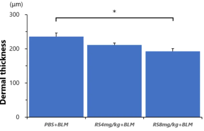
On examination of H&E staining, all PBS + bleomycin mice showed definite dermal sclerosis. On the contrary, mice treated with RS-504393 + bleomycin showed suppressed induction of dermal sclerosis (Figure 1. a–c). Furthermore, Elastica-Masson staining showed thickening of the dermal collagen bundles and decreased deposition of collagen fibers (Figure 1. d–f). Compared with PBS- treated mice, RS-504393-treated mice showed a 10% decrease in dermal thickness in those dosed with 4 mg/kg, and 18% in those dosed with 8 mg/kg (Figure 2.) (RS-504393 [4 mg/kg] + bleomycin: 211.4
± 5.21 μm vs PBS + bleomycin: 235.5 ± 10.28 μm, p =0.260; RS-504393 [8 mg/kg] + bleomycin:
192.5 ± 8.05 μm vs PBS + bleomycin: 235.5 ± 10.28 μm, p = 0.043). No statistically significant dose
dependence was observed in RS-504393-induced inhibition. The amount of collagen in the skin sites
of RS-504393 +bleomycin showed tendency to decrease compared with PBS + bleomycin (Figure 3.)
(RS-504393 [4 mg/kg] + bleomycin: 11.1 ± 1.55 μg/ml vs PBS + bleomycin: 24.6 ± 5.32 μm p =
0.054; RS-504393 [8 mg/kg] + bleomycin: 10.7 ± 2.33 μg/ml vs PBS+ bleomycin: 24.6 ± 5.32 μm, p
9
= 0.054).
Injection of CCR2 antagonist reduces mast cell infiltration in scleroderma lesions induced by bleomycin
On examination of toluidine blue staining, all PBS + bleomycin mice showed increased intradermal infiltration of mast cells. RS-504393 + bleomycin mice showed suppressed infiltration of mast cells (Figure 4. a–c). The number of mast cells in the RS-504393-treated skin sites was significantly lower compared with the with PBS-treated skin sites (Figure 4 d) (RS-504393 [4 mg/kg] + bleomycin: 98.9
± 14.76/10 high-power field [HPF]vs PBS + bleomycin: 132.8 ± 14.34/ HPF, p = 0.116; RS-504393[8 mg/kg] + bleomycin: 78.8 ± 8.60/10 HPF vs PBS + bleomycin: 132.8 ± 14.34/ HPF, p = 0.024). No statistically significant dose dependence was observed in RS-504393-induced reduction.
Injection of CCR2 antagonist reduces myofibroblast accumulation in bleomycin- induced scleroderma skin
We compared the number of α-SMA-positive myofibroblasts in the dermis of RS-504393 + bleomycin mice with PBS + bleomycin mice (Figure 5. a–c). The number of myofibroblasts in the RS-504393 8mg/kg-treated skin sites was significantly lower compared with PBS-treated skin sites (Figure 5. d) (RS-504393 [4 mg/kg] + bleomycin: 43.8 ± 7.91/10 HPF vs PBS + bleomycin: 79.2 ± 7.45/10 HPF, p =0.072; RS-504393 [8 mg/kg] + bleomycin: 40.8.0 ± 6.00/10 HPF vs PBS + bleomycin: 79.2 ± 7.45/10 HPF, p = 0.023). No statistically significant dose dependence was observed in RS-504393-induced reduction.
CCR2 antagonist inhibits lung fibrosis induced by bleomycin
On histopathological examination of the lungs in PBS + bleomycin mice, significant inflammation and fibrosis were observed in the lung tissue, including alveolar inflammation, thickening of the alveolar septae, and fibrous obliteration of the peribronchiolar and parenchymal regions. The lung tissue of the RS-504393 + bleomycin mice showed moderate inflammation and fibrosis compared with PBS + bleomycin mice (Figure 6.). Semiquantitative histopathological scoring showed the inhibition of fibrosis (Figure 7.) (RS504393 [4 mg/kg] + bleomycin: 3.6± 0.53 vs PBS + bleomycin: 4.2 ± 0.22, p = 0.260; RS504393 [8 mg/kg] + bleomycin: 2.9 ± 0.59 vs PBS + bleomycin: 4.2 ± 0.22, p =0.170).
The amount of collagen in the lung of RS-504393-treated mice was 31% (4 mg/kg) and 40% (8 mg/kg)
lower, showing a significant difference in 8mg/kg, compared with PBS treated mice (Figure 8.)
(RS504393 [4 mg/kg] + bleomycin: 8.1 ± 0.93 μg/ml vs PBS + bleomycin: 11.7 ± 1.10 μg/ml, p =
0.072; RS-504393 [8 mg/kg] + bleomycin: 7.1 ± 0.73 μg/ml vs PBS + bleomycin: 11.7 ± 1.10 μg/ml,
p = 0.043).
10
Discussion
The purpose of this study was to investigate the efficacy of CCR2 antagonist RS-504393 in a mouse model of scleroderma induced by bleomycin. We demonstrated that RS-504393 inhibited dermal sclerosis and lung fibrosis and reduced the numbers of mast cells and myofibroblasts in the dermis.
These results suggest the effects of CCR2 antagonist on skin and lung fibrosis, and that blocking MCP- 1/CCR2 signaling pathway may have the potential to be a pharmaceutical target for the treatment of SSc.
It has been demonstrated that blocking MCP-1/CCR2 signaling pathway, such as administration of an anti-MCP-1 neutralizing antibody, reduced bleomycin-induced dermal sclerosis (4). Similar to that study, we demonstrated that CCR2 antagonist inhibited dermal sclerosis in the same mouse model.
Monocytes/macrophage are one of the major sources of TGF-β, which is a key cytokine in fibrosis, in SSc and in bleomycin-induced mice (25). In addition, CCR2 expression increase on the infiltrating mononuclear cells in bleomycin-induced mice (4). Accumulation of inflammatory infiltrates such as macrophages, mast cells and, Th2 cells are common in early stages of SSc and bleomycin-induced dermal sclerosis (22). From these results, the assumed mechanism of CCR2 antagonist inhabitation for dermal sclerosis is that CCR2 antagonist prevents MCP-1 from binding to CCR2 and regulates TGF-β expression in monocytes/macrophage, results in decrease of collagen production from fibroblasts. On the other hand, CCR2 antagonist did not completely inhibit dermal sclerosis in our study. As other chemokines may contribute to sclerosis in this model, mononuclear cells TGF-β expression was not completely inhibited by CCR2 blocking.
Mast cells are increased in the skin of human SSc and bleomycin-induced mice (26). They are also one of the major sources of TGF-β in SSc and may play a role in accelerating the formation of dermal sclerosis for stimulating fibroblasts to produce collagen in bleomycin-induced mice (26).
Stem cell factor, a growth factor of mast cells, promotes increased production of MCP1 from mast cells, thereby promoting collagen production from fibroblasts (11). In addition, MCP-1 provokes mast cell aggregation and induces histamine release in rat models (27). In this study, the CCR2 antagonist significantly inhibited mast cell infiltration in the dermis of the bleomycin-induced scleroderma model. On the other hand, some past studies suggested that bleomycin-induced skin sclerosis was demonstrable in the mast cell-deficient genetic ablation mice (26). Although mast cells may not have main roles in experimental mouse models of bleomycin-induced scleroderma, our results suggested that CCR2 inhibition could mediate direct or indirect decreases in mast cell infiltration and contribute to the suppression of skin fibrosis in the experimental mouse models.
Myofibroblasts play a major role in the pathogenesis of SSc (28). They secrete increased amounts
of extracellular matrix components, including collagen, and are predominant in areas of increased
collagen deposition in scleroderma lesional skin, where the number of myofibroblasts correlates with
the severity of fibrosis (25). Myofibroblasts are also increased in the mouse model of bleomycin-
11
induced scleroderma (29). In the current study, the number of myofibroblasts in the RS-504393- treated skin sites was significantly lower. A previous study suggested that myofibroblasts could be recruited in an MCP-1-dependent manner in the pathogenesis of oral submucous fibrosis (13). Thus, antifibrotic effects of CCR2 inhibition may have resulted from the inhibition of myofibroblast accumulation in bleomycin-induced scleroderma models.
Although in bleomycin induced scleroderma model, lung fibrosis is also induced preceding the cutaneous changes (22), the its exact mechanism is unknown. Lung fibrosis induced by bleomycin is not only induced by bronchial administration but also by intravenous administration or intraperitoneal administration (30). Possible mechanism of lung fibrosis in bleomycin induced scleroderma model may be that bleomycin arriving at lung intravenously caused inflammation characterized by macrophages and fibrosis at their lungs (31). We demonstrated that CCR2 antagonist inhibited lung fibrosis in bleomycin induced scleroderma model. Assumed mechanism is that CCR2 antagonist which arrived at the lung intravenously suppressed inflammation and fibrosis induced by bleomycin at the lung. To prove this mechanism, it is necessary to examine the blood concentration of CCR2 antagonist and bleomycin.
We acknowledge that there are several limitations in this study. First, as we administrated RS- 504393 4mg/kg or 8mg/kg via intradermal injection before bleomycin, it has not been able to fully demonstrate its potential as a therapeutic agent. Although we have tried various experimental systems, based on past administration methods (20-21, 32-33), such as administering RS-504393 and bleomycin at the same time, orally administering RS-504393 before bleomycin, or administering RS- 504393 1mg/kg to 8mg/kg, only the current method was effective. We need to investigate efficacy of RS-504393 after skin fibrosis is completed. However, since the course of SSc is slowly progressive with a variable degree, if we can diagnose SSc on early stage, RS-504393 might be effective to stop progressing SSc. Second, we only observed the condition of skin and lung four weeks after the start of administration. As in bleomycin-induced scleroderma model, total mast cell number is gradually increased as the sclerotic lesions are induced (22), it is necessary to observe the skin and lungs over time, for example one week, two weeks after starting RS-504393 and bleomycin administration. Third, although we described the possible role of MCP-1/CCR2 pathway in SSc using bleomycin induced scleroderma model and RS-504393, we did not analyze the expression level of cytokines which contribute to sclerosis. Future studies are also needed to evaluate the mRNA expression of those cytokines.
In conclusion, we demonstrated the efficacy of CCR2 antagonist RS-504393 in the scleroderma
model mice. Our data also suggested that MCP-1/CCR2 signaling may play an important role in the
pathogenesis of sclerosis induced by bleomycin, and CCR2 antagonist may have a potential as a
therapeutic agent for SSc.
12
Acknowledgments
We are grateful to Tomoko Okada and Naoko Suzuki for their technical supports.
13
References
1. Orlandi M, Barsotti S, Lepri G, et al. One year in review 2018: systemic sclerosis. Clin Exp Rheumatol. 2018; 36 Suppl 113: 3-23.
2. Tashiro M, Izumikawa K, Yoshioka D, et al. Lung fibrosis 10 years after cessation of bleomycin therapy. Tohoku J Exp Med. 2008; 216: 77-80.
3. Querfeld C, Eckes B, Huerkamp C, et al. Expression of TGF-beta 1, -beta 2 and -beta 3 in localized and systemic scleroderma. J Dermatol Sci. 1999; 21: 13-22.
4. Chujo S, Shirasaki F, Kondo-Miyazaki M, et al. Role of connective tissue growth factor and its interaction with basic fibroblast growth factor and macrophage chemoattractant protein-1 in skin fibrosis. J Cell Physiol. 2009; 220: 189-95.
5. Yamamoto T, Nishioka K. Role of monocyte chemoattractant protein-1 and its receptor, CCR-2, in the pathogenesis of bleomycin-induced scleroderma. J Invest Dermatol. 2003; 121: 510-6.
6. Leonard EJ, Yoshimura T. Human monocyte chemoattractant protein-1 (MCP-1). Immunol Today.
1990; 11: 97-101.
7. Hasegawa M, Sato S, Takehara K. Augmented production of chemokines (monocyte chemotactic protein-1 (MCP-1), macrophage inflammatory protein-1alpha (MIP-1alpha) and MIP-1beta in patients with systemic sclerosis: MCP-1 and MIP-1alpha may be involved in the development of pulmonary fibrosis. Clin Exp Immunol. 1999; 117: 159-65.
8. Zhang Y, McCormick LL, Desai SR, et al. Murine sclerodermatous graft-versus-host disease, a model for human scleroderma: cutaneous cytokines, chemokines, and immune cell activation. J Immunol. 2002; 168: 3088-98.
9. Yamamoto T, Katayama I, Nishioka K. Expression of stem cell factor in the lesional skin of systemic sclerosis. Dermatology. 1998; 197: 109-14.
10. Kihira C, Mizutani H, Asahi K, et al. Increased cutaneous immunoreactive stem cell factor expression and serum stem cell factor level in systemic scleroderma. J Dermatol Sci. 1998; 20: 72-8.
11. Yamamoto T, Hartmann K, Eckes B. Role of stem cell factor and monocyte chemoattractant
14
protein-1 in the interaction between fibroblasts and mast cells in fibrosis. J Dermatol Sci. 2001; 26:
106-11.
12. Baghestanian M, Hofbauer R, Kiener HP, et al. The c-kit ligand stem cell factor and anti-IgE promote expression of monocyte chemoattractant protein-1 in human lung mast cells. Blood. 1997;
90: 4438-49.
13. Sarode G, Sarode SC, Deshmukh R, et al. Myofibroblasts could be recruited in a chemokine (C-C motif) ligand 2-dependent manner in pathogenesis of oral submucous fibrosis. J Oral Pathol Med.
2017; 46: 443-447.
14. Hasegawa M. The roles of chemokines in the development of systemic sclerosis. Jpn. J. Clin.
Immunol. 2008; 31: 23-36.
15. Gharaee-Kermani M, Denholm EM, Phan SH. Costimulation of fibroblast collagen and transforming growth factor beta1 gene expression by monocyte chemoattractant protein-1 via specific receptors. J Biol Chem. 1996; 271: 17779-84.
16. Carulli MT, Ong VH, Ponticos M, et al. Chemokine receptor CCR2 expression by systemic sclerosis fibroblasts: evidence for autocrine regulation of myofibroblast differentiation.
Arthritis Rheum. 2005; 52: 3772-82.
17. Gu L, Tseng S, Horner RM, et al. Control of TH2 polarization by the chemokine monocyte chemoattractant protein-1. Nature. 2000; 404: 407-11.
18. Distler JH, Jüngel A, Caretto D, et al. Monocyte chemoattractant protein 1 released from glycosaminoglycans mediates its profibrotic effects in systemic sclerosis via the release of interleukin- 4 from T cells. Arthritis Rheum. 2006; 54: 214-25.
19. Okuma T, Terasaki Y, Kaikita K, et al. C-C chemokine receptor 2 (CCR2) deficiency improves bleomycin-induced pulmonary fibrosis by attenuation of both macrophage infiltration and production of macrophage-derived matrix metalloproteinases. J Pathol. 2004; 204: 594-604.
20. Kitagawa K, Wada T, Furuichi K, et al. Blockade of CCR2 ameliorates progressive fibrosis in
kidney. Am J Pathol. 2004; 165: 237-46.
15
21. Furuichi K, Wada T, Iwata Y, et al. CCR2 signaling contributes to ischemia-reperfusion injury in kidney. J Am Soc Nephrol. 2003; 14: 2503-15.
22. Yamamoto T, Takagawa S, Katayama I, et al. Animal model of sclerotic skin. I: Local injections of bleomycin induce sclerotic skin mimicking scleroderma. J Invest Dermatol. 1999; 112: 456-62.
23. Ashcroft T, Simpson JM, Timbrell V. Simple method of estimating severity of pulmonary fibrosis on a numerical scale. J Clin Pathol. 1988; 41: 467-70.
24. Cutolo M, Ruaro B, Montagna P, et al. Effects of selexipag and its active metabolite in contrasting the profibrotic myofibroblast activity in cultured scleroderma skin fibroblasts. Arthritis Res Ther.
2018; 20: 77.
25. Ayers NB, Sun CM, Chen SY. Transforming growth factor-β signaling in systemic sclerosis. J Biomed Res. 2018; 32: 3-12.
26. Yamamoto T, Takahashi Y, Takagawa S, et al. Animal model of sclerotic skin. II. Bleomycin induced scleroderma in genetically mast cell deficient WBB6F1-W/W(V) mice. J Rheumatol. 1999;
26: 2628-34.
27. Conti P, Boucher W, Letourneau R, et al. Monocyte chemotactic protein-1 provokes mast cell aggregation and [3H]5HT release. Immunology. 1995; 86: 434-40.
28. Kissin EY, Merkel PA, Lafyatis R. Myofibroblasts and hyalinized collagen as markers of skin disease in systemic sclerosis. Arthritis Rheum. 2006; 54: 3655-60.
29. Yamamoto T, Nishioka K. Animal model of sclerotic skin. V: Increased expression of alpha-smooth muscle actin in fibroblastic cells in bleomycin-induced scleroderma. Clin Immunol. 2002; 102: 77-83.
30. Adamson IY, Bowden DH. Origin of ciliated alveolar epithelial cells in bleomycin-induced lung injury. Am J Pathol. 1977; 87: 569-80.
31. Williamson JD, Sadofsky LR, Hart SP. The pathogenesis of bleomycin-induced lung injury in animals and its applicability to human idiopathic pulmonary fibrosis Exp Lung Res. 2015; 41: 57-73.
32. Kang YS, Lee MH, Song HK, et al. CCR2 antagonism improves insulin resistance, lipid
16
metabolism, and diabetic nephropathy in type 2 diabetic mice. Kidney Int. 2010; 78: 883-94.
33. Awad AS, Kinsey GR, Khutsishvili K, et al. Monocyte/macrophage chemokine receptor CCR2
mediates diabetic renal injury. Am J Physiol Renal Physiol. 2011; 301: F1358-66.
㻝㻣㻌 㻌
Figures
㻌Figure 1. Histopathologic evaluation of the lesional skin of mice treated with bleomycin and either RS-504393 or PBS.
(a-c) Mice were treated with either bleomycin (BLM) and RS-504393 or PBS for 4 weeks (H&E staining). Mice treated with RS-504393 showed suppressed induction of dermal sclerosis.
(d–f) Elastica-Masson staining showed thickening of the dermal collagen bundles and decreased
deposition of collagen fibers in mice treated with RS-504393. Original magnification × 100. Scale
bar 300 µm.
㻝㻤㻌 㻌
Figure 2. Dermal thickness in mice treated with bleomycin and either RS-504393 or PBS.
Dermal thickness was measured on photomicrographs of the H&E-stained sections, measuring the distance between the epidermal-dermal junction and the dermal-fat junction at ten randomly-selected sites, that were between rete ridges, per section under a magnification of × 100. Dermal thickness of RS-504393 8mg/kg + bleomycin (BLM) mice significantly decreased compared with PBS + BLM mice. * p =0.043.
Figure 3. Amount of the collagen in mice treated with bleomycin and either RS-504393 or PBS.
Collagen deposition was estimated by determining the total collagen content of the cryopreserved skin
using Sircol Soluble Collagen Assay Kit. The amount of collagen in RS-504393 8mg/kg +
bleomycin (BLM) mice and that in RS-504393 4mg/kg + BLM showed tendency to decrease
compared with PBS + BLM mice.
㻝㻥㻌 㻌
(d)
Figure 4. Histopathological examination of toluidine blue staining in mice treated with bleomycin and either RS-504393 or PBS.
(a-c) Mice were treated with either bleomycin (BLM) and RS-504393 or PBS for 4 weeks (toluidine blue staining). Mice treated with RS-504393 showed suppressed infiltration of mast cells.
Original magnification × 400. Scale bar 100 µm.
(d) The number of mast cells in RS-504393 8mg/kg + BLM mice were significantly lower compared
with PBS + BLM mice. * p = 0.024.
㻞㻜㻌 㻌
(d)
Figure 5. Immunobiologic examination of myofibroblasts in mice treated with bleomycin and either RS-504393 or PBS.
(a-c) Mice were treated with either bleomycin (BLM) and RS-504393 or PBS for 4 weeks. Alpha- Smooth muscle actin (α-SMA) is used as a marker for a subset of myofibroblasts. Mice treated with RS-504393 showed suppressed accumulation of myofibroblasts. Original magnification × 400.
Scale bar 100 µm.
(d) The number of α-SMA-positive myofibroblasts in RS-504393 8mg/kg + BLM mice was
significantly lower compared with PBS + BLM mice. * p = 0.023.
㻞㻝㻌 㻌
㻌
Figure 6. Histopathologic evaluation of the lungs of mice treated with bleomycin and either RS-504393 or PBS.
(a-c) Mice were treated with local dermal injection of bleomycin (BLM) and either RS-504393 or PBS for 4 weeks (H&E staining). On histopathological examination of the lungs in PBS + BLM mice, significant inflammation and fibrosis were observed in the lung tissue, including alveolar inflammation, thickening of the alveolar septae, and fibrous obliteration of the peribronchiolar and parenchymal regions. RS-504393 + BLM mice showed moderate inflammation and fibrosis compared with PBS + BLM mice. Original magnification × 100. Scale bar 300 µm.
Figure 7. Semiquantitative histopathological scoring of the lungs of mice treated with bleomycin and either RS-504393 or PBS.
The severity of lung fibrosis was determined by semiquantitative histopathological scoring using light microscopy at a magnification of × 100 (Ashcroft score). Lung fibrosis was scored on a scale of 0–
8 by examining 45 randomly chosen fields for each mouse. The lung tissue of RS-504393
+bleomycin (BLM) mice showed the inhibition of fibrosis compared with the lung tissue of PBS
+BLM mice.
㻞㻞㻌 㻌
Figure 8. The amount of collagen in the lung of mice treated with bleomycin and either RS- 504393 or PBS.
Collagen deposition was estimated by determining the total collagen content of the cryopreserved lung using Sircol Soluble Collagen Assay Kit. The amount of collagen in RS-504393 8mg/kg + bleomycin (BLM) mice significantly decreased compared with PBS + BLM mice. * p = 0.043.
㻌