Fukushima Medical University
This document is downloaded at: 2021-11-07T23:52:58Z
Title Henoch-schönlein purpura nephritis in childhood:
pathogenesis, prognostic factors and treatment
Author(s) Kawasaki, Yukihiko; Ono, Atsushi; Ohara, Shinichiro; Suzuki, Yuichi; Suyama, Kazuhide; Suzuki, Junzo; Hosoya, Mitsuaki
Citation Fukushima Journal of Medical Science. 59(1): 15-26
Issue Date 2013
URL http://ir.fmu.ac.jp/dspace/handle/123456789/360
Rights © 2013 The Fukushima Society of Medical Science
DOI 10.5387/fms.59.15
Text Version publisher
HSPN IN CHILDHOOD 15 Fukushima J. Med. Sci.,
Vol. 59, No. 1, 2013
15
Corresponding author : Yukihiko Kawasaki E-mail address : [email protected] https://www.jstage.jst.go.jp/browse/fms http://www.fmu.ac.jp/home/lib/F-igaku/
[Review]
HENOCH
-SCHÖNLEIN PURPURA NEPHRITIS IN CHILDHOOD : PATHOGENESIS, PROGNOSTIC FACTORS
AND TREATMENT
YUKIHIKO KAWASAKI, ATSUSHI ONO, SHINICHIRO OHARA, YUICHI SUZUKI, KAZUHIDE SUYAMA, JUNZO SUZUKI and MITSUAKI HOSOYA
Department of Pediatrics, Fukushima Medical University School of Medicine
Abstract: Henoch-Schönlein purpura (HSP) is a systemic disorder characterized by leukocytoclas- tic vasculitis involving the capillaries and the deposition of IgA immune complexes. Renal involve- ment is the principal cause of morbidity and mortality in children with HSP. Thus, it is important to clarify the onset mechanism as well as the prognostic factors of Henoch- Schönlein purpura nephri- tis (HSPN) and to identify the most appropriate treatment. We herein review the pathogenesis, the prognostic factors and treatment of HSPN. As to the pathogenesis, several studies suggest that galactose-deficient IgA1 (Gd-IgA1) is recognized by anti-glycan antibodies, leading to the formation of circulating immune complexes and their mesangial deposition, thereby inducing renal injury. With regard to the prognostic factors, a number of factors have been suggested including nephrotic syndrome, decreased factor XIII activity, hypertension, severe renal injury, high renal accumulation of activated macrophage, alpha-smooth muscle actin, and high serum myeloid-related protein levels.
For the treatment of severe HSPN, aggressive therapies including multiple drug combination ther- apy and plasmapheresis have been shown to be effective in ameliorating proteinuria and histological severity. Nevertheless, detailed investigation into the pathogenesis of HSPN and double-blind ran- domized control studies on children with HSPN are still necessary.
Key words: HSPN, Pathogenesis, urokinase pulse therapy, cyclophosphamide, mizoribine
INTRODUCTION
Henoch-Schönlein purpura (HSP) was first rec- ognized by Heberden in 1801 and first described as an association between purpura and arthritis by Schönlein in 1837. Henoch added descriptions of gastrointestinal involvement in 1874 and renal involvement in 1899. HSP is a small vessel vascu- litis, the major manifestations of which include arthritis, nonthrombocytopenic purpura, abdominal pain, and renal disease. HSP is one of the most common vasculitides of childhood and is considered to be self-limiting. One manifestation of HSP that can result in lifelong problems is renal involve- ment1,2). Approximately 40% of pediatric patients develop nephritis within 4 to 6 weeks of the initial
presentation. A majority of children with HSPN present with only hematuria and/or low-grade pro- teinuria, or both, and have a good chance of recov- ery. However, patients with massive proteinuria at onset frequently show a progressive course. In specialized centers, the proportion of children with HSPN who progress to renal failure or end-stage renal disease varies from 1% to 17%2-8).
It is therefore important to clarify the onset mechanism, clinical manifestations of and ascertain the most appropriate treatment for HSPN. Herein we review the literature on the pathogenesis, clini- cal manifestations, prognostic factors, and treatment of HSPN.
INCIDENCE OF DISEASE
Gardner-Medwin et al. examined the frequency of and ethnic variations in childhood vasculitides in the West Midlands region of the United Kingdom6). Their survey was completed using monthly ques- tionnaires sent to consultants and a single question- naire sent to family doctors, along with a review of case notes with diagnostic codes for vasculi- tis. The annual incidence of HSP in the study was 22.1 cases per 100,000 children, which was higher than previous estimates of 13.5-18.0 cases per 100,000 children7,8). The authors postulated that a higher incidence of HSP may lead to increases in the incidences of renal disease and need for renal medi- cal treatment. Stewart et al evaluated a total of 270 patients with HSP from a total pediatric population of 155,000 over a 13-year period and showed that the incidence of HSPN was 2.7 cases per 100,000 children7). The mean incidence of HSPN in Asian children, however, has been reported to be 4.9 cases per 100,000 children per year, and over a 22-year period in Japan, Kawasaki et al. reported that the mean number of HSPN cases per 100,000 children per year to be 3.6+/−1.0 (Figure 1)9).
PATHOGENESIS
The pathogenesis of HSP remains unknown ; however, HSP is generally believed to be an immune complex-mediated disease characterized by the presence of polymeric IgA1 (pIgA1)-containing immune complexes predominantly in the dermal,
gastrointestinal and glomerular capillaries10-14). The pathognomonic granular IgA and C3 deposits in the mesangium are indistinguishable from those seen in IgA nephropathy, and similar immunohisto- logic findings have been observed in the kidneys of patients with liver cirrhosis, dermatitis herpetifor- mis, celiac disease, and chronic inflammatory dis- ease of the lung.
Although the pathogenic mechanisms of HSPN have not been fully elucidated, perturbations in the immune system, elevations in the serum levels of IgA1, IgA1-containing circulating immune com- plexes, circulating IgA-antineutrophil cytoplasmic antibodies (ANCA), and IgA-rheumatoid factors have been documented for patients with HSP15-19). Coppo et al. also reported elevated serum levels of IgA and IgA-containing immune complexes in patients with HSPN20). In addition, it was reported that all HSP patients have IgA1-containing circulat- ing immune complexes of small molecular mass, but only those with nephritis have additional IgA1-IgG- containing circulating immune complexes of large- molecular-mass21). Furthermore, using GalNAc- specific lectin from Helix aspersa, Lau et al. reported that the serum levels of galactose- deficient IgA1 (Gd-IgA1) were higher in children with HSPN than in healthy controls and patients with C1q nephropathy22). However, the median levels of serum Gd-IgA1 in children with HSP with- out nephritis did not significantly differ from those in healthy controls. These data corroborate a potential pathogenic role for Gd-IgA1 in HSPN.
Both increased IgA synthesis and diminished
3
2
1
0 c-Met
Staining score in glomeruli
First biopsy Second biopsy
**
*
10
5
0 AI
First biopsy Second biopsy
* *
10
5
0 CI
First biopsy Second biopsy
* *
3
2
1
0 Alpha-SMA
Staining score in glomeruli
First biops Second biopsy
**
3
2
1
0 Alpha-SMA
Staining score in interstitial lesion
First biopsy Second biopsy
**
Group 1 Group 2 Group 3 p<0.01 p<0.05
***p<0.01 p<0.05
***
* *
*
Figure 1
Fig. 1. Renal expression of alpha-smooth muscle actin and c-Met in children with Henoch-Schönlein purpura nephritis.
The mean scores for glomerular and interstitial α-SMA staining in Groups 2 and 3 were significantly higher than those in Group 1. At the first renal biopsy, none of the patients in Group 1 and all patients in Groups 2 and 3 clearly expressed c-Met in the glomeruli. At the first renal biopsy, AI and Cl in Groups 2 (p<0.01) and 3 (p<0.01) were higher than those in Group 1.
HSPN IN CHILDHOOD 17
clearance have been implicated in the pathogenesis of IgA immune complex deposition. Increased polymeric IgA production by the mucosal immune system in response to a mucosally presented anti- gen, such as bacteria, viruses, or fungi, has been hypothesized as a potential mechanism for the development of HSP9). Hyper-reactivity of both B and T cells in response to specific antigenic stimuli in vitro has been reported in patients with HSP, resulting in increased polymeric IgA production, including Gd-IgA in mucosal and tonsillar cells18). Gd-IgA1, in particular, is currently assumed to have a pivotal role in the pathogenesis of HSPN22).
The mechanisms of renal injury by the Gd-IgA1 immunocomplex in HSPN are as follow : 1) The Gd-IgA1 immunocomplex in the mesangial areas activates a complement pathway, such as the alter- nate or lectin pathways23). Deposition of C3 and properdin without C1q or C4 is typical, suggesting alternate pathway activation. Despite the demon- stration of complement components in skin and renal biopsies, questions remain regarding the role of the complement system in the pathogenesis of HSP21-25). However, Hisano et al. found that com- plement activation through the lectin pathway may contribute to the development of advanced glomeru- lar injuries and prolonged urinary abnormalities in patients with HSPN25). 2) The Gd-IgA1 immuno- complex in the mesangial areas activates mesangial cells, which results in the proliferation of cells, such as macrophages and lymphocytes, and the produc- tion of inflammatory and profibrogenic cytokines and chemokines, suggesting a pivotal role in mesangial cell proliferation, matrix expansion, and inflamma- tory cell recruitment26). Kawasaki et al. reported that the accumulation of macrophages in the glomer- uli was a predictor of poor prognosis in HSPN patients27).
On the other hand, there have been several reports on endothelial cell dysfunction in HSPN28,29). Fujieda et al. showed that endothelial cells are dam- aged in cases of HSPN, and high IgA antiendothelial cell antibody titers and elevated serum thrombo- modulin levels may be clinically useful markers of renal involvement in patients with HSPN28). Kawasaki et al. reported that serum E-selectin con- centrations at the time of the first biopsy in patients with HSPN were higher than those at the time of the second biopsy and could be used to evaluate glo- merular endothelial dysfunction in HSPN29).
CLINICAL AND LAbORATORy FINDINGS
(1) Skin
The characteristic rash is purpuric and is sym- metrically distributed over the extensor surfaces of the lower legs and arms and over the sides of the buttocks. It is nearly always present in the area of the lateral malleolus and at times is present only there. It usually begins as a red maculopapular rash that then becomes purpuric and eventually takes on a fawn color as it fades. The patches of purpura may range from tiny to very large. Some- times the rash does not have a purpuric stage. It does not itch. In children under five years of age, the illness may start with a generalized urticarial rash, which may later become purpuric. Edema of the scalp and face and of the dorsa of the hands and feet is common. Subcutaneous bleeding may occur anywhere and is often seen in the scrotum, eyelids, and conjunctivae30).
(2) Joints
Pain, with or without swelling or tenderness, predominantly affects the ankles and knees. Other joints of the hands and feet may also affected. Peri- arti cular edema of short duration is observed, but there is no residual injury of the joints.
(3) Gastrointestinal tract
Gastrointestinal involvement occurs in approxi- mately two-thirds of cases of HSP, and usually mani- fests itself as abdominal pain, and symptoms precede the rash in 14% to 36% of patients. Vomiting, diar- rhea, periumbilical pain mimicking appendicitis, and bloody stool are the main abdominal symptoms.
Major gastrointestinal complications develop in about 5% of patients, with intussusception the most common. Bowel ischemia and infarction, necrosis, intestinal perforation, fistula formation, late ileal stricture, acute appendicitis, massive upper gastro- intestinal hemorrhage, pancreatitis, hydrops of the gallbladder, and pseudomembranous colitis are seen infrequently.
(4) Renal manifestations
a. Incidence of renal involvement
The proportion of patients reported to suffer renal involvement varies between 20% and 80%1-8). Part of this variation is attributable to the differ- ences in criteria used to define renal involvement, as well as to the differences in methods used to detect microscopic hematuria. Urinary abnormali-
ties may be transient, and unless repeated checks are performed, may be missed. A study of the sur- veys referred to above suggests that 20% to 30% of children have macroscopic hematuria, whereas 30%
to 70% have albuminuria, or microscopic hematuria, or both, that persists for more than a week.
However, increased rates of red cell excretion in urine have been found in all children with HSPN30). b. Renal presentation
Just as skin, joint, and gut symptoms may occur in any order at any time over a period of several days or weeks, so too may renal manifestations occur at any time. In general, the first urinary abnormality is noted after other symptoms, but hematuria may occasionally be the initial feature.
In 30-80% of children with urinary abnormalities, the first abnormality is detected within 4 weeks of onset of the illness. In most of the remainder of cases, urinary abnormalities develop within the next 8 weeks, and a small minority of affected children are found to have urinary abnormalities several months later30).
The non-renal manifestations of illness fluctu- ate over a period of days or weeks before disappear- ing. Recurrences are common and appear to be particularly common in those in whom severe renal damage has occurred. Meadow et al. found that 22 (25%) of 88 children with HSPN suffered a late relapse of the syndrome two months or more after the initial episode. Relapses may occur in associa- tion with upper respiratory tract infections30).
The most common urinary abnormalities are albuminuria and microscopic hematuria. A smaller number of patients have macroscopic hematuria.
Acute nephritic syndrome occurs in the more severe cases and may lead to nephrotic syndrome or renal insufficiency. Both of these may develop indepen- dently and insidiously, but they are much more likely to develop in children who experience an acute nephritic stage during the course of the illness.
c. Pathologic Changes Immunofluorescence
In contrast to the frequently focal and segmen- tal nature of the glomerular lesions observed under light microscopy, one of the more striking features noted in immunofluorescence studies is the wide- spread involvement of glomeruli. These abnormal- ities are granular deposits of IgA and, to a lesser extent, IgG or IgM1-8,30). The later-acting compo- nents of the complement sequence, C3 and proper- din, are more frequently found than C1q or C4.
The deposits are largely mesangial in distribution, with an occasional segmental paramesangial capil-
lary deposit. Fibrin-related antigens are frequently deposited in the mesangial areas.
Light Microscopy
The basic pattern of glomerular involvement is that of mesangial injury or mesangial proliferative glomerulonephritis with varying degrees of hyper- cellularity, similar to the lesions seen in IgA nephropathy. Segmental capillary thrombosis, pos- sibly related to the development of necrosis and crescents, is often present1-8,30). For classification of the degree of involvement and its correlation with clinical manifestations and prognostic indices, the glomerular changes were graded according to the classification devised by the pathologists of the International Society of Kidney Disease (ISKDC) in children. Patients with grade II and IIIa histological findings tend to have better outcomes, with either return of normal renal function or persistent micro- scopic hematuria and proteinuria, whereas patients with grade IIIb, IV, and V have persistent proteinuria and hematuria or progress to terminal renal failure30). A few patients develop rapidly progressive renal fail- ure accompanied by exuberant crescent formation.
DIAGNOSTIC INvESTIGATION
Diagnosis is made instead on the basis of clini- cal suspicion, and laboratory tests are mainly directed toward excluding other diagnostic possibili- ties and assessing the extent of renal involvement.
Renal biopsy is particularly useful in distinguishing HSP from other disorders, and in assessing progno- sis and indicating the need for treatment for the patients with urinary protein excretion more than 0.5 g/day at 1-2 months after the onset of HSPN.
COURSE AND CLINICOPATHOLOGICAL CORRELATIONS
Although HSP is generally a benign, self-limit- ing disorder, there may be episodic and recurrent bouts of rash, arthralgia, gastrointestinal symptoms, and hematuria for several months or even years after the initial onset.
In patients with focal and segmental prolifera- tive glomerular lesions, the overall mortality is less than 10% at 5 and 10 years after onset31-33). In a large series of patients seen by Meadow et al. at 2 years or more after diagnosis, 55% were entirely normal, 22% had residual urinary abnormalities but normal GFR, 10% had both abnormal urine sedi- ment and reduced GFR, and 8% had a severe reduc- tion in GFR, were receiving dialysis, or had died of
HSPN IN CHILDHOOD 19
renal failure33).
Kawasaki et al. investigated the cases of HSPN for whom long-term follow-up was available and enrolled 114 patients who had been diagnosed with HSPN between 1974 and 199727). These patients were divided into 2 groups based upon features at the last follow-up. One group, designated “favor- able”, consisted of 69 patients with normal urine and 25 patients with minor urinary abnormalities, and the second group, designated “unfavorable”, con- sisted of 15 patients with active renal disease and 5 patients with renal failure. The clinical features, laboratory data and pathological findings were inves- tigated for the 2 groups (Tables 1 and 2).
Nephrotic syndrome, decreased factor XIII activity, hypertension and renal failure at onset were more frequent in the “unfavorable” than in the “favorable”
group. The rate of glomeruli with crescents, mac- rophage infiltrations, tubulointerstitial changes and acute exacerbation in the “unfavorable” group were
higher than those in the “favorable” group. There were 5 cases with renal insufficiency and the renal survival rate for 15 years or over was 95.6%.
These results suggest that the abovementioned risk factors play an important role in the prognosis of patients with active renal disease and renal failure.
As to the other prognostic factors, Kawasaki et al. reported that the expression of renal alpha-SMA was a predictor of poor prognosis in HSPN patients34). Alpha-smooth muscle actin (alpha-SMA) is the pre- dominant actin isoform within vascular smooth- muscle cells and plays an important role in fibrogen- esis. On the other hand, c-Met is the receptor for hepatocyte growth factor (HGF), which plays a role in protection from injury and has anti-fibrogenetic effects. Kawasaki et al. evaluated the renal expres- sion of alpha-SMA and c-Met in HSPN patients (Figure 1). Patients were divided into three groups. Group 1 consisted of eight patients (male : female 4 : 4) classified as International Study of Table 1. Comparison of clinical manifestations, laboratory data and pathological findings at onset in both groups.
“Favorable”
(n=94) “Unfavorable”
(n=20) Clinical manifestation
Purpura 114 (100%) 94 (100%) 20 (100%) NS
Abdominal pain 29 ( 25%) 23 ( 24%) 6 ( 30%) NS
Arthralgia 11 ( 10%) 8 ( 9%) 3 ( 15%) NS
Quincke edema 9 ( 8%) 7 ( 7%) 2 ( 10%) NS
AGN 9 ( 8%) 3 ( 3%) 6 ( 40%) p<0.01
NS 17 ( 15%) 9 ( 10%) 8 ( 60%) p<0.01
RPGN 4 ( 4%) 0 ( 0%) 4 ( 25%) p<0.05
School urinary screeing 4 ( 4%) 4 ( 4%) 0 ( 0%) NS
Intussusception 1 ( 1%) 1 ( 1%) 0 ( 0%) NS
Laboratory data
Proteinuria (mg/m2/h) 58 (71) 146 (104) p<0.05
Hematuria 92 (98%) 20 (100%) NS
Serum albumin (g/dl) 36±10 26±8 p<0.05
Serum creatinine (mg/l) 4.9±2.8 8.8±5.0 p<0.05
24-h creatinine clearance
(ml/min/1.73 m2) 96.9 (29.9) 70.8 (20.4) p<0.05
Mean blood pressure (mmHg) 80±10 93±14 p<0.05
ISKDC classification
Type II 31 ( 33%) 0 ( 0%) p<0.05
Type IIIa 24 ( 26%) 1 ( 5%) NS
Type IIIb 21 ( 22%) 6 ( 30%) NS
Type IVb 16 ( 17%) 7 ( 35%) NS
Type Vb 2 ( 2%) 3 ( 15%) p<0.05
Type VI 0 ( 0%) 3 ( 15%) p<0.01
NS=not significant
Kidney Disease in Children (ISKDC) grade II or lower, Group 2 consisted of 20 patients (male : female 11 : 9) with ISKDC grade III or greater and a good prognosis, and Group 3 consisted of seven patients (male : female 3 : 4) with ISKDC grade III or greater and poor prognosis. Renal biopsy find- ings, including c-Met and alpha-SMA staining, were investigated for each group. At first biopsy, the mean scores for renal alpha-SMA and glomerular c-Met in Groups 2 and 3 were higher than those in Group 1, while there were no differences in mean scores for renal alpha-SMA and glomerular c-Met between Groups 2 and 3. At second biopsy, the mean scores for renal alpha-SMA staining in Group 3 were higher than those in Group 2, and mean score for glomerular c-Met staining in Group 3 was lower than that in Group 2. In Groups 2 and 3, the mean scores for glomerular and interstitial alpha- SMA staining at first biopsy were correlated with the chronicity index (CI) at second biopsy, but the mean score for glomerular c-Met staining at first biopsy was not correlated with either the activity index (AI) or CI at the first or second biopsies in any group. Our findings suggest that the expression of renal alpha-SMA may be associated with the pro-
gression of renal injury in HSPN.
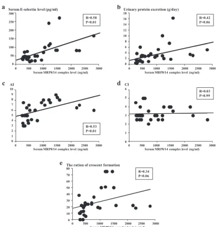
In addition, Kawasaki et al. evaluated whether serum MRP8/14 complex, which is a marker of monocyte and neutrophil activation, is associated with the clinical manifestations and pathological findings of HSPN35). Patients were divided into two groups based on serum MRP8/14 complex levels at renal biopsy. Group 1 consisted of 18 HSPN patients with lower than median (670 ng/ml) MRP8/14 complex levels, and Group 2 of 12 HSPN patients with greater than median levels. Clinical manifestations, laboratory findings and serum E-selectin levels, as a marker of vascular endothelial cell dysfunction, as well as histological and immuno- histochemical findings were investigated for both groups. Kawasaki et al. also measured MRP8/14 complex levels in disease control and healthy control children. Urinary protein excretions, serum MRP8/14 complex levels, and serum E-selectin lev- els were all higher in Group 2 than in Group 1 patients. Serum MRP8/14 complex levels were higher in HSPN patients than in the con- trols. Serum MRP8/14 complex levels were strongly associated with serum E-selectin levels (Figure 2). Pathological findings revealed that the Table 2. Comparison of immunofluorescence and light microscopic findings in both groups at the first and second
biopsies.
Pathological finding First biopsy Second biopsy
“Favorable” “Unfavorable” p “Favorable” “Unfavorable” p
IF findings IgG 0.3 (0.3) 0.6 (0.5) NS 0.6 (0.2) 1.8 (0.5) p<0.01
IgA 2.5 (0.6) 2.7 (0.7) NS 2.3 (0.5) 2.1 (0.8) NS
IgM 0.3 (0.4) 0.2 (0.3) NS 0.5 (0.2) 1.2 (0.4) p<0.05
C1q 0.1 (0.2) 0.1 (0.2) NS 0.0 (0.0) 0.1 (0.2) NS
C3 1.2 (0.8) 1.4 (0.9) NS 1.5 (0.7) 1.1 (1.2) NS
C4 0.2 (0.3) 0.0 (0.0) NS 0.0 (0.0) 0.0 (0.0) NS
Fib 2.5 (0.6) 1.8 (1.1) NS 1.8 (1.7) 2.2 (0.8) NS
Acute findings
Mesangial proliferation 2.0±0.5 1.8±1.1 NS 1.2±0.8 1.3±1.3 NS
Cellular crescent 1.0±1.0 2.1±1.7 p<0.01 0.4±0.4 0.5±0.9 NS
Macrophage infiltration in glomeruli 3.8±1.3 11.4±6.1 p<0.05 0.6±0.5 2.2±0.6 p<0.05 Interstitial mononuclear inflammation 0.9±0.6 2.2±0.5 p<0.01 0.9±0.5 1.8±0.8 NS
Acute index 7.7±3.4 17.5±9.4 p<0.01 3.1±2.2 5.8±3.6 p<0.01
Chronic findings
Glomerular sclerosis 0.6±0.4 1.4±0.9 p<0.05 0.6±0.4 1.4±0.9 p=0.07
Fibrous crescent 0.3±0.5 0.4±0.9 NS 0.7±0.7 1.8±1.1 p<0.05
Tubular atropy 0.5±0.7 1.5±0.5 p<0.05 0.7±0.4 2.1±0.9 p<0.05
Interstitial fibrosis 0.3±0.4 1.2±0.8 p<0.05 0.7±0.6 1.9±0.7 p<0.05
Chronicity index 1.7±2.0 4.5±3.1 p<0.01 2.7±2.1 7.2±3.6 p<0.01
NS=not significant
HSPN IN CHILDHOOD 21
proportions of patients with ISKDC grades III, IV and V in Group 2 were higher than in Group 1. Our findings suggest that serum MRP8/14 complex lev- els might be associated with the severity of renal injury and endothelial cell dysfunction in HSPN patients.
In a long-term follow-up of 78 patients with an average observation period of 23 years after the onset of HSPN in childhood, Goldstein et al. noted that 44% of patients presenting with nephrotic syn- drome or acute nephritis had persisting hyperten- sion or a progressive decline in GFR, whereas 82%
of those who presenting with hematuria alone were normal. Sixteen of 44 full-term pregnancies were also complicated by proteinuria and/or hypertension, even in the absence of active renal disease32). Subsequent deterioration in clinical status was observed in approximately 20 to 25% of patients, even after initial and apparently full recovery, indi- cating the need for the long-term follow-up of patients with HSP32).
TREATMENT OF HSPN
The extrarenal manifestations of HSPN are managed by appropriate symptomatic measures.
Severe skin lesions may require oral corticoste- roids13,37-39), which may also improve abdominal pain and protein-losing enteropathy37). Severe gastroin- testinal complications may occasionally require sur- gical intervention30).
As for the treatment of HSPN, there have been many reports dealing with the use of corticosteroids and multiple combined agents, including immuno- suppressive drugs.
(1) The efficacy of steroids
A majority of patients with HSPN have either no clinical renal involvement or demonstrate micro- hematuria, mild proteinuria, and normal renal func- tion. These patients do not require steroid therapy, and the disease is usually managed symptomati- cally. Niaudet et al.39) reported that MPT is effec-
33
300 250 200 150 100 50 0
0 500 1000 1500 2000 2500 3000 Serum E-selectin level (pg/ml)
Serum MRP8/14 complex level (ng/ml) R=0.58 P<0.01
0 500 1000 1500 2000 2500 3000
0 500 1000 1500 2000 2500 3000 0 500 1000 1500 2000 2500 3000 Serum MRP8/14 complex level (ng/ml)
Serum MRP8/14 complex level (ng/ml) Serum MRP8/14 complex level (ng/ml)
0 500 1000 1500 2000 2500 3000 Serum MRP8/14 complex level (ng/ml)
Urinary protein excretion (g/day) 18
16 14 12 10 8 6 4 2 0
R=0.42 P=0.06
AI
R=0.53 P<0.01 10
9 8 7 6 5 4 3 2 1 0
CI
R=0.03 P=0.99 6
5 4 3 2 1 0
80 70 60 50 40 30 20 10 0
The ration of crescent formation
R=0.34 P=0.06
a b
c d
e
Figure 2
Fig. 2. Relationships among serum E-selectin level, urinary protein excretion, AI, CI, the degree of crescent forma- tion, and serum MRP8/14 complex level.
a. Serum E-selectin level was associated with serum MRP8/14 complex level (R=0.58, p<0.01).
b. Urinary protein excretion tended to be associated with serum MRP8/14 complex level (R=0.42, p=0.06) c. AI was associated with serum MRP8/14 complex level (R=0.53, p<0.01).
d. CI was not associated with serum MRP8/14 complex level (R=0.03, p=0.99).
e. The degree of crescent formation tended to be weakly associated with serum MRP8/14 complex level (R=0.34, p=0.06), AI activity index, CI chronicity index.
tive for patients at the risk of progression of nephropathy. Another study appeared to confirm that the early administration of prednisone is useful in preventing the development of HSPN40). In an uncontrolled study, Kawasaki et al. performed a long-term observation of the clinical manifestations and prognosis of 56 patients undergoing methylpred- nisolone and urokinase pulse therapy (MUPT).
The mean urinary protein excretion after 6 months of treatment was found to have decreased signifi- cantly compared with the “pre-MUPT” level.
Hyper coagulant status “after the completion of uro- kinase pulse therapy” was also improved compared with the “pre-MUPT” status. First renal biopsies were performed in all patients, and second biopsies were performed in 27 patients. The activity index decreased significantly from 4.1 +/− 1.9 at the first biopsy to 2.5 +/− 1.7 at the second biopsy, whereas there were no differences in chronicity index between the first and second biopsies. No patients showed any renal insufficiency and the renal sur- vival rate was 100% for the decade. These results suggested that MUPT is effective for those patients at risk of progression of nephropathy, particularly if started early in the course of the disease before the crescents become fibrous. Urokinase is a plasmin- ogen activator derived from fresh human urine that first attracted attention as a therapeutic agent for thrombotic diseases such as cardiovascular diseases or cerebral thrombosis. The rationale for such treatment was as follows : 1) stronger defibrinating activity was observed with urokinase administration than with anti-coagulant drug administration, 2) spe- cific accumulations of urokinase were seen in the kidney and liver despite a very short turn-over rate, and 3) adverse effects were very rare, even when urokinase was administered for a long period41). (2) The efficacy of multiple-drug therapy
A prospective study of 12 patients with HSP who presented with rapidly progressive glomerulo- nephritis suggested benefits could be derived from intensive multiple-drug therapy42). Clinical improvement associated with combined corticoste- roid and azathioprine therapy was also suggested by another study of 21 children with severe HSPN43). Iijima et al. showed that multiple combined therapy including prednisolone, cyclophosphamide, heparin/
warfarin, and dipyridamole was effective in the treatment of histologically severe HSN44). In addi- tion, Flynn et al.45) reported that treating children with HSPN with high-dose corticosteroids plus oral cyclophosphamide is safe and, as in nephrotic syn-
drome, appears to significantly reduce proteinuria.
This study, however, was not a controlled study.
Therefore, Kawasaki et al. evaluated the efficacy of methylprednisolone and urokinase pulse therapy combined with cyclophosphamide for patients with HSPN. They studied 37 patients who had been diagnosed with HSPN of at least ISKDC grade IVb. Of them, 20 (Group A) were treated with methylprednisolone and urokinase pulse therapy, and 17 (Group B) were treated with methylpredniso- lone and urokinase pulse therapy combined with cyclophosphamide. Methylprednisolone and uroki- nase pulse therapy combined with cyclophospha- mide, but not methylprednisolone and urokinase pulse therapy alone, was found to significantly reduce urinary protein excretion (Figure 3) and pre- vent any increase in crescentic or sclerosed glomer- uli in HSPN patients with at least ISKDC grade IV HSPN. At the most recent follow-up, none of the patients treated with methylprednisolone and uroki- nase pulse therapy combined with cyclophospha- mide were observed to have persistent nephropathy or renal insufficiency46). Shin et al. suggest that CyA therapy is also effective in reducing proteinuria, which is a known risk factor for the development of renal insufficiency in HSPN and may lead to a regression in renal pathology in patients with nephrotic-range proteinuria47).
However, some problems, such as anemia, leu- kopenia, alopecia, hemorrhagic cystitis, carcinogene- sis, and hypogonadism, remain with the use of the above immunosuppressive drugs. Thus, Kawasaki et al. evaluated whether methylprednisolone and urokinase pulse therapy combined with mizoribine, which has only mild side effects and is compara- tively safe, (MUPM) was effective in children with severe HSPN. They studied 12 patients who had been diagnosed with HSPN of at least ISKDC grade III. All patients were treated with MUPM and clin- ical features, pathological findings, and prognosis were investigated prospectively. Ten patients (responders ; nine with ISKDC grade IIIb and one with grade IVb) were treated with MUPM, whereas MUPM was discontinued due to a lack of response in two patients (non-responders ; both with grade IVb). Among the responders, urinary protein excretion had decreased significantly from 99.7±37.8 to 25.9±33.4 mg/m2 per hour after 3 months of ther- apy. The acute index and tubulointerstitial scores decreased significantly from 5.8±1.5 and 3.8±0.6 at the first biopsy to 2.3±1.3 and 1.0±0.8 at the sec- ond biopsy, respectively. At the most recent fol- low-up, eight of the responders had normal urine,
HSPN IN CHILDHOOD 23
and two had minor urinary abnormalities. The non-responders demonstrated continued high levels of urinary protein excretion after 3 months of ther- apy, and MUPM was discontinued. These results suggests that MUPM is effective in ameliorating proteinuria and the histological severity of HSPN in patients with <50% crescents, but is not so effec- tive for HSPN in patients with >50% crescents48).
On the other hand, Ninchoji et al. retrospec- tively reported that patients with moderately severe HSPN (histological grade I-III and serum albumin [Alb] >2.5 g/dl), who were treated with angiotensin- converting enzyme inhibitors and/or angiotensin receptor blockers, showed resolution of proteinuria without renal dysfunction during the observation period (3.76±0.37 years). Thus, aggressive thera- pies, particularly combination therapies, are unnec- essary for moderate-severe HSPN49).
(3) Plasmapheresis
There have been a number of reports on plas- mapheresis (PP) for HSPN in childhood50,51). Hattori et al. retrospectively evaluated the clinical courses of nine children with a rapidly progressive type of HSPN who were treated with plasmaphere- sis (PP) as the sole therapy. All patients had nephrotic-range proteinuria (4.9+/−2.5 g/m2/d, mean+/−SD) and decreased glomerular filtration rate (GFR) (46.5+/−9.5 mL/min/1.73 m2) at the time of the initiation of PP. Biopsy specimens taken before PP showed large crescents involving more than 50% of the glomerular circumference in 56.8+/−6.9% of the glomeruli examined. The mean interval between disease onset and initiation of PP was 39.1+/−22.1 days. The PP regimen consisted of thrice-weekly treatment for 2 weeks, then weekly treatment for 6 weeks. All patients responded promptly to PP with improvement in renal function, reduction of proteinuria, and subsid- ence of purpuric rash and abdominal pain. Six of nine patients showed further improvements without any other treatment ; four recovered completely, and two had only microscopic hematuria at the latest observation (follow-up period, 9.6+/−4.3 years).
The remaining three patients showed a rebound increase in proteinuria after the completion of PP ; two of whom progressed to end-stage renal failure at 14.1 years and 1.8 years, respectively, after dis- ease onset. These results suggests that PP as the sole therapy is effective in improving the prognosis of patients with rapidly progressive HSP nephritis, particularly if instituted early in the course of the disease50).
On the other hand, Kawasaki et al. reviewed the cases of six Japanese children with rapidly pro- gressive HSPN who received multiple drug therapy combined with PP. After five courses of PP, multi- ple drug therapy, including methylprednisolone and urokinase pulse therapy, oral prednisolone, cyclo- phophamide, dipyridamole, and warfarin, was given.
At presentation, urine protein excretion and histo- logical indices of the mean activity and chronicity were 245+/−101 mg/m2 per hour, 6.6+/−1.2, and 1.5+/−1.3, respectively. After 6 months of ther- apy, urinary protein excretion had decreased signifi- cantly (P<0.001). The activity index was also decreased significantly at the second renal biopsy performed at a mean interval of 4.3 months after the first biopsy (2.8+/−1.4, P<0.05), whereas there was no change in the chronicity index. At the most recent observation, all patients showed clinical improvement. Two patients had normal urine, three had proteinuria of <20 mg/m2 per hour, one had proteinuria of >20 mg/m2 per hour, and none showed any renal insufficiency. Although this case series was examined without controls, this treat- ment protocol may be of benefit to children with rap- idly progressive HSPN51).
The benefits of the abovementioned treatments for treating HSPN deserve to be assessed further in larger randomized controlled trials.
(4) Other types of treatment
The use of intravenous immunoglobulin (IVIg) for the treatment of HSP is anecdotal and has been advocated as effective for abdominal pain and other gastrointestinal symptoms52).
Some studies have reported that tonsillectomy was effective for patients with severe HSPN53-55). Kawasaki et al. reported an 11-year-old boy with HSPN accompanied by recurrent purpura and per- sistent nephropathy despite conventional therapy such as prednisolone, methylprednisolone pulse therapy and mizoribine. The patient was treated with tonsillectomy plus methylprednisolone pulse therapy. This treatment decreased proteinuria, induced the disappearance of microscopic hematuria, and improved renal pathological findings. This case suggests that tonsillectomy plus methylprednisolone pulse is an effective and useful therapy for some children with recurrent purpura and persistent nephropathy54). Furthermore, Ohara et al. reported a 13-year-old girl with HSPN of ISKDC grade VI and persistent nephrotic syndrome despite receiving conventional therapy such as prednisolone, methyl- prednisolone and urokinase pulse therapy and plas-
mapheresis (PP). The patient was treated with tonsillectomy, which subsequently decreased pro- teinuria, induced the disappearance of microscopic hematuria, and improved renal pathological findings.
A regimen of methylprednisolone and urokinase pulse therapy plus PP with tonsillectomy may be an effective and useful therapy for some children with severe HSPN children of ISKDC grade VI and per- sistent nephrotic syndrome55).
(5) Transplantation
HSPN may recur after transplantation, and rates of recurrence are increased in recipients of liv- ing-related transplantations56,57). Meulders et al.
reported the actuarial risks for renal recurrence and graft loss due to recurrence to be 35 and 11%, respectively, at 5 years after transplantation56). Recurrence appeared to be associated with shorter duration of the original episode of disease, occur- rence despite delays of more than 1 year between the disappearance of purpura and transplantation, and was not prevented by a triple immunosuppres- sive regimen that included CyA.
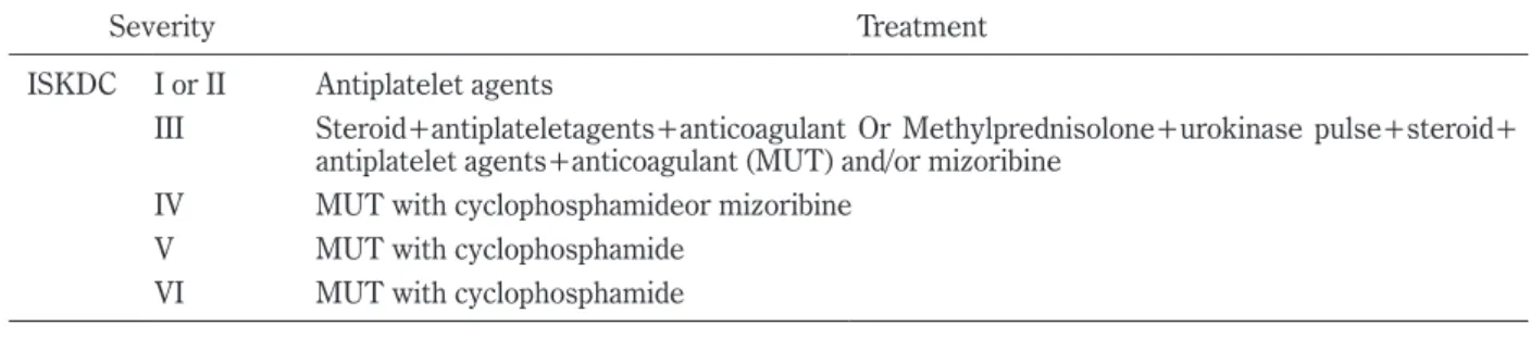
Finally, our recommendations for the treatment of HSPN in our hospital are shown in Table 3. We try to perform renal biopsy at 1-2 months after the onset of HSPN for patients with a urinary protein excretion of more than 0.5 g/day, and provide aggres- sive therapy according to the severity of pathological lesions. We believe these treatments are effective in improving the prognosis for HSPN.
CONCLUSION
We have reviewed the pathogenesis, clinical manifestations, prognostic factors of and treatment for HSPN, including multiple drug combination ther- apies. Further detailed investigation of HSPN pathogenesis and treatment is necessary to identify
the most appropriate treatment.
REFERENCES
1. Mills JA, Michel BA, Bloch DA, Calabrese LH, Hunder GG, Arend WP, et al. The American College of Rheumatology 1990 criteria for the clas- sification of Henoch-Schonlein purpura. Arthritis Rheum, 33(8) : 1114-1121, 1990.
2. Saulsbury FT. Epidermiology of Henoch- Schonlein purpura. Cleve Clin J Med, 69: 187- 189, 2002.
3. Bunchman TE, Mauer SM, Sibley RK, Vernier RL. Anaphylactoid purpura : characteristics of 16 patients who progressed to renal failure. Pediatr Nephrol, 2: 393-397, 1988.
4. Counahan R, Winterborn MH, White RH, Heaton JM, Meadow SR, Bluett NH, Swetschin H, Cameron JS, Chantler C. Prognosis of Henoch- Schönlein nephritis in children. Br Med J, 2: 11- 14, 1977.
5. Yoshikawa N, Ito H, Yoshiya K, Nakahara C, Yoshiara S, Hasegawa O, Matsuyama S, Matsuo T. Henoch-Schoenlein nephritis and IgA nephropathy in children : a comparison of clinical course. Clin Nephrol, 27: 233-237, 1987.
6. Gardner-Medwin JM, Dolezalova P, Cummins C, Southwood TR. Incidence of Henoch-Schonlein purpura, Kawasaki disease, and rare vasculitides in children of different ethnic origins. Lancet, 360: 1197-1202, 2002.
7. Stewart M, Savage JM, Bell B, McCord B. Long term renal prognosis of Henoch-Schönlein purpura in unselected childhood population. Eur J Pediatr, 147: 113-115, 1988.
8. Nielsen HE. Epidemiology of Schonlein-Henoch Purpura. Acu Pathol Scand, 77: 125-131, 1988.
9. Kawasaki Y, Suyama K, Yugeta E, Katayose M, Suzuki S, Sakuma H, et al. The incidence and severity of Henoch-Schönlein purpura nephritis over a 22-year period in Fukushima Prefecture, Japan. Int Urol Nephrol, 42: 1023-1029, 2010.
Table 3. Treatment of HSPN in our hospital.
Severity Treatment
ISKDC I or II Antiplatelet agents
III Steroid+antiplateletagents+anticoagulant Or Methylprednisolone+urokinase pulse+steroid+
antiplatelet agents+anticoagulant (MUT) and/or mizoribine IV MUT with cyclophosphamideor mizoribine
V MUT with cyclophosphamide VI MUT with cyclophosphamide
Patients with rapidly progressive HSPN MUCT with plasmapheresis Patients with ISKDC III a presenting as nephroticsyndrome MUT
HSPN IN CHILDHOOD 25
10. Vogler C, Eliason SC, Wood EG. Glomerular membranopathy in children with IgA nephropathy and Henoch-Schoenlein purpura. Pediatr Dev Pathol, 2: 227-235, 1999.
11. Kerr MA. The structure and function of human IgA. Biochem J, 271: 285-296, 1990.
12. Yoo EM, Morrison SL. IgA : an immune glyco- protein. Clin Immunol, 116: 3-10, 2005.
13. Jones CL, Powell HR, Kincaid-Smith P, Roberton DM. Polymeric IgA and immune complex con- centrations in IgA-related renal disease. Kidney Int, 38: 323-331, 1990.
14. Allen AC, Willis FR, Beattie TJ, Feehally J.
Abnormal IgA glycosylation in Henoch-Schoenlein purpura restricted to patients with clinical nephri- tis. Nephrol Dial Transplant, 13: 930-934, 1998.
15. Egmond M, Damen CA, Spriel AB, Vidarsson G, Garderen E, Winkel JGJ. IgA and IgA Fc receptor.
TRENDS in immunology, 22: 205-211, 2001.
16. Moja P, Quesnel A, Resseguier V, Lambert C, Freycon F, Berthoux F, et al. Is there IgA from gut mucosal origin in the serum of children with Henoch-Schoenlein purpura? Clin Immunol Immunopathol, 86: 290-297, 1998.
17. Silva FG. IgA nephropathy and Henoch- Schoenlein Syndrome. In Jennette JC, Olson JL, Schwartz MM, Silva FG(eds) : Heptinstall’s Pathology of the Kidney. 15th ed. Lippincott- Raven, Philadelphia, 479-540, 1998.
18. Allen A, Harper S, Feehally J. Origin and struc- ture of pathogenic IgA in IgA nephropathy.
Biochem Soc Trans, 25: 486-490, 1997.
19. Jones CL, Powell HR, Kincaid-Smith P, Roberton DM. Polymeric IgA and immune complex con- centrations in IgA-related renal disease. Kidney Int, 38: 323-331, 1990.
20. Coppo R, Basolo B, Bulzomi MR, Roccatello D, Carbonara AO, Barbiano di Belgiojoso G, et al.
IgA1 and IgA2 immune complexes in primary IgA nephropathy and Henoch-Schoenlein purpura nephritis. Clin Exp Immunol, 57: 583-590, 1984.
21. Levinsky RJ, Barratt TM. IgA immune com- plexes in Henoch-Schoenlein purpura. Lancet, 24: 1100-1103, 1979.
22. Leu KK, Wyatt RJ, Moldoveanu Z, Tomana M, Julian BA, Hogg RJ, et al. Serum levels of galac- tose-deficient IgA in children with IgA nephropa- thy and Henoch-Schoenlein purpura. Pediatr Nephrol, 22: 2067-2072, 2007.
23. Oortwijn BD, Eijgenraam JW, Rastaldi MP, Roos A, Daha MR, van Kooten C. The role of secretary IgA and complement in IgA nephropathy. Semin Nephrol, 28: 58-65, 2008.
24. Endo M, Ohi H, Ohsawa I, Fujita T, Matsushita M.
Complement activation through the lectin pathway in patients with Henoch-Schönlein purpura nephri-
tis. Am J Kidney Dis, 35: 401-407, 2000.
25. Hisano S, Matsushita M, Fujita T, Iwasaki H.
Activation of the lectin complement pathway in Henoch-Schönlein purpura nephritis. Am J Kidney Dis, 45: 295-302, 2005.
26. Moura IC, Benhamou M, Launay P, Vrtovsnik F, Blank U, Monteiro RC. The glomerular response to IgA deposition in IgA nephropathy. Semin Nephrol, 28: 88-95, 2008.
27. Kawsaki Y, Suzuki J, Nemoto K, Nozawa R, Suzuki S, Suzuki H. Clinical and pathological features of children with Henoch-Schoenlein purpura nephri- tis : risk factors associated with poor prognosis.
Clin Nephrol, 60: 153-160, 2003.
28. Fujieda M, Oishi N, Naruse K, Hashizume M, Nishiya K, Kurashige T, Ito K. Soluble thrombo- modulin and antibodies to bovine glomerular endo- thelial cells in patients with Henoch-Schoenlein purpura. Arch Dis Child, 78: 240-244, 1998.
29. Kawasaki Y, Suzuki J, Nozawa R, Sakai N, Tannji M, Isome M, et al. FB21, a monoclonal antibody that reacts with a sialic-acid-dependent carbohydrate epitope, is a marker for glomerular endothelial cell injury. Am J Kidney Dis, 44: 239-249, 2004.
30. Kawasaki Y, Suzuki H, Henoch-Schoenlein Nephritis. In Geary DF, Schaefer F (eds) : Com- pre hensive Pediatric Nephrology, Mosby, Elsevier.
pp. 343-351, 2008.
31. Algoet C, Proesmans W. Renal biopsy 2-9 years after Henoch Schönlein purpura. Pediatr Nephrol, 18: 471-473, 2003.
32. Goldstein AR, White RH, Akuse R, Chantler C.
Long-term follow-up of childhood Henoch-Schoen- lein nephritis. Lancet, 339: 280-282, 1992.
33. Meadow SR, Glasgow EF, White RH, Moncrieff MW, Cameron JS, Ogg CS. Schoenlein-Henoch nephritis. Q J Med, 41: 241-245, 1972.
34. Kawasaki Y, Imaizumi T, Matsuura H, Ohara S, Takano K, Suyama K, et al. Renal expression of alpha-smooth muscle actin and c-Met in children with Henoch-Schönlein purpura nephritis.
Pediatr Nephrol, 23: 913-919, 2008.
36. Kawasaki Y, Ohara S, Abe Y, Watanabe M, Suyama K, Sato M, Hashimoto K, Hosoya M. The role of serum myeloid-related protein 8/14 complex in Henoch-Schoenlein purpura nephritis, Pediatr Nephrol, 27: 65-71, 2012.
37. Reif S, Jain A Santiago J Rossi T. Protein-losing enteropathy as a manifestation of Henoch-Schoen- lein purpura. Acuta Paediatr Scand, 80: 482-485, 1991.
38. Cull DL, Rosario V, Lally KP, Ratner I, Mahour GH.
Surgical implications of Henoch-Schoenlein pur- pura. J Pediatr Surg, 25: 741-743, 1990.
39. Niaudet P, Habib R. Methylprednisolone pulse therapy in the treatment of severe forms of
Schoenlein-Henoch purpura nephritis. Pediatr Nephrol, 12: 238-243, 1998.
40. Mollica F, Li Volti S, Garozzo R, Russo G. Effec- tive ness of early prednisone treatment in prevent- ing the development of nephropathy in anaphylac- toid purpura. Eur J Pediatr, 151: 140-144, 1992.
41. Kawasaki Y, Suzuki J, Nozawa R, Suzuki S, Suzuki H. Efficacy of methylprednisolone and urokinase pulse therapy for severe Henoch-Schoenlein nephritis. Pediatrics, 111: 785-789, 2003.
42. Oner A, Tinaztepe K, Erdogan O. The effect of triple therapy on rapidly progressive type of Henoch-Schoenlein purpura nephritis to cortico- steroid and azathioprine therapy. Clin Nephrol, 49: 9-14, 1998.
43. Bergstein J, Leiser J, Andreoli SP. Response of crescentic Henoch-Schoenlein purpura nephritis to corticosteroid and azathioprine therapy. Clin Nephrol, 49: 9-14, 1998.
44. Iijima K, Ito-Kariya S, Nakamura H, Yoshikawa N.
Multiple combined therapy for severe Henoch- Schoenlein nephritis in children. Pediatr Nephrol, 12: 244-248, 1998.
45. Flynn JT, Smoyer WE, Bunchman TE, Kershaw DB, Sedman A. Treatment of Henoch-Schoenlein purpura Glomerulonephritis in Children with High-Dose Corticosteroids plus Oral Cyclo phos- phamide. Am J Nephrol, 21: 128-133, 2001.
46. Kawasaki Y, Suzuki J, Suzuki H. Efficacy of meth- ylprednisolone and urokinase pulse therapy com- bined with or without cyclophosphamide in severe Henoch-Schoenlein nephritis : a clinical and histo- pathological study. Nephrol Dial Transplant, 19: 858-864, 2004.
47. Shin JI, Park JM, Shin YH, Kim JH, Lee JS, Jeong HJ. Henoch-Schönlein purpura nephritis with nephrotic-range proteinuria : histological regres- sion possibly associated with cyclosporin A and steroid treatment. Scand J Rheumatol, 34: 392- 395, 2005.
48. Kawasaki Y, Suyama K, Hashimoto K, Hosoya M.
Methylprednisolone pulse plus mizoribine in chil- dren with Henoch-Schoenlein purpura nephritis.
Clin Rheumatol, 30: 529-535, 2011
49. Ninchoji T, Kaito H, Nozu K, Hashimura Y, Kanda K, Kamioka I, Shima Y, Hamahira K, Nakanishi K, Tanaka R, Yoshikawa N, Iijima K, Matsuo M.
Treat ment strategies for Henoch-Schoenlein pur- pura nephritis by histological and clinical sever- ity. Pediatr Nephrol, 26: 563-569, 2011.
50. Hattori M, Ito K, Konomoto T, Kawaguchi H, Yoshioka T, Khono M. Plasmapheresis as the sole therapy for rapidly progressive Schoenlein-Henoch purpura nephritis in children. Am J Kidney Dis, 33: 427-433, 1999.
51. Kawasaki Y, Suzuki J, Murai M, Takahashi A, Isome M, Nozawa R, et al. Plasmapheresis ther- apy for rapidly progressive Henoch-Schoenlein nephritis. Pediatr Nephrol, 19: 920-923, 2004.
52. Heldrich FJ, Minkin S, Gatdula CI. Intravenous immunoglobulin in Henoch-Schoenlein purpura : A case study. Md Med J, 42: 577-579, 1993.
53. Suguyama H, Watanabe N, Onoda T, Kikumoto Y, Yamamoto M, Maeta M, et al. Successful treat- ment of progressive Henoch-Schoenlein purpura nephritis with tonsillectomy and steroid pulse therapy. Intern Med, 44: 611-615, 2005.
54. Kawasaki Y, Suyama K, Matsumoto A, Takano K, Hashimoto K, Suzuki S, et al. Efficacy of tonsil- lectomy plus methylprednisolone pulse therapy for a child with Henoch-Schoenlein purpura nephritis.
Tohoku J Exp Med, 211: 291-295, 2007.
55. Ohara S, Kawasaki Y, Matsuura H, Oikawa T, Suyama K, Hosoya M. Successful therapy with tonsillectomy for severe ISKDC grade IV Henoch- Schoenlein purpura nephritis and persistent nephrotic syndrome. Clin Exp Nephrol, 15: 749- 753, 2011.
56. Meulders Q, Pirson Y, Cosyns JP, Squifflet JP, van Ypersele de Strihou C. Course of Henoch- Schoenlein nephritis after renal transplantation : Report on ten patients and review of the literature.
Transplantation, 58: 1179-1186, 1994.
57. Ramos EL. Recurrent diseases in the renal allo- graft. J Am Soc Nephrol, 2: 109-121, 1991.