Epidemiological studies on infectious diseases among carnivores
Laboratory of Veterinary Microbiology
The United Graduate School of Veterinary Science
Yamaguchi University
Presented by
Shohei MINAMI
1. General introduction 1
1.1. Ferret coronavirus 2
1.1.1. History 2
1.1.2. Virus properties 2
1.1.3. Epidemiology 3
1.1.4. Diseases and pathogenesis 4
1.1.5. Diagnosis 5
1.1.6. Prevention and control 5
1.2. Pseudorabies virus 7
1.2.1. History 7
1.2.2. Virus properties 7
1.2.3. Epidemiology 9
1.2.4. Diseases and pathogenesis 10
1.2.5. Diagnosis 12
1.2.6. Prevention and control 12
2. CHAPTER 1 Establishment of serological test to detect antibody against ferret coronavirus. 15
2.1. Abstract 16
2.2. Introduction 17
2.3. Materials and methods 19
2.3.1. Samples from domestic ferrets 19
2.3.2. Amplification of N genes 19
2.3.3. Construction of expression plasmids 19
2.3.4. Expression and purification of glutathione-S transferase (GST)-fusion proteins, 20
2.3.5. SDS-PAGE analysis of recombinant proteins 21
2.3.6. Quantification of recombinant proteins 21
2.3.7. ELISA 21 2.3.8. Immunoblot analysis 22 2.3.9. Sequence analysis 22 2.3.10. Phylogenetic analysis 23 2.3.11. Statistical analysis 23 2.4. Results 24
2.4.1. Antigenic comparison of GST fused recombinant proteins, N (1-179) and
GST-N (180-374) 24
(180-2.4.3. Seroprevalence of FRCoV infection in ferrets in Japan 25
2.5. Discussion 26
2.6. Figure legends 27
2.7. Figures and tables 28
3. CHAPTER 2 Detection of novel ferret coronaviruses and evidence of recombination among ferret
coronaviruses. 32
3.1. Abstract 33
3.2. Introduction 34
3.3. Materials and methods 35
3.3.1. Reverse transcription-polymerase chain reaction (RT-PCR) 35
3.3.2. Sequence analysis 35
3.3.3. Phylogenetic analysis and homology search 36
3.4. Results 37
3.4.1. Detection of FRCoV RdRp gene from ferrets in Japan 37
3.4.2. Phylogenetic and sequence analyses 37
3.4.3. Homology search 38
3.5. Discussion 39
3.6. Figure legends 40
3.7. Figures and tables 41
4. CHAPTER 3 Interspecies transmission of pseudorabies virus among wild animals. 45
4.1. Abstract 46
4.2. Introduction 47
4.3. Materials and methods 49
4.3.1. Samples from wild animals 49
4.3.2. Cells 49 4.3.3. Viruses 49 4.3.4. Virus isolation 50 4.3.5. Nested PCR 50 4.3.6. Virus-neutralization (VN) test 50 4.3.7. Sequence analysis 51 4.3.8. Phylogenetic analysis 51 4.3.9. Accession numbers 52 4.4. Results 53
4.4.2. Nested PCR for detection of PRV 53
4.4.3. Seroprevalence of PRV 53
4.4.4. Complete genome of PRV RC1 strain 53
4.4.5. Phylogenetic analysis of PRV 54
4.5. Discussion 55
4.6. Figure legends 56
4.7. Figures and tables 57
5. General conclusion 61
6. Acknowledgments 64
7. References 65
1.1. Ferret coronavirus
1.1.1. History
Ferret coronavirus (FRCoV) is a novel coronavirus which causes various diseases to ferrets (Mustela putorius furo). In 1993, FRCoV was first reported as a causative agent of epizootic catarrhal enteritis (ECE) in the U.S.A. and coronavirus-like particles were detected from the feces and jejunum of the ferret (Williams et al., 2000). In 2006, a novel coronavirus was detected by reverse transcription-polymerase chain reaction (RT-PCR) from feces of a ferret and designated as ferret enteric coronavirus (FRECV) due to possessing the similarity to the other coronaviruses, such as feline coronavirus (FCoV) and canine coronavirus (CCoV) (Wise et al., 2006). Since 2002, ferrets with feline infectious peritonitis (FIP)-like symptoms have been reported and antigen or partial gene of coronavirus was detected from them (Martinez et al., 2006; Garner et al., 2008; Martinez et al., 2008; Perpiñán and López, 2008). In 2010, the antigen of coronavirus was also detected from ferrets with FIP-like symptoms in Japan by immunohistochemistry using anti-FIP antibody (Michimae et al., 2010). This virus was designated as ferret systemic coronavirus (FRSCV) and was considered as FRCoV different from FRECV (Wise et al., 2010). Finally, the complete genome has been determined in 2017 (Li et al., 2017b). However, pathogenicity of FRCoV has been still unknown.
1.1.2. Virus properties
FRCoV is a single-strand positive-strain RNA virus. FRCoV belongs to order Nidovirales, suborder Cornidovirineae, family Coronaviridae, subfamily Orthocoronavirinae, genus
Alphacoronavirus (Walker et al., 2019). The size of viral particle is from 100 to 140 nm. FRCoV has
predicted 9 open reading frames (ORFs), 1a, 1b, spike (S), envelope (E), membrane (M), nucleocapsid (N), 3c, 3x and 7b (Li et al., 2017b). The pseudoknot tertiary structure which is commonly found
among Coronaviridae (Brierley et al., 1987; Brierley et al., 1989; Thiel et al., 2003) exists between ORF1a and 1b. S, E, M and N proteins are structural proteins and 3c, 3x and 7b are nonstructural proteins. ORF1a and 1b encode several proteins including RNA-dependent RNA polymerase (RdRp) (Le Poder, 2011). These genes are initially translated when the viral genome enters the host cells and the translated proteins were two polyproteins, pp1a and pp1ab. Subsequently, these two polyproteins are cleaved by viral proteases and cleaved proteins possess various functions such as interferon antagonist (Huang et al., 2011), protease (Neuman et al., 2008), RNA-binding (Egloff et al., 2004), RdRp (Le Poder, 2011), endoribonuclease (Bhardwaj et al., 2006), exonuclease (Chen et al., 2009), methyltransferase (Chen et al., 2009) and so on. S protein is the receptor binding glycoprotein (Delmas and Laude, 1990) and induces cell-cell fusion (Collins et al., 1982). However, the receptor of FRCoV is still unknown. E protein is the component of viral envelope (Godet et al., 1992; Schoeman and Fielding, 2019). M protein is the supportive protein and contribute to keep the shape of viral particle with E protein (Sturman, 1977). M protein interacts with S protein to conserve the structure of S protein in ER-Golgi intermediate compartment (ERGIC) (Fehr and Perlman, 2015). N protein is the phosphoprotein which binds to the viral genome and composes the nucleocapsid (King and Brian, 1982). 3c, 3x and 7b proteins are called accessary proteins whose function is still unclear. 3c protein may be related to the virulence and viral replication (Hora et al., 2016; Hsieh et al., 2013; Pedersen et al., 2009). 7b proteins may prevent the host immune response (Lemmermeyer et al., 2016). The function of 3x protein is unclear except translation from the genome of CCoV and FRCoV (Horsburgh et al., 1992; Li et al., 2017b). Moreover, 3x protein of FRCoV possesses only 23.9 % of similarity to that of CCoV (Wise et al., 2006). The isolation of FRCoV has never been reported until now. Therefore, the functions of these proteins, especially nonstructural proteins, are still unclear.
FRCoV was detected in the U.S.A., Peru, Europe and Japan by immunohistochemistry or RT-PCR. The epidemiological studies of FRCoV were performed in the Netherlands and Japan (Provacia et al., 2011; Smits et al., 2013; Terada et al., 2014b), resulting that 42%-72% of ferrets were positive for anti-FRCoV antibody or FRCoV genes. The case reports were also reported in Peru (Lescano et al., 2015), Japan (Fujii et al., 2015; Michimae et al., 2010), France (Gnirs et al., 2016), the U.S.A. (Lindemann et al., 2016), Spain (Dominguez et al., 2011) and United Kingdom (Graham et al., 2012). However, these case reports were described about FIP-like systemic diseases. The case reports on ECE were not so many. The epidemiological study have not been conducted. FRCoV must spread among pet ferrets in the world. In the U.S.A., the population of domestic ferret is increasing (Jurek, 1998). Further epidemiological studies of FRCoV will be required.
1.1.4. Diseases and pathogenesis
FRCoV infection is divided into two biotypes of diseases, ECE and FIP-like systemic disease. ECE is caused by FRECV infection. The affected ferrets initially showed lethargy, inappetence and vomiting and subsequently excreted high mucus containing bright green diarrhea. This main symptom of ECE was mild enteritis and its mortality was low (<5%) (Williams et al., 2000). Another biotype, systemic disease was caused by FRSCV infection. This biotype was characterized by various diseases, such as lethargy, inappetence, diarrhea, abdominal mass and neurologic disorder. Especially, the mass was detected in various organs, such as lymph node, liver, kidney, spleen and lung (Garner et al., 2008; Martínez et al., 2006; Perpiñán et al., 2008). Recently, the antigens of FRCoV were detected from pancreas of a ferret and FRCoV may also cause pancreatitis (Wills et al., 2018). FRSCV infection was severe and its mortality was high. These symptoms caused by FRSCV infection are similar to the dry type of FIP. FCoV infection is also divided into two biotypes of diseases, feline enteric coronavirus (FECV) infection and FIP. FIPV was emerged from FECV by genetic mutation or
recombination. In the case of emerging FIPV by genetic mutation, S and 3c gene are significantly mutated (Hora et al., 2016; Licitra et al., 2013; Pedersen et al., 2009). The mutations of S and the deletion of 3c genes were also found in FRSCV (Wise et al., 2010). These genes may be closely related to the pathogenicity of FRCoVs. Type II FCoV is emerged by the recombination between type I FCoV and type II CCoV (Terada et al., 2014a). A novel FRCoV was generated by resembling recombination event (Lamers et al., 2016). However, the pathogenicity of FRCoV has been still unclear.
1.1.5. Diagnosis
For diagnosis, viral gene detection from feces of ferret is recommended FRCoV-specific
primers: FRCoV RdRp- -GTT GGT TGC TGC ACA CAT AG- -R1
-GGA GAA GTG CTT ACG CAA ATA- for detection of FRCoV (Terada et al., 2014b). The genotyping using primers targeting S gene may not be related with pathotype and severity of diseases. Since antibodies against FIPV cross-react to the antigens of FRCoV, immunohistochemical staining using anti-FIPV antibody is one of the methods for diagnosis of FRCoV (Autieri et al., 2015; Fujii et al., 2015; Doria-Torra et al., 2016; Gnirs et al., 2016; Graham et al., 2012; Lescano et al., 2015; Lindemann et al., 2016; Wills et al., 2018; Wise et al., 2006). CoVs including FRCoV were easily identified and characterized by electron microscopy because of its characteristic particle like a corona (Drosten et al., 2003; Kheyami et al., 2010; Williams et al., 2000). Unfortunately, although many researchers tried to isolate FRCoV, the virus have never been isolated yet. Virus isolation and the subsequent identification are not suitable method for the diagnosis of FRCoV. High mucus containing bright green diarrhea and the abdominal mass are known as the characteristic symptoms of ECE and FIP-like systemic disease, respectively.
The treatment specific for FRCoV has not been established yet. The antiviral drug which inhibits 3c protein is developing (Perera et al., 2018). However, it has not been available in clinical use. Treatment of FRCoV infection is conducted by supportive therapy if the ferret shows symptoms. Monitoring the condition of the host ferret and treatment of other basic disease will contribute to improve FRCoV infection. It is recommended that the FRCoV-positive ferret should be separated from the FRCoV-negative ferrets to prevent the transmission via fecal-oral route. Since FRCoV is the enveloped virus, various disinfectants efficiently reduce the infectivity of FRCoV.
1.2. Pseudorabies virus
1.2.1. History
In 1902, Dr. Aladar Aujeszky in Hungary first described the disease AD) caused by pseudorabies virus
infection (Aujeszky, 1902). This disease was named as pseudorabies due to the clinical resemblance to rabies (Shope, 1931). In the 1970s, more than 2,000 cases of PR were recorded in America (Lee and Wilson, 1979). Since the early 1980s, pseudorabies virus which is the causative agent of PR might have been spread all over the world (Müller et al., 2011). Government of each countries tried to control PRV infection due to serious economic losses in pig farms. The attenuated live vaccines with deletion of glycoprotein E (gE) and thymidine kinase (TK) genes were commonly used in the program of vaccination with DIVA (differentiating infected from vaccinated animals) strategy (Freuling et al., 2017). The eradication of PRV achieved in several countries (Hahn et al., 2010; MacDiarmid, 2000; Müller et al., 2003; Müller et al., 2011). In Japan, PRV infection among domestic pigs was firstly reported in 1981. However, the first invasion of PRV was expected before 1979 by the result of epidemiological studies (Shimizu, 1983). Until now, Japanese government has attempted to complete the eradication of PRV. The eradication of PRV have achieved in almost prefectures. However, the eradication of PRV have not completely achieved yet (Ministry of Agriculture, Forestry and Fisheries, 2019). In 2019, dogs bitten by a wild boar were dead by PRV infection in Miyazaki prefecture in Japan (Minamiguchi et al., 2019). Miyazaki prefecture is one of the PRV-free prefectures among domestic pigs. To achieve the complete eradication of PRV, the prevalence of PRV among wild boars must be monitored and the new counterplot against the transmission of PRV is required.
PRV is a double-strand DNA virus. PRV belongs to order Herpesvirales, family
Herpesviridae, subfamily Alphaherpesvirinae, genus Varicellovirus (Carstens and Ball, 2009). The
particle of PRV is composed of the genome, capsid, tegument and the envelope and its size is from 120 to 200 nm. The genome of PRV is composed of approximately 150,000 nucleotides and encodes 70 genes. The genome of PRV contains high concentration of G + C (73%) and is divided into two unique regions, the unique long (UL) and the unique short (US). US genes are surrounded by the inverted regions, internal repeat sequence (IRS) and terminal repeat sequence (TRS). The capsid is composed of 6 proteins, UL6, UL18, UL19, UL25, UL35 and UL38 proteins. The tegument which functions as a matrix between the capsid and the envelope is composed of 14 proteins, UL11, UL13, UL16, UL21, UL36, UL37, UL41, UL46, UL47, UL48, UL49, UL51, US2 and US3 proteins. The envelope is composed of two groups of the proteins, the glycoproteins and the membrane proteins. The glycoproteins which include the receptor binding proteins are composed by UL1, UL10, UL22, UL27, UL44, UL49.5, UL53, US4, US6, US7 and US8 proteins, generally called as gL, gM, gH, gB, gC, gN, gK, gG, gD, gI and gE, respectively. The other envelope proteins, UL20 protein and possibly UL24, UL43 and US9 proteins function as membrane proteins (Klupp et al., 2004; Mettenleiter, 1991; Mettenleiter 2000; Pomeranz et al., 2005). A few genes, such as US1 and the large latency transcript (LLT), are spliced (Pomeranz et al., 2005). MicroRNAs (miRNAs) are small RNA which regulates the gene expression. Some viruses produce miRNA to gain the benefit in their replication (Umbach and Cullen, 2009). Herpesvirus also produce miRNAs and the production of miRNA is related to the latent infection (Jurak et al., 2011; Umbach et al., 2008). Five miRNAs of PRV have already been reported (Anselmo et al., 2011). However, the target genes of PRV miRNAs are still unclear. The glycoproteins are related to the entry of PRV. PRV gC binding to the cellular heparin sulfate proteoglycans mediates gD binding to nectin 1, nectin 2 or CD155 (Mettenleiter, 2002a; Spear et al., 2000). Most herpesviruses infect in the host specific manner. However, since PRV infects various mammals, PRV has three kinds
of receptors (Nauwynck et al., 2007). After gD binding to the receptor, gB, gD and gH/gL complex mediate to fuse between cellular membrane and viral envelop (Klupp et al., 1997, Rauh and Mettenleiter 1991). Once the genome of PRV enters the host cells, the genes are transcribed and the viral proteins are expressed by the translation of viral mRNA. The models of herpesvirus assembly and egress are well studied (Mettenleiter et al., 2009). Firstly, the capsid is assembled and encloses the genome in the host nucleus. The capsid is enveloped with the host inner nuclear membrane (INM). Subsequently, the primary envelop fused with the host outer nuclear membrane (ONM) and the capsid is released into the cytosol (Mettenleiter et al., 2013). The released capsid surrounded by the trans-Golgi vesicles including various viral proteins (Fuchs et al., 1997). Thus the capsid is enveloped by the secondary envelop in the cytosol (Owen et al., 2015). Viral particles bud from the host cell surface by exocytosis or spread to the neighbor cells at cell junctions (Johnson and Baines, 2011; Mettenleiter, 2002b; Owen et al., 2015; Smith, 2017). gB, gK and gH/gL complex are necessary for this cell-to-cell spread. Although gM and gE/gI complex modulate the cell-to-cell spread, they are not necessary (Mettenleiter, 2000).
1.2.3. Epidemiology
PRV infection among domestic pigs are monitored by many countries. However, the natural host of PRV is not only domestic pigs, but also wild boars. A number of epidemiological studies of PRV infection among wild boars are reported in Croatia, Czech Republic, France, Germany, Italy, Russia, Slovenia, South Korea, Spain, Switzerland, the U.S.A. and Japan (Albina et al., 2000; Chiari et al., 2015; Corn et al., 2009; Gortázar et al., 2002; Gresham et al., 2002; Jeoung et al., 2015; Köppel et al., 2007; Leuenberger et al., 2007; Mahmoud et al., 2011; Müller et al., 2011; Müller et al., 1998; Pannwitz et al., 2012; Pirtle et al., 1989; Ruiz-Fons et al., 2007; Sedlak et al., 2008; van der Leek et al., 1993; Vengust et al., 2006; Vicente et al., 2005; Wyckoff et al. In
late 2011, PRV infections among vaccinated pigs were reported in China. The isolated PRV were considered as novel PRV variants because the vaccination did not protect half of the challenged sheep from PRV infection (An et al., 2013). These PRV variants are still active in China (Gu et al., 2018; He et al., 2019; Wu et al., 2018; Zhai et al., 2019). The lethal acute infections of PRV among various mammals, cats (Hara et al., 1991), cattle (Matsuoka et al., 1987), dogs (Minamiguchi et al., 2019; Pedersen et al., 2018; Serena et al., 2018; Zhang et al., 2015), foxes (Jin et al., 2016), minks (Liu et al., 2017; Wang et al., 2018a) and non-human primates (Hurst, 1936), were also reported. Recently, long term epidemiological study of PRV infection among wild boars was reported in Spain (Casades-Marti et al., 2019). The study showed that there was no significant difference between sex and PRV infection among juvenile wild boars and a high proportion of seropositive wild boars increases the PRV transmission. However, the dynamics of PRV transmission among wild animals are complicated and remain the unknown factors.
1.2.4. Diseases and pathogenesis
PRV infection causes different diseases between pig and the other hosts (Shope, 1931). In pigs, the clinical symptoms are depend on their age (Shimizu, 1983; Verpoest et al., 2016). Adult pigs showed milder symptoms than piglets or asymptomatic. Piglets which are less than five weeks old are the most susceptible to PRV. PRV infects to the host pig and initially replicates in upper respiratory tract. After initial replication, the propagated virus spread to the brain via nerves and the several organs including reproductive organs via lymph and blood (Nauwynck et al., 2007). The PRV-infected pigs show neurologic disorder including muscular trembling, incoordination, ataxia, posterior paresis, opisthotonus, epileptiform convulsions, head pressing, circling motion and jaw champing (Lee and Wilson, 1979; Verpoest et al., 2016). The reproductive disorders are also caused by PRV infection, which are main cause of economic losses. The other animals show increased salivation, depression,
lethargy, dyspnea, vomiting, various neurologic disorders, anorexia, diarrhea and pruritus (Monroe, 1989; Zhang et al., 2015). PRV possesses the typical latency-associated transcript (LAT) locus which achieves the latent infection in pigs (Klupp et al, 2004). During the latent infection, the genome of PRV circularizes and persists as a stable episome in the neurons of trigeminal ganglia (Gutekunst et al., 1980; Priola et al., 1990). In addition, all PRV-infected animals except for pig are dead and the pruritus is characteristic in diseased animals except for pig. The possibility of PRV infection to human is previously suspected. From 1983 to 1986, the antibodies against PRV were detected from three patients (Mravak et al., 1987). Moreover, the receptor binding protein of PRV, gD, recognizes human nectin-1 (Li et al., 2017a). Recently, a few PRV sequences were detected from cerebrospinal fluid of patients with several symptoms such as encephalitis and acute retinal necrosis (ARN) (Wang et al., 2019b; Yang et al., 2019). These reports lacked the critical evidences of PRV infection in human (Wong et al., 2019). However, these reports suggested the possibility of PRV infection to human. Some genes of PRV are artificially deleted in attenuated live vaccine. The genes of gE and TK are generally deleted for the attenuation (Freuling et al., 2017). PRV expresses immediate-early (IE) protein, IE180, which is encoded in IRs. The promoter of IE180 is immediately activated when the virus infects to the host and strongly works in cerebral cortex and cerebellum in mice. The transgenic mice expressing IE180 showed severe neurologic symptoms due to the reduction of their cerebellum (Ono et al., 2007). gD and US3 proteins down-regulate CD112 and up-regulate CD300a, respectively, resulting in the suppression of natural killer (NK)-mediated cell lysis by the recognition of gB (De Pelsmaeker et al., 2019; Grauwet et al., 2014; Grauwet et al., 2015). Moreover, US3 protein degrades the Bcl-2 associated transcription factor 1 (Bclaf1), resulting in the inhibition of type I interferon (IFN) response (Qin et al., 2019). UL21 protein is an inner tegument protein and may relate the retrograde axonal transport and neuroinvasion of PRV (Yan et al., 2019). UL50 protein of PRV which is identified as dUTPase inhibits type I IFN signaling by the degradation of IFN alpha/beta receptor 1 (IFNAR1) (Zhang et al.,
2017). These proteins are closely related to the pathogenicity of PRV.
1.2.5. Diagnosis
For diagnosis of PRV infection, detection of viral gene (Mengeling et al., 1992; Ren et al., 2018; Sayler et al., 2017; Wang et al., 2018b) and antigen (Li et al., 2015) and virus isolation are performed from the sample of suspected animals. Detection of antibody against PRV is also available (Mahmoud et al., 2011; Sun et al., 2016). One of the typical histopathological observation among herpesviruses is the intranuclear inclusions. The cells infected with PRV also make the intranuclear inclusions (Kaplan and Vatter, 1959). The observations of released herpesvirus in electron microscopy are the high electron-dense capsid, low electron-dense tegument and the rough envelope. The capsid of PRV were observed in the center of the virion without any significant polarity. During the replication in the host cells, primary and secondary envelopes are also observed (Mettenleiter et al., 2009). Since PRV possesses the lipid envelop, the virus particle is sensitive for ether and chloroform. 5-iodo-2-deoxyuridine (IUDR) inhibits the replication of DNA viruses (Markham et al., 1982). The sensitivity test using these materials contribute to identify the isolated PRV. The vaccination using live attenuated PRV are widely introduced among domestic pigs (Freuling et al., 2017) and detection of antibodies against deleted proteins were available for differentiation of antibodies induced by wild PRV infection and vaccination. PRV affected non-pig hosts showed severe neurologic disorder resembling the symptoms of rabies virus infection (Fooks et al., 2017). In diagnosis of rabies, the methods described above are also available (World Health Organization, 2013). Differential diagnosis of rabies is one of the most important for diagnosis of PRV infection.
1.2.6. Prevention and control
al., 2019; Sui et al., 2010; Zhao et al., 2017). As described above, PRV is the enveloped virus. Various disinfectants efficiently reduce the infectivity of PRV. However, there is no treatment specific for PRV infection. On the other hand, vaccination is available for decrease of clinical symptoms and the virus shedding (Freuling et al., 2017; McFerran and Dow, 1975; Müller et al., 2011; Sun et al., 2016; van Oirschot et al., 1996). In Japan, the program for eradication of PRV has been administered by Ministry of Agriculture, Forestry and Fisheries. According to the program, the vaccination is allowed in the prefecture where wild type PRV strain is detected among domestic pigs. A classical attenuated vaccine described above is commonly used. The antibodies induced by this classical vaccine can neutralize the recent PRV variant (Wang et al., 2019a). To develop more efficient vaccines, many types of novel PRV vaccines are studied (Brun et al., 2008; Dong et al., 2014; Freuling et al., 2017). Recombinant PRVs (rPRVs) which express other antigenic proteins such as E1 or E2 glycoprotein of classical swine fever virus (CSFV) (Hooft van Iddekinge et al., 1996; Peeters et al., 1997), GP5 protein of porcine reproductive and respiratory syndrome virus (PRRSV) (Qiu et al., 2005), the major immunodominant surface antigen 1 (TgSAG1) of Toxoplasma gondii (Liu et al., 2008), glycoprotein of rabies virus (Yuan et al., 2008), NS1 protein of Japanese encephalitis virus (JEV) (Xu et al., 2004), HA glycoprotein of swine influenza virus (SIV) (Tian et al., 2006), VP1 protein of foot-and-mouth disease virus (FMDV) (Qian et al., 2004) and ORF1 and ORF2 proteins of porcine circovirus type 2 (PCV2) (Ju et al., 2005) were used in order to vaccinate against multiple pathogens. In contrast, other virus vectors, recombinant porcine adenovirus and bovine herpesvirus-1, expressing the proteins of PRV such as gC and gD are also studied (Hammond et al., 2001; Kit et al., 1992). DNA vaccines encoding gB, gC or gD are also studied and pigs inoculated with three plasmids acquired the immune response against PRV glycoproteins (Dory et al., 2005; Dufour and De Boisséson, 2003). Recombinant gD and gB fused into the mouse IgG Fc as adjuvants induced better immune response to mice than gD and gB without IgG Fc (Li et al., 2019). However these new vaccines have not been available in clinical use
in Japan. Monitoring PRV infection among domestic pigs and other species including wild animals is the most important for the prevention of PRV epidemics.
2. CHAPTER 1 Establishment of serological test to detect antibody
against ferret coronavirus.
2.1. Abstract
Since there is no available serological methods to detect antibodies to ferret coronavirus (FRCoV), an enzyme-linked immunosorbent assay (ELISA) using recombinant partial nucleocapsid (N) proteins of the ferret coronavirus (FRCoV) Yamaguchi-1 strain was developed to establish a serological method for detection of FRCoV infection. Many serum samples collected from ferrets recognized both a.a. 1 179 and a.a. 180 374 of the N protein, but two serum samples did not a.a. 180 374 of the N protein. This different reactivity was also confirmed by immunoblot analysis using the serum from the ferret. Therefore, the a.a. 1 179 of the N protein was used as an ELISA antigen. Serological test was carried out using sera or plasma of ferrets in Japan. Surprisingly, 89% ferrets in Japan had been infected with FRCoV. These results indicated that our established ELISA using a.a. 1 179 of the N protein is useful for detection of antibody to FRCoV for diagnosis and seroepidemiology of FRCoV infection.
2.2. Introduction
Epizootic catarrhal enteritis (ECE), a new enteric disease of domestic ferrets (Mustelo putorius furo), was first described in the United States in the early 1990s (Williams et al., 2000). A novel alphacoronavirus, ferret coronavirus (FRCoV), was detected as the causative agent of ECE in 2000 and designated as ferret enteric coronavirus (FRECV) (Williams et al., 2000; Wise et al., 2006). Ferrets with ECE show general clinical signs including lethargy, anorexia and vomiting, and characteristic signs with foul-smelling, green mucous-laden diarrhea (Wise et al., 2006). FRCoV was also reported as the causative agent of feline infectious peritonitis (FIP)-like disease in 2006, and the virus was designated as ferret systemic coronavirus (FRSCV) (Martínez et al., 2006; Garner et al., 2008; Graham et al., 2012). Ferrets with FIP-like disease show characteristic clinical signs of large palpable intraabdominal masses like dry type of FIP (Martínez et al., 2006; Garner et al., 2008; Graham et al., 2012). FRCoVs were divided into two genotypes, I and II, based on differences in the spike (S) gene, and it was suggested that genotype I was associated with FIP-like disease and genotype II was with ECE (Wise et al., 2010). However, we previously showed that there was no significant relationship between the genotypes of FRCoV and disease in Japan (Terada et al., 2014b). In addition, genotype I FRCoV was also detected from many asymptomatic ferrets in the Netherlands (Provacia et al., 2011). The relationship between genotypes of FRCoV and clinical symptoms remains unclear.
Although FRCoV genes were detected in ferrets by reverse transcription-polymerase chain reaction (RT-PCR), there is no method to detect antibodies to FRCoV. We attempted to isolate FRCoV using feline cell lines and our newly established ferret cell line (manuscript in preparation), but the virus has not yet been isolated. Because the nucleocapsid (N) is conserved between coronaviruses and used as an antigen to detect antibody (Motokawa et al., 1996; Saijo et al., 2005), the N protein of FRCoV was one of the most likely antigen candidates to detect antibody to FRCoV. In this study, an enzyme-linked immunosorbent assay (ELISA) using recombinant N proteins was established and
2.3. Materials and methods
2.3.1. Samples from domestic ferrets
From animal hospitals in Japan, 9 serum and 26 plasma samples were collected from domestic ferrets between Aug 1st, 2012 and Feb 4th, 2014 and used for ELISA and immunoblot analysis. We analyzed and reported the results for 79 of the feces samples in our previous study (Terada et al., 2014b). One fecal sample from a ferret in our animal facility was used to amplify the N gene of the FRCoV Yamaguchi-1 strain.
2.3.2. Amplification of N genes
RNA of the Yamaguchi-1 strain was extracted from feces using a QIAamp Viral RNA Mini
Yamaguchi-1 strain was amplified by RT-PCR using TaKaRa RNA LA PCRTM Kit (AMV) Ver. 1.1 (TaKaRa, Otsu, Japan). RT was performed using random 9-mer oligonucleotide primers, and PCR was
-TTA CAT ATG GTA TAA GAA CTA AAC-
-CGA TGT AGG AAC CTT CAA AAT
A-extracted using a QIAEX II Gel Extraction Kit (QIAGEN).
2.3.3. Construction of expression plasmids
Yamaguchi- -TGG GAT CCA
TGG CTG GAA ACG GAC CAC- -GAC TCG AGT TAG TTA TTG GAT CTA TTG 537 encoding a.a. 1 -TGG GAT CCA TTA ACA GTA ACA GTG GTG ATA T- -GAC TCG AGT TAG TTT AGT TCA TCA ATA ATT TCA-for nt 538 1125 encoding a.a. 180 374. These TCA-forward and reverse primers contained BamHI and
-end, respectively. Fragments were purified using a MinElute PCR purification Kit (QIAGEN) and digested with restriction enzymes, BamHI and XhoI. Two fragments of the Yamaguchi-1 strain were electrophoresed on a 0.8% gel and extracted using a QIAEX II Gel Extraction Kit (QIAGEN). Fragments were then cloned into BamHI and XhoI sites of the expression plasmid pGEX-6P-1 vector (GE Healthcare, Piscataway, NJ, U.S.A.) using a DNA Ligation Kit Ver.
Japan).
2.3.4. Expression and purification of glutathione-S transferase (GST)-fusion proteins
Two N protein fragments, N1-179 and N180-374, were expressed as fusion proteins with GST, GST-N (1-179) and GST-N (180-374), respectively. E. coli containing recombinant or control plasmid was cultured in 2 × yeast extract and tryptone (YT) medium (1.6% tryptone, 1% yeast extract and 0.5% NaCl, pH 7.0) containing 50 µg ampicillin ml-1. Expression of recombinant proteins was -D-1- thiogalactopyranoside (Wako, Osaka, Japan) for 4 hr. The bacterial cells were suspended in sonication buffer (50 mM Tris HCl, pH 8.0, 50 mM NaCl, 1 mM EDTA and 1 mM dithiothreitol) and lysed using a Multi-beads shocker (YASUI KIKAI, Osaka, Japan). After centrifugation, supernatants were mixed with Triton X-100 at a final concentration of 1% for 30 min and then centrifuged at 20,630 × g at 4°C for 30 min. The supernatants were collected, mixed with glutathione sepharose 4B beads (GE Healthcare) and incubated at 4°C for 30 min. After centrifugation, beads were washed four times with phosphate-buffered saline (PBS) containing 0.5% Triton X-100 and once with sonication buffer. The beads were mixed with 300 µl of 10 mM glutathione and incubated at 4°C for 1 hr. After incubation, supernatants were harvested as purified recombinant proteins and used for ELISA and immunoblot analysis. The purified proteins were confirmed to be single bands by coomassie-brilliant blue (CBB) staining after sodium dodecyl sulfate-polyacrylamide
gel electrophoresis (SDS-PAGE) analysis.
2.3.5. SDS-PAGE analysis of recombinant proteins
Purified recombinant proteins were mixed in equal volumes of 2 × sample buffer (125 mM Tris HCl, pH 6.8, 40% glycerol, 4% SDS, 0.002% bromophenol blue and 10% 2-mercaptoethanol) and boiled for 3 min. Samples were electrophoresed by SDS-PAGE and stained with CBB.
2.3.6. Quantification of recombinant proteins
Concentration of purified proteins was measured using Bio-Rad Protein Assay Dye Reagent Concentrate
(BIO-standard curve was constructed using bovine serum albumin (Sigma-Aldrich, St. Louis, MO, U.S.A.). The absorbance was measured using a spectrophotometer (BIO-RAD) at 595 nm.
2.3.7. ELISA
The concentration of purified recombinant proteins was adjusted to 5 µg ml-1 with adsorption buffer (0.05 M carbonate-bicarbonate buffer, pH 9.6). GST was used as a control at 5µg ml-1. One hundred microliters of purified recombinant proteins and GST were added to 96-well microplates (Maxisorp; Nunc, Roskilde, Denmark). After incubation at 37°C for 2 hr, plates were placed at 4°C overnight. The wells were washed three times with PBS containing 0.05% Tween 20 (PBS-T) and then incubated with 200 µl of 1% Block Ace (Dainippon Pharmaceutical, Osaka, Japan) in PBS at 37°C for 30 min. After washing three times with PBS-T, 100 µl of diluted sera or plasma were added to duplicate wells and incubated at 37°C for 30 min. Sera or plasma was diluted to 1:100 or 1:500 with PBS-T containing 0.4% Block Ace. Subsequently, wells were washed three times with PBS-T before 100 µl of peroxidase-conjugated anti-ferret immunoglobulin (ROCKLAND, Limerick,
PA, U.S.A.) diluted with PBS-T containing 0.4% Block Ace was added and incubated at 37°C for 30 min. Following three washes with PBS-T, 100 µl of Horseradish Peroxidase Substrate (BIO-RAD) was added to each well. After incubation at room temperature for 30 min, the enzymatic reaction was stopped by adding 100 µl of 2% oxalic acid to each well. The absorbance was measured using a spectrophotometer (BIO-RAD) at 415 nm. All results were subtracted from the value for GST, and the cut-off value was arbitrarily set at 0.5.
2.3.8. Immunoblot analysis
Recombinant proteins mixed with 2 × sample buffer were electrophoretically separated by SDS-PAGE and then transferred to polyvinylidene difluoride membranes (Millipore, Bedford, MA, U.S.A.). After transferring, the membranes were incubated with Tris-buffered saline (TBS) (20 mM Tris-HCl and 150 mM NaCl, pH 7.5) containing 3% gelatin (BIO-RAD) at 37°C for 45 min. After washing three times with TBS containing 0.05% Tween 20 (T-TBS), membranes were incubated with 2 ml of ferret serum or plasma diluted to 1:1,000 in T-TBS containing 1% gelatin (BIO-RAD) at 37°C for 45 min. After three washes with T-TBS, membranes were incubated with 2 ml of peroxidase-conjugated anti-ferret immunoglobulins with T-TBS containing 1% gelatin at 37°C for 45 min. The membranes were washed three times with T-TBS and then three times with TBS. The reaction was
-diaminobenzidine tetrahydrochloride (Wako).
2.3.9. Sequence analysis
Nucleotide sequences were determined using a BigDye Terminator Ver. 3.1 Cycle
instructions. The deduced amino acid sequences of the N protein were compared with FRECV strain MSU-2 (GenBank accession no. GU338457), FRECV strain MSU-1 (DQ340562) and FRSCV strain
MSU-1 (GU338456). The nucleotide sequences of N gene of the Yamaguchi-1 strain were deposited into DDBJ (accession no. LC029423).
2.3.10. Phylogenetic analysis
A phylogenetic tree was constructed using the program MrBayes Ver. 3.2.2 (Ronquist and Huelsenbeck, 2003) for MrModeltest analysis with a WAG substitution matrix (Whelan and Goldman, 2001). We referred to the following sequences to construct the phylogenetic tree of N protein sequences; FRECV strain MSU-2 (GU338457), FRECV strain MSU-1 (DQ340562), FRSCV strain MSU-1 (GU338456), mink CoV strain WD1127 (HM245925), mink CoV strain WD1133 (HM245926), CCoV type II strain fc1 (AB781790), FCoV type II strain M91-267 (AB781788), FCoV type I strain C3663 (AB535528), SARS-CoV strain BJ182-12 (EU371564) and FRCoV strain Yamaguchi-1 (LC029423). The tree was represented graphically using FigTree Ver. 1.4.2 (Andrew 2009).
2.3.11. Statistical analysis
Significant differences were statistically analyzed using Chi-probability tests. P values of <0.05 were considered to be statistically significant.
2.4. Results
2.4.1. Antigenic comparison of GST fused recombinant proteins, GST-N (1-179) and GST-N (180-374)
Nucleotide sequence of the Yamaguchi-1 strain N gene (1,125 bp) was determined, and the deduced amino acid sequence of N protein (374 amino acids) was phylogenetically analyzed (Fig. 2-1). Two recombinant N proteins, GST-N (1-179) and GST-N (180-374), based on the Yamaguchi-1 strain were expressed as GST fusion proteins in E. coli and used as ELISA antigens with 7 sera and 15 plasma samples from ferrets. Although most samples reacted to both recombinant proteins, the plasma of ferret No.10 and serum of ferret No.22 only reacted to GST-N (1-179) and did not recognize GST-N (180-374) (Fig. 2-2). These results indicated that GST-N (1-179) was suitable for detection of antibodies to FRCoVs. Therefore, we decided to use GST-N (1-179) in the subsequent investigation. In addition, a cut-off value was arbitrarily set at OD=0.5.
2.4.2. Comparison of the antigenic differences between GST-N (1-179) and GST-N (180-374) by immunoblot analysis
The plasma of No.10 and serum of ferret No.22 showed different reactivities from the other samples in ELISA (Fig. 2-2). To confirm the different antigenicity, immunoblot analysis was carried out using serum of ferret No.22. Plasma of ferret No.48 was used to compare with serum of ferret No.22. The purified proteins were confirmed to be single bands by CBB staining after SDS-PAGE analysis and used (Fig. 2-3 A). Plasma of ferret No.48 and serum of ferret No.22 reacted with recombinant protein GST-N (1-179), but only plasma of ferret No.48 also reacted with GST-N (180-374) (Fig. 2-3 B and 2-3 C). The results of the immunoblot analysis were consistent with those of the ELISA.
2.4.3. Seroprevalence of FRCoV infection in ferrets in Japan
ELISA using GST-N (1-179) was carried out with 1:100 dilutions of nine sera and 26 plasma samples from domestic ferrets in 12 animal hospitals in five prefectures in Japan. The results showed that 31 of the 35 (89%) ferrets were seropositive for FRCoV infection. There was no significant difference between seropositivity and age or sex (Table 2-1).
2.5. Discussion
In this study, we attempted to clarify the seroprevalence of FRCoV in Japan and developed an ELISA using two Yamaguchi-1 strain recombinant N proteins, GST-N (1-179) and GST-N (180-374). More ferret serum samples recognized GST-N (1-179) than GST-N (180-374) (Fig. 2-2). In addition, identities of N (1-179) between Yamaguchi-1 and the other FRCoVs (96.6-98.3%) were higher than those of N (180- 374) (90.7-93.8%) (data not shown). Therefore, we selected GST-N (1-179) as the ELISA antigen for our serosurvey. Surprisingly, we found that 89% (31/35) of domestic ferrets were seropositive to this antigen by ELISA (Table 2-1). There are reports of FRCoV gene detection in 56%-61% of ferrets in Japan and the Netherlands (Provacia et al., 2011; Terada et al., 2014b). These data indicate that FRCoV has already spread within the ferret population and that many ferrets may be persistently infected with FRCoV. However, there was no significant difference between seropositivity and symptoms, age or sex. Further studies are required to clarify the pathogenesis of FRCoV in ferrets.
Plasma from ferret No.10 and serum from ferret No.22 showed different reactivities from those of other ferret samples in ELISA, reacting only with GST-N (1-179), but not with GST-N (180-374) (Fig. 2-2). The different reactivity of ferret No. 22 serum was also confirmed by immunoblot analysis using GST-N (180-374) (Fig. 2-3 C). These results indicated that GST-N (1-179) is a better choice of antigen for ELISA than GST-N (180-374). ELISA using GST-N (1-179) will be useful for serological surveys for FRCoV. In future studies, this FRCoV infected with ferret No.22 should be analyzed closely.
In conclusion, a new ELISA system using the recombinant N protein of FRCoV, GST-N (1-179), was established. This ELISA will be useful for diagnosis and epidemiological studies on FRCoV infection in ferrets.
2.6. Figure legends
Fig. 2-1. Phylogenetic tree based on the N protein amino acid sequences. We referred to the following
sequences to construct a phylogenetic tree of N proteins: FRECV strain MSU-2 (GU338457), FRECV strain MSU-1 (DQ340562), FRSCV strain MSU-1 (GU338456), mink CoV strain WD1127 (HM245925), mink CoV strain WD1133 (HM245926), CCoV type II strain fc1 (AB781790), FCoV type II strain M91-267 (AB781788), FCoV type I strain C3663 (AB535528), SARS-CoV strain BJ182-12 (EU371564) and FRCoV strain Yamaguchi-1 (LC029423). Posterior probabilities are indicated above the branches. The sequences analyzed in this study are listed in boldface.
Fig. 2-2. ELISA using two recombinant proteins, GST-N (1-179) and GST-N (180-374). Seven sera
and 15 plasma samples collected from domestic ferrets in Japan were diluted to 1:500. Peroxidaseconjugated anti-ferret immunoglobulin at 1:2,000 was used as the secondary antibody. The absorbance was measured using a spectrophotometer at 415 nm. Horizontal and vertical axes indicate the ELISA absorbances using GST-N (1-179) and GST-N (180-374), respectively. White circles ( ) indicate ferrets No.10 and No.22 with low reactivities to GST-N (180-374).
Fig. 2-3. Immunoblot analysis using recombinant proteins. Three purified proteins, GST (lane 1),
GST-N (1-179) (lane 2) and GST-N (180-374) (lane 3), were used as antigens. The result of CBB staining after SDS-PAGE analysis is shown in A. Immunoblot analysis was performed using plasma of ferret No.48 (B) and serum of No.22 (C) diluted to 1:1,000. Peroxidase-conjugated anti-ferret immunoglobulin was diluted to 1:1,000 and used as a secondary antibody. The reaction was visualized with 3,3 -diaminobenzidine tetrahydrochloride.
Table 2-1 Detection of antibody to FRCoV from ferrets in Japan
Age Sex Total
<1y 1y 2y 3y 3y< Unknown Male Female Number of examined animals 2 3 7 6 16 1 19 16 35 Number of antibody-positive animals 2 2 6 6 14 1 16 15 31 Percentage of antibody-positive animals 100% 67% 86% 100% 88% 100% 84% 94% 89%
3. CHAPTER 2 Detection of novel ferret coronaviruses and
evidence of recombination among ferret coronaviruses.
3.1. Abstract
In an epidemiological study of ferret coronaviruses (FRCoVs), novel FRCoV strains (Saitama-1 and Aichi-1) were detected by reverse transcription-polymerase chain reaction (RT-PCR) and nucleotide sequence analysis of partial RNA-dependent RNA polymerase (RdRp) genes. Phylogenetic analysis indicated that these strains belonged to different clusters from other FRCoV strains. Next, the nucleotide sequence of the 3 -terminal region of Saitama-1 (8271 bases) strain was determined and compared with those of the other FRCoVs, indicating that the Saitama-1 strain differed from the previously reported MSU-1 and MSU-2 strains in the regions encoding spike (S) protein, nucleocapsid, and open reading frame 7b. Furthermore, the results of SimPlot analysis indicated that FRCoV (MSU-2 strain) emerged via a recombination event of S protein between the MSU-1 and Saitama-1 strains. This mechanism is similar to that responsible for the emergence of type II feline coronavirus. This information will be useful for understanding the pathogenesis of FRCoV in ferrets.
3.2. Introduction
Epizootic catarrhal enteritis (ECE) in ferrets (Mustelo putorius furo) was first reported in the United States in the early 1990s as a new enteric disease (Williams et al., 2000). A novel alphacoronavirus, ferret coronavirus (FRCoV), was detected as the causative agent of ECE in 2000, and was designated as ferret enteric coronavirus (FRECV) (Williams et al., 2000; Wise et al., 2006). General clinical signs of ECE include vomiting, lethargy, anorexia, and foul-smelling, green mucous-laden diarrhea (Wise et al., 2006). FRCoV was also reported as the causative agent of a feline infectious peritonitis (FIP)-like disease in 2006. This FRCoV was designated as ferret systemic coronavirus (FRSCV) (Martínez et al., 2006; Garner et al., 2008; Graham et al., 2012). The characteristic clinical sign of FIP-like disease is large palpable intra-abdominal masses, like dry-type FIP (Martínez et al., 2006; Garner et al., 2008; Graham et al., 2012). In addition, there are two genotypes of FRCoV based on differences in the spike (S) gene. This suggests that genotype I is associated with FIP-like disease and genotype II is associated with ECE (Wise et al., 2010). However, we previously showed that there was no significant relationship between the genotypes of FRCoV and disease in Japan (Terada et al., 2014b). In addition, genotype I FRCoV was also detected in numerous asymptomatic ferrets in the Netherlands (Provacia et al., 2011). The relationship between FRCoV genotype and clinical symptoms thus remains unclear.
Reverse transcription-polymerase chain reaction (RT-PCR) was used for epidemiological study of FRCoV infection. FRCoV gene detection in 56 61 % of ferrets in Japan and the Netherlands has been reported (Provacia et al., 2011; Terada et al., 2014b). Recently, an enzyme-linked immunosorbent assay (ELISA) using recombinant nucleocapsid (N) protein of FRCoV had been established, indicating that many ferrets possess antibodies against FRCoV (Minami et al., 2016). However, FRCoV has not yet been isolated.
3.3. Materials and methods
3.3.1. Reverse transcription-polymerase chain reaction (RT-PCR)
In animal hospitals throughout Japan, 201 fecal samples were collected from domestic ferrets between Aug 1, 2012 and Dec 8, 2015 and were examined by RT-PCR. RNA was extracted from fecal samples using QIAamp Viral RNA Mini Kit (QIAGEN, Hilden, Germany) and RT-PCR was performed using a QIAGEN OneStep RT-PCR Kit (QIAGEN, Hilden, Germany). For specific and sensitive detection of FRCoV, FRCoV-specific primers, FRCoV RdRp-F1 (5 -GTT GGT TGC TGC ACA CAT AG-3 ) and FRCoV RdRp-R1 (5 -GGA GAA GTG CTT ACG CAA ATA-3 ) were used (Terada et al., 2014b). Coronavirus (CoV)-consensus primers, IN-6 (5 -GGT TGG GAC TAT CCT AAG TGT GA-3 ) and IN-7 (5 -CCA TCA TCA GAT AGA ATC ATC ATA-3 ), were also used for the detection of coronaviruses (Poon et al., 2005). Both primer pairs target partial RNA-dependent RNA polymerase (RdRp) gene.
3.3.2. Sequence analysis
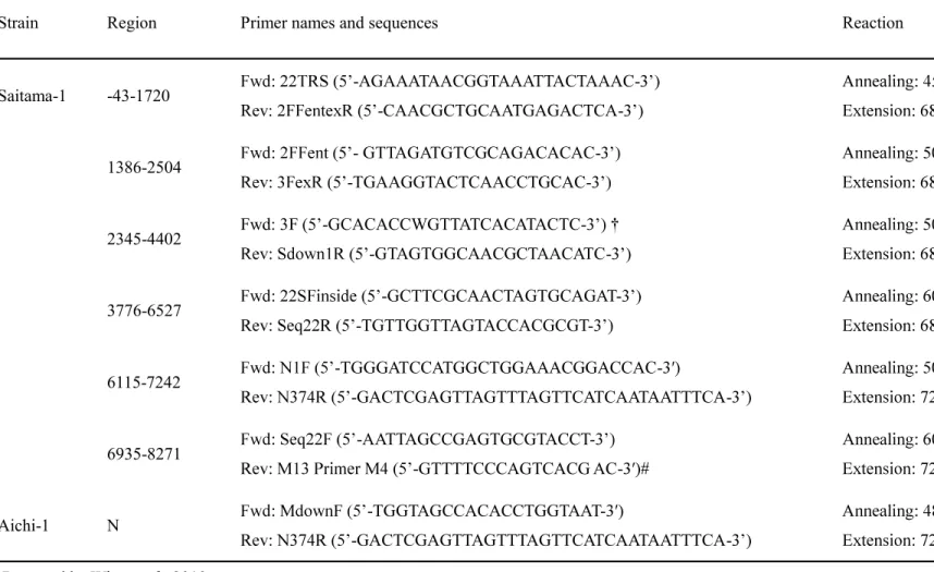
The nucleotide sequences of partial RdRp genes were determined using a BigDye Terminator Ver. 3.1 Cycle Sequencing Kit (Applied Biosystems, Foster City, CA) according to the In order to determine the 3 -terminal region of the Saitama-1 genome, further sequence analysis was performed. QIAGEN OneStep RT-PCR Kit (QIAGEN, Hilden, Germany) and TaKaRa RNA LA PCR Kit (AMV) Ver. 1.1 (TaKaRa, Shiga, Japan) were used to amplify each fragment of the Saitama-1 and Aichi-1 strains using the primer pairs listed in Table 3-2. The 3 -terminal region, nt 6935-8271, of the Saitama-1 strain was amplified by 3 -RACE using TaKaRa RNA LA PCR Kit (AMV) Ver. 1.1 (TaKaRa) according to th
of Aichi-1 strain [1128 bases, 375 amino acids (a.a.)] were determined. All sequences were deposited in the DDBJ.
3.3.3. Phylogenetic analysis and homology search
A phylogenetic tree was constructed using the program MrBayes Ver. 3.2.2 (Ronquist and Huelsenbeck, 2003) for MrModeltest analysis with a general time reversible (GTR) or WAG substitution matrices (Whelan and Goldman, 2001). All trees were graphically represented using FigTree Ver. 1.4.2 (Rambaut, 2009). The nucleotide sequence from the S gene to poly A was compared among FRECV MSU-2, FRSCV MSU-1, and Saitama-1 strain using SimPlot Ver. 3.5.1 (Lole et al., 1999).
3.4. Results
3.4.1. Detection of FRCoV RdRp gene from ferrets in Japan
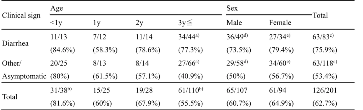
The results showed that 126 ferrets (62.7 %) in Japan were positive (Table 3-1). Significant differences were statistically analyzed using
Chi-of <0.05 were considered to be statistically significant. The ratio Chi-of detection Chi-of FRCoV between diarrhea (75.9 %) and other symptoms (53.4 %) was significantly different. In particular, the difference was marked in ferrets aged more than 3 years, suggesting that FRCoV is the primary pathogen or exacerbating factor for diarrhea in ferrets. In addition, most ferrets aged less than 1 year (81.6 %) were infected with FRCoV, regardless of their clinical symptoms, suggesting that most ferrets are infected with FRCoV soon after birth. Importantly, some strains, Saitama-1 strain from ferret no. 22 and Aichi-1 strain from ferret no. Aichi-160, were detected by RT-PCR using CoV-consensus primers, but not FRCoV-specific primers, even though the latter can detect FRCoV with more sensitivity than CoV-consensus primers (Terada et al., 2014b).
3.4.2. Phylogenetic and sequence analyses
The phylogenetic tree based on the partial RdRp genes showed that the FRCoV Saitama-1 and Aichi-1 strains belonged to a different cluster from other FRCoV strains (Fig. 3-1 A). The Saitama-1 strain had open reading frames (ORFs) encoding S (4308 bases, Saitama-1435 a.a.), ORF3 (744 bases, 247 a.a.), envelope (E) (249 bases, 82 a.a.), membrane (M) (798 bases, 265 a.a.), N (1128 bases, 375 a.a.), 3x-like (225 bases, 74 a.a.), and ORF7b (609 bases, 202 a.a.). The consensus transcription-regulating sequences (TRSs) of coronaviruses, 5 -CTAAAC-3 (Spaan et al., 1988), were observed upstream of each ORF, except ORF7b. Although we could not detect typical TRS of ORF7b, a possible TRS 5 -CTAAAG-3 was observed upstream of ORF7b.
Phylogenetic analysis was performed based on the N-terminal one-third of the S protein (a.a. 1-438 of 1 S protein) and the C-terminal two-thirds of the protein (a.a. 439-1435 of 1 S protein), indicating that the MSU-2 strain was more similar to the MSU-1 strain than the 1 strain in the N-terminal one-third of the S protein (Fig. 3-1 B) and was more similar to the Saitama-1 strain than the MSU-Saitama-1 strain in the C-terminal two-thirds (Fig. 3-Saitama-1 C). Furthermore, a phylogenetic tree based on the N protein indicated that Saitama-1 and Aichi-1 strains differed more from the other FRCoVs than from mink coronaviruses (Fig. 3-1 D).
3.4.3. Homology search
SimPlot analysis showed that the Saitama-1 strain was different in some regions, including the N, 3x-like, and ORF7b genes, when compared with other strains (data not shown). Surprisingly, the 3 -terminal in two-thirds of S genes in the MSU-2 strain was similar to that in the Saitama-1 strain, but one-third of S genes in the MSU-2 strain was similar to that in the MSU-1 strain (Fig. 3-2 A). Alignment of S proteins among Saitama-1, MSU-1, and MSU-2 strains also indicated that the MSU-2 strain was similar to the MSU-1 strain in the N-terminal one-third of S protein and to the Saitama-1 strain in the C-terminal two-thirds of the protein (Fig. 3-2 B). In addition, a low similarity in nucleotide sequences between the MSU-1 and MSU-2 strains and the Saitama-1 strain was observed in the N, 3x-like, and ORF7b genes, but the E and M genes were highly conserved among all strains (data not shown).
3.5. Discussion
The results of phylogenetic analysis and homology search indicate that the Saitama-1 strain possesses different S, N, 3x-like, and ORF7 genes than other FRCoVs, and that the E and M genes are conserved among FRCoVs. Therefore, we consider the Saitama-1 and Aichi-1 strains to comprise a novel FRCoV genotype. In addition, these results indicate that the MSU-2 strain emerged by recombination of the S protein between the MSU-1 and Saitama-1 strains. These recombination events often occurred among CoVs, resulting in the diversity of CoV genomes. Especially, the recombination of S protein caused the cross-species transmission or change of pathogenesis of SARS-CoV (Graham and Baric, 2010), porcine transmissible gastroenteritis virus (TGEV), canine coronavirus (CCoV) type II (Decaro et al., 2009), and feline coronavirus (FCoV) (Terada et al., 2014a). Novel CoVs must emerge and drastically evolve by these recombination events. Further investigation will thus be required to determine the evolution of CoV including FRCoV.
In conclusion, novel FRCoV strains (Saitama-1 and Aichi-1) were detected in Japan. These FRCoVs appear to have emerged by recombination events among other FRCoVs. This information will be useful for understanding the pathogenesis of FRCoV in ferrets.
3.6. Figure legends
Fig. 3-1 Phylogenetic relationship among ferret coronaviruses. Phylogenetic trees were constructed
based on nucleotide sequences of the partial RdRp gene (394 bp) (A), N-terminal one-third of S protein (a.a. 1-438 of 1 S protein) (B), C-terminal two-thirds of S protein (a.a. 439-1435 of Saitama-1 S protein) (C) and N protein (D). Posterior probabilities are indicated above branches in all trees. Sequences reported in this study are shown in bold.
Fig. 3-2 Recombination between Saitama-1 and the other FRCoV. Schematic diagram of MSU-2 and
SimPlot analysis of MSU-2 with Saitama-1 and MSU-1 (A). Green and red boxes indicate regions similar to Saitama-1 and MSU-1, respectively. Arrows indicate TRS (5 -CTAAAC-3 ). SimPlot analysis was carried out using the nucleotide sequence of the Saitama-1 strain from the S gene to poly A (8263 bases), the MSU-1 strain (8025 bases), and the MSU-2 strain (8309 bases). Green and red lines indicate Saitama-1 and MSU-1, respectively. Alignments of S proteins of Saitama-1, MSU-1, and MSU-2 (B). Green and red boxes indicate regions similar to Saitama-1 and MSU-1, respectively.
Table 3-1. Detection of FRCoV genes in ferrets
Clinical sign Age Sex Total
<1y 1y 2y 3y Male Female
Diarrhea 11/13 (84.6%) 7/12 (58.3%) 11/14 (78.6%) 34/44a) (77.3%) 36/49d) (73.5%) 27/34e) (79.4%) 63/83c) (75.9%) Other/ Asymptomatic 20/25 (80%) 8/13 (61.5%) 8/14 (57.1%) 27/66a) (40.9%) 29/58d) (50%) 34/60e) (56.7%) 63/118c) (53.4%) Total 31/38 b) (81.6%) 15/25 (60%) 19/28 (67.9%) 61/110b) (55.5%) 65/107 (60.7%) 61/94 (64.9%) 126/201 (62.7%) Number of positives / Number of examined samples.
a) : P<0.001 b), c) : P<0.01 d), e): P<0.05
Table 3-2. List of primer pairs for amplification of each fragment
Strain Region Primer names and sequences Reaction
Saitama-1 -43-1720 -AGAAATAACGGTAAATTACTAAAC-Rev: -CAACGCTGCAATGAGACTCA-1386-2504 - GTTAGATGTCGCAGACACAC- -TGAAGGTACTCAACCTGCAC-2345-4402 Fwd: 3F -GCACACCWGTTATCACATACTC- -GTAGTGGCAACGCTAACATC-3776-6527 -GCTTCGCAACTAGTGCAGAT- -TGTTGGTTAGTACCACGCGT-6115-7242 -TGGGATCCATGGCTGGAAACGGACCAC- -GACTCGAGTTAGTTTAGTTCATCAATAATTTCA-6935-8271 -AATTAGCCGAGTGCGTACCT--GTTTTCCCAGTCACG AC-Anneal Aichi-1 N -TGGTAGCCACACCTGGTAAT- -GACTCGAGTTAGTTTAGTTCATCAATAATTTCA-et al., 2010.
4. CHAPTER 3 Interspecies transmission of pseudorabies virus
among wild animals.
4.1. Abstract
In February 2016, pseudorabies viruses (PRV) were isolated from two raccoons found dead in the western part of Japan. PRV was detected using PCR on oral swab and serum samples from one raccoon. Analysis of the complete genome indicated that the PRV contained the gene encoding glycoprotein E which is deleted in the PRV vaccine strain available in Japan. In addition, PRV antibodies were found in 11.7% of wild boars, but not in raccoons, sampled in the same region. In conclusion, PRV was the cause of death in two raccoons, and the virus is still active in a region where domestic pigs are PRV-free. This is the first report of raccoon deaths by natural infection and of interspecies transmission of PRV among wildlife.
4.2. Introduction
Pseudorabies virus (PRV) belongs to the order Herpesvirales, the family Herpesviridae, the subfamily Alphaherpesvirinae and the genus Varicellovirus (King et al., 2012). PRV is the causative ase in newborn piglets and other mammals, resulting in serious economic losses. For this reason, PRV is a candidate for eradication by the pork industry in Japan, and many prefectures have been successful in eradicating PRV from domestic pig populations. Now, an attenuated PRV vaccine, in which the glycoprotein E (gE) gene has been deleted, remains in use among domestic pigs in a few prefectures (Ministry of Agriculture, Forestry and Fisheries, 2019).
Wild boar (Sus scrofa) is the natural host for PRV. Although PRV-infected wild boars show tremors, incoordination and fever, their condition is generally good and they do not show symptoms commonly observed in non-pig hosts, such as pruritus and self-mutilation (Gortázar et al., 2002). Reports of seroprevalence of PRV infection among wild boars in Croatia, Czech Republic, France, Germany, Italy, Russia, Slovenia, South Korea, Spain, Switzerland and the U.S.A. suggest that wild boars can survive PRV infection (Albina et al., 2000; Chiari et al., 2015; Corn et al., 2009; Gortázar et al., 2002; Gresham et al., 2002; Jeoung et al., 2015; Köppel et al., 2007; Leuenberger et al., 2007; Müller et al., 2011; Müller et al., 1998; Pannwitz et al., 2012; Pirtle et al., 1989; Ruiz-Fons et al., 2007; Sedlak et al., 2008; van der Leek et al., 1993; Vengust et al., 2006; Vicente et al., 2005; Wyckoff et al.,
pigs and vaccines have not been applied, some wild boars have shown antibodies against PRV (Mahmoud et al., 2011).
PRV causes a fatal disease with neurological disorders in various mammals, including cats (Hara et al., 1991), cattle (Matsuoka et al., 1987), dogs (Zhang et al., 2015), foxes (Jin et al., 2016), minks (Liu et al., 2017) and non-human primates (Hurst, 1936). Experimental infection with PRV in
raccoons (Procyon lotor) induced death, indicating that raccoons are also a dead-end host for the virus (Kirkpatrick et al., 1980; Platt et al., 1983; Thawley and Wright, 1982; Wright and Thawley, 1980). However, there have been no reports of death due to natural PRV infection in raccoons.
In February 2016, two raccoons were found to have died as a result of PRV infection in the western part of Japan, and the virus was genetically and serologically analyzed.
4.3. Materials and methods
4.3.1. Samples from wild animals
isolation and viral gene detection. Oral and rectal swabs and sera were collected from the two dead raccoons. Sera were also collected from 111 wild boars and 61 raccoons captured from 2011 to 2016 in the same area where the two dead raccoons were found. Under local government programs for prevention from bird and animal damage, the wild boars were hunter-harvested or culled, and wild raccoons were captured and culled for invasive pest control. No animals were killed solely for this study. Hunters received permission from local governments. All collected sera were stored at -20°C until analysis.
4.3.2. Cells
Vero cells (Vero 9013, JCRB number; JCRB9013) purchased from the Health Science Research Resource Bank (HSRRB, Osaka, Japan) were cultured in Eagle's minimum essential medium (EMEM; Thermo Fisher Scientific, MA, U.S.A.) containing 5% fetal calf serum (FCS), 100 U/ml
humidified 5% CO2atmosphere at 37 °C.
4.3.3. Viruses
PRV RC1 strain isolated from the serum of raccoon No. 1 in this study was propagated in Vero cells. Viral titers of the RC1 strain was measured by plaque assay using Vero cells as described previously (Mahmoud et al., 2011).
4.3.4. Virus isolation
Swab samples were mixed with 2 ml of EMEM containing 1% antibiotic-antimycotic (Thermo Fisher Scientific), and then centrifuged at 2,000 × g for 15 min at 4 °C. Supernatants were -X® centrifuge tube filters (Corning, NY, U.S.A.). Filtrates and sera were inoculated onto Vero cells. Cells were incubated with EMEM containing 2% FCS and 1% antibiotic-antimycotic at 37 °C. Cells were maintained and passaged until cytopathic effects (CPE) were observed. After five blind passages, samples without CPE were judged as negative for virus isolation.
4.3.5. Nested PCR
For detection of PRV, DNA was extracted from samples using the DNeasy Blood & Tissue Kit (QIAGEN, Hilden, Germany). PCR for the target gene, glycoprotein B (gB), was carried out using
-ATG GCC ATC TCG CGG TGC- -ACT CGC
GGT CCT CCA GCA-
-ACG GCA CGG GCG TGA TC- -GGT TCA GGG TCA CCC
GC-(Mengeling et al., 1992).
4.3.6. Virus-neutralization (VN) test
VN titer against the RC1 strain was measured using Vero cells by the 80% plaque reduction neutralizing test (PRNT80). The complement in sera was inactivated by incubation at 56 °C for 30 min. The RC1 strain was diluted to 1×10³ PFU/ml with EMEM containing 2% FCS, and mixed with an equal volume of serum diluted with EMEM containing 2% FCS. The mixture was incubated at 37 °C for 1 hr. Fifty microliter aliquots of the mixture were applied to duplicate wells in 24-well plates
(Sumitomo Bakelite Co., Ltd., Tokyo, Japan) and the plates were incubated at 37 °C for 1 hr. After incubation, the cells were washed twice with EMEM without FCS, and overlaid with EMEM containing 0.8% SeaPlaque GTG Agarose (Lonza, Basel, Swizerland) and 6.7% FCS. After coagulation of the agarose, the plate was incubated at 37 °C until CPE was observed. The cells were fixed using 5% phosphate-buffered formalin and stained by crystal violet. Numbers of plaques were counted and the highest dilution showing less than 80% of that in control wells was taken as the VN titer (Mahmoud et al., 2011).
4.3.7. Sequence analysis
In order to determine the complete nucleotide sequence of the RC1 strain, the supernatants of Vero cells infected with the RC1 strain were collected and mixed with a final concentration of 8% polyethylene glycol 6,000 and 0.5 mol/L sodium chloride. After incubation for 4 h at 4 °C, the mixture was centrifuged at 5,870 × g for 30 min at 4 °C to precipitate the virus. DNA was extracted from the pellets and analyzed using the MiSeq Reagent Nano Kit v2 (Illumina, Inc., San Diego, CA) according
polymerase (TaKaRa, Shiga, Japan) using specific primers and determined using the BigDye
instructions.
4.3.8. Phylogenetic analysis
Phylogenetic trees based on the complete genome and amino acid sequences of glycoprotein C (gC) were constructed using a distance-based (neighbor-joining) method with MEGA 7.0 software (Kumar et al., 2016).
4.3.9. Accession numbers
The complete nucleotide sequence of the RC1 strain (141,757 bp) was deposited into the DNA Databank of Japan (DDBJ) under accession no. LC342744.
4.4. Results
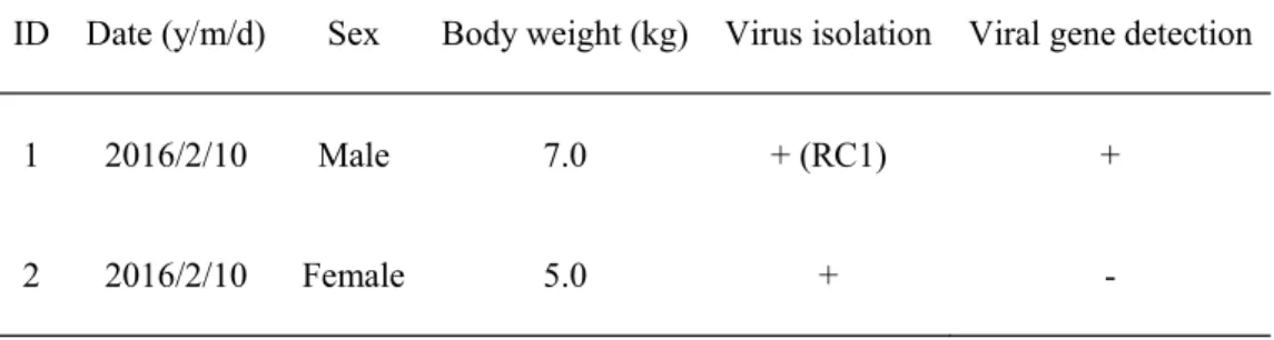
4.4.1. Virus isolation
PRVs were isolated from oral swab and serum samples from raccoon No. 1 and the oral swab from raccoon No. 2 (Table
4-RC1.
4.4.2. Nested PCR for detection of PRV
In order to detect PRV, nested PCR was performed. The PRV viral gene was detected from the oral swab from raccoon No. 1, but not from raccoon No. 2 samples. The nucleotide sequences of all PCR products from isolates and oral swabs were identical (data not shown).
4.4.3. Seroprevalence of PRV
The results from PRNT80 using PRV showed that 13 wild boars (11.7%), but no raccoons, were seropositive for PRV (Table 4-2).
4.4.4. Complete genome of PRV RC1 strain
Comparing the complete genome sequence of the RC1 strain to those of the LA and Bartha strains, the nucleotide identities were 97.8% and 96.2%, respectively. Similar comparisons for the amino acid identities of the gB protein were 98.8% and 96.8%, respectively. The RC1 strain contained the gE gene, which is deleted in the vaccine strain used in Japan. When compared with those of the LA, Yangsan and P-PrV strains, the amino acid identities of the gE protein of the RC1 strain were 97.9%, 97.9% and 97.6%, respectively. When compared with those of the LA, Bartha and Yamagata S-81 strains, the amino acid identities of the gC were 96.1%, 91.6% and 90.9%, respectively.
4.4.5. Phylogenetic analysis of PRV
A phylogenetic tree was constructed based on the complete genome and amino acid sequences of gC. This suggested that the RC1 strain is similar to recent Chinese isolates including LA, Fa and Ea, but not to the previous Japanese isolate, Yamagata S-81, which was isolated in northern Japan in 1981 (Figs. 4-1 and 4-2).
4.5. Discussion
In this study, we identified PRV causing a lethal disease in raccoons. Until now, there have been no reports of such lethal PRV pathogens in raccoons in Japan. Notably, this is the first report of natural PRV infection in raccoons not only in Japan, but also worldwide.
PRV infection is fatal in many mammals, but is non-lethal in pigs, wild boars and humans. Although the Japanese government is attempting to eradicate PRV in the domestic pig population (Ministry of Agriculture, Forestry and Fisheries, 2019), complete eradication has not yet been achieved. We isolated PRV from two dead raccoons in an area where PRV has been eradicated in domestic pigs (Table 1). Thirteen PRV-positive wild boars were detected in the same area (Table 4-2), indicating that PRV is still present in the wild boar population, and that the virus may have spilled over from wild boar to raccoon. PRV infection is a differential diagnosis for rabies in raccoons.
The phylogenetic tree based on gC suggested that the RC1 strain was genetically related to recent Chinese PRVs, but not to the previous Japanese PRV Yamagata S-81 (Fig. 4-2). It is not known whether this PRV strain has been maintained for a long period among wild boars, or whether it recently invaded the region. Thus, further study to detect PRV in other regions of Japan is necessary.
In conclusion, this is the first report of lethal PRV infection in raccoons anywhere in the world, and of possible interspecies transmission of PRV from wild boars to raccoons. Populations of raccoons and wild boars have been gradually increasing in Japan (Ministry of the Environment, 2015). As encounters become more likely, the opportunity for interspecies transmission of PRV might also increase. Because interspecies transmission of viruses among wild animals may induce the emergence of new pathogenic viruses, it is important to monitor such transmission among wildlife.
4.7. Figure legends
Fig.4-1 Phylogenetic tree based on nucleotide sequences of complete PRV genome. Bootstrap values
are indicated above each branch. GenBank accession numbers of nucleotide sequences are shown in parentheses. The sequence analyzed in this study is shown in bold.
Fig. 4-2 Phylogenetic tree based on amino acid sequences of PRV gC. Bootstrap values are indicated
above each branch. GenBank accession numbers of nucleotide sequences are shown in parentheses. The sequence analyzed in this study is shown in bold.
Table 4-1. Isolation and detection of PRV in raccoons.
ID Date (y/m/d) Sex Body weight (kg) Virus isolation Viral gene detection
1 2016/2/10 Male 7.0 + (RC1) +
Table 4-2. Seroprevalence of PRV infections in wild boars and raccoons.
Species Number of examined sera
Number of PRV-positive sera (% of PRV-positive sera)
Wild boar 111 13 (11.7%)