Title
Analysis of protocadherin-a in mouse behavioral
regulation
Author(s)
福田, 絵美
Citation
Issue Date
Text Version ETD
URL
http://hdl.handle.net/11094/49304
DOI
rights
Note
Osaka University Knowledge Archive : OUKA
Osaka University Knowledge Archive : OUKA
https://ir.library.osaka-u.ac.jp/repo/ouka/all/
Analysis of protocadherin-a. in mouse behavioral regulation マウス行動制御における多様化膜分子群 protocadherin-a. の機能解析 FukudaEmi KOKORO-Biology Group, Laboratories for Integrated Biology, Graduate School of Frontier Bioscience, Osaka University 2008.3
要旨 脳はきわめて多数の神経細胞から構成され、複雑であるが組織化された神経回路を形成してい る。脳の重要な構成要素であるニューロンは多数の樹状突起を伸ばし、周囲のニューロンと結合 しで情報を受け取っている。そしてそれらの情報を神経回路が正確に処理することにより、脳が 正常に機能する。組織化した神経回路形成には、個々の神経細胞を認識し、結合する分子が重要 な役割を担っていることが知られている。私は、細胞認識分子が、行動を制御する分子機構の解 明を目指し、多様化膜分子群 protocadherin (Pcdh)-a ファミリーに注目して、マウス個体レベルに おける学習・記憶を中心とした行動制御での役割について解析を行った。 多様化膜分子群である Pcdh-a ファミリーは、神経細胞で発現するカドヘリン様膜タンパク質で あり、神経軸索、シナプス領域に局在していることが示唆されている。この Pcdh-a ファミリーは、 ゲノム染色体上で遺伝子クラスター構造をもち、多様性をもつ細胞外領域は複数の可変領域エク ソンにコードされ、共通の細胞内領域は定常領域エクソンに由来している。 Pcdh-a ファミリーは、 個々の神経細胞ごとに異なった発現パターンをもつことが明らかとなっており、神経細胞の認識 に関わる分子群であることが示唆されている。 Pcdh-a ファミリーの行動制御に関わる分子的役割 を明らかにするため、私は Pcdh-a遺伝子改変マウスを用いて実験を行った。 マウス Pcdh-a ファミリーの全てのメンバーに共通する定常領域には選択的スプライシングに より細胞内領域の異なる Pcdh-aA 型、 B 型アイソフォームが存在する。まず私は本研究室で作製
された Pcdh-al!.Bneo/1!.Bneo マウスを用いた。この Pcdh-al!.Bneo/1!.Bneo マウスは、 Pcdh-aB 型アイソフォーム
が欠損し、同時に Pcdh-aA 型アイソフォームが野生型に比べ 20% まで減少していた。このマウス は外見の異常は認められず、繁殖も可能であった。しかし、 Pcdh-al!.Bneo/l!.Bneo マウスに対して、運 動、感覚、情動、学習・記憶などについての網羅的行動解析を行ったところ、運動、感覚、情動 のテストにおいては有意な差が認められなかったものの、恐怖条件付け学習テストにおいて、 Pcdh-al!.Bneo/l!.Bneo マウスでは空間学習能力の有意な九進が認められた。また空間作業能力の指標と なる 8 方向放射状迷路においても異常が認められた。これら空間学習の異常は、 Pcdh-aB 型のみ が欠損し Pcdh-aA 型の発現レベルが正常な Pcdh-al!.B/1!.B マウスでは有意な差は認められなかった。 これらの結果から、 Pcdh-aA 型が空間学習を制御する重要な役割を担っていることが明らかにな った。更に、 Pcdh-a△Bneo/1!.Bneo マウスは、感覚運動ゲーティング制御の障害を調べるプレパルス抑 制テストで、抑制がかかりにくい有意な異常が認められた。 次に、 Pcdh-a タンパク質の減少と行動異常との関連性を探る為に、行動の制御に関連すること が知られているモノアミン量について高速液体クロマトグラフィ一を用いた解析を行った。その 結果、 Pcdh-a△Bneo/1!.Bneo マウスの海馬体では、野生型に比ベセロトニン量が有意に増加していた。 ー方、 Pcdh-al!.B/1!.B マウスの海馬体では有意な差は見られなかった。また、 Pcdh-a△Bneo/1!.Bneo マウスの 海馬体 CAI 領域において長期増強 (LTP) が顕著に減少していることが判明した。 これらの結果から、 Pcdh-a ファミリーが学習・記憶の過程、感覚運動ゲーティング制御に重要 な役割を持つことが明らかになった。またこれらの行動異常には、海馬体でのセロトニン量の異 常な増加が関与している可能性が示唆された。ー方、 Pcdh-a ファミリーの減少は海馬体のシナプ ス伝達効率を変化させることから、 Pcdh-a ファミリーがシナプス機能、神経回路網に直接的な役 割を持っていることが示唆された。
General Introduction
CHAPTER 1 Summary Introduction
Materials and Methods Results
Discussion
CHAPTER2
Summary Introduction
Materials and Methods Results Discussion Acknowledgments References Appendix 1i}f,E~-Table of Contents 2 4 5 8 14
33
37
38
39
4149
51 52 5964
General introduction
The brain is composed enormous number of neurons, and organized complex neural networks systematically. The normal behavior is highly associated with a network of neural connections. During development, neurons extend dendrites and axons, and they generated complex arborizations. These branch patterns are at least assembled from developmental programs underling genetic-code that give rise to number of neurons, location of projection, the morphology of dendrites and axons, and the synaptic connections. It is thought to be that neurons are labeled with cell-surface recognition proteins so that they project to target sites and make synaptic connections. I have a lively interest in questions that how neurons distinguish the enormous number of different cells, and how the neural connections affect the behavior.
Neuronal interaction and recognition are thought to be mediate, in part, by cell surface proteins. The cadherin superfamily is a group of cell surf ace proteins that is composed more than 100 members in vertebrates. This family suggested from their molecular structures and diversity that they regulate the cell-cell interactions or transmit signaling from outside to inside of the cell. Extracellular domain contains cadherin repeats, which contain sequences that are involved in calcium binding (Overdiun et al., 1995; Shapiro et al., 1995). Several subgroup of cadherin can be defined based on shared propertied and sequence, classic cadherins, demosomal cadherins, protocadherins (Pcdhs), Flamingo/CELSRs and FAT. Among of these subgroups, Pcdhs are the largest subgroup, more than sixty members, in the cadherin superfamily (Nollet et al., 2000).
The Pcdhs family has been identified more than 70 different Pcdhs genes. The Pcdhs family can be divided into two groups based on their genomic structure; clustered Pcdhs and nonclustered Pcdhs (Radies et al., 2005). Clustered Pcdhs are consist of the Pcdh-a, f3 and y family, which give rise to over 50 members of proteins by diverse splicing in a small genomic locus (Hirayama & Yagi, 2006). Most of Pcdhs are predominantly expressed in neurons, and some of them are localized in synapses and have led to combinatorial expression patterns. (Kohmura et al., 1998; Phillips et al., 2003; Morishita et al., 2004). The features of the Pcdh-a have suggested the hypothesis that the diversity of the Pcdh-a potentially contributes to the neuronal recognition (Serafani, 1999; Shapiro & Colman, 1999; Yagi & Takeichi, 2000; Hamada & Yagi,
2001). However, the molecular functions of Pcdhs are still poorly understood. To elucidate the molecular function of Pcdhs in vivo, my laboratory generated the Pcdhs gene-targeting mice. In this study, to address the roles of Pcdhs in regulating behavior, we subjected these mutant mice to behavioral analysis. Furthermore, we approached to reveal the mechanisms underlying behavioral abnormalities through physiological, anatomical, and neurochemical phenotypes.
CHAPTERl
Summary
The brain is composed of enormous numbers of neuron, and complex neuronal networks are organized systematically. During the neural development, axons choose specific dendrites for synapse formation by the cell recognize molecules. It is important to understand that how the molecular mechanisms organized the neuronal networks for perceiving the behavior function such as learning and memory. The clustered protocadherins (Pcdhs) family is the subgroup of the diverse cadherin superfamily and they are encoded as a large cluster (composed of a,
13
and y clusters) in the genome. Pcdhs are predominantly localized in the brain and Pcdhs are up-regulate during the neuronal development. Then it is possible that they contribute to generate sophisticated neural networks, and to regulate brain function. To address the molecular roles of Pcdhs in regulating individual behavior, here we generated knockdown mice of Pcdh-o: proteins and examined their behavioral abnormalities. In Pcdh-o: proteins, there are two alternative splicing variants of the constant region, Pcdh-o: A and B isoforms, with different cytoplasmic tails. Pcdh-a:!iBneotABneo mice, which lacked a splicing variant of Pcdh-o: B and were down-regulated in Pcdh-o: A isoforms to approximately 20%, displayed enhancement of contextual fear conditioning and disparities in the eight-arm radial maze. These learning abnormatities were, however, normalized in Pcdh-o:ABIAB mice (removing the neomycin-resistant gene cassette from Pcdh-o:ABneotABneo alleles), which recovered the expression level of Pcdh-o: A isoforms but completely lacked the Pcdh-o: B isoforms in their brains. Furthermore, Pcdh-o:ABneotABneo mice showed a reduced of prepulse inhibition of the acoustic startle response. In addition, the amount of 5-hydroxytryptamine (5-HT) specifically increased in the hippocampus of Pcdh-o:ABneotABneo mutant mice. These results suggested that the Pcdh-o: family had significant roles for regulating learning and memory function, sensorimoter gating and the amount of 5-HT in the hippocampus.Introduction
The formation of neural connection with their intended targets is controlled by complex cell-cell interactions. During the development of the nervous system, cell recognition molecules play roles in organizing neuronal networks by regulating neural migration,
fasciculation, synaptogenesis and intracellular signaling (Dodd & Jessell, 1988;
Edelman & Crossin, 1991; Fields & ltoh, 1996). A large number of the cadherin superfamily has been identified in the CNS and contributes to neural development, including the compartmentalization of the brain during development (Inoue et al.,
2001), the formation of neuronal connectivity, synapse formation, stabilities (Togashi et al., 2002; Abe et al., 2004) and synaptic plasticity (Tang et al., 1998; Bozdagi et al.,
2000; Manabe et al., 2000).
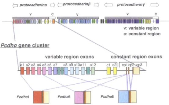
The protocadherin-a (Pcdh-a) family was originally identified as cadherin-related neuronal receptors (CNRs) (Kohmura et al., 1998). The Pcdh-a cluster is followed by the Pcdh-(3 and Pcdh-y clusters, which are arranged in tandem on human chromosome 5 and mouse chromosome 18. The mouse Pcdh-a gene cluster is composed of 14 variable exons and a set of three constant region exons (Sugino et al., 2000; Wu &
Protocadherin (PcdhJ family gene clusters
mouse (Chromosome1883)
¢:::J protocadherlna
q
t_;:Jprotocadherinpq
¢=:J protocadherinyq
V C
Pcdha gene cluster
\
variable region exons
1 a2 a3 a4 a5 a6 a7 a8 a9 a10a11 a12
....
Pcdha1 Pcdha5
aJ
Pcdha8V C
v: variable region c: constant region
Maniatis, 2000). Mature Pcdh-a mRNAs are generated from one of these variable exons encoding alternate extracellular, transmembrane, and a set of constant region exons, which encode short cytoplasmic domains (Fig. 1). In the Pcdh-a 3rd
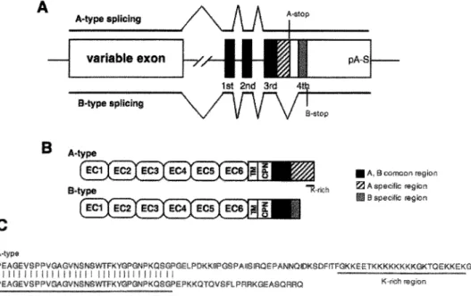
constant exon, two kinds of alternative splicing variants, Pcdh-a A and B isoforms, are produced in all Pcdh-a transcripts (Sugino et al., 2000) (Fig. 2). Diverse Pcdh proteins appear to be exclusively expressed in the brain, especially in the olfactory, hippocampus and cerebellum. Pcdh proteins, coupled with the complex formation of the proteins, located in axons and partially at the synapses during neuronal development (Kohmura et al., 1998; Phillips et al., 2003; Morishita et al., 2004), and have led to combinatorial expression patterns. This molecular diversity of Pcdhs has led to the speculation that Pcdhs might underlie the precise formation of the neuronal network (Serafani, 1999; Shapiro & Colman, 1999; Yagi & Takeichi, 2000; Hamada & Yagi, 2001). A A>-type splicing IMype splicing S.type EC1 EC2 C A-stop
•
A, El oomoon region !2As;,ecijJcIii e specif., region
Figure 2. (A) Alrernative splicing of Pcdh-aA and B isoforms in constant Pcdh-aA- and
sequences are derived from different exons in the constant Black boxes repr-ese11t Pcdh-aA- and common stripe boxes represent the specific region of A-type and gray boxes represent the specific region B-type. A-type splicing had three, and B-type splicing had four exons. Termination of both transcripts is a common poly A signal (pA-S). (B) Diagram of the protein structure of Pcdh-a A and B isoforms. Each protein has a common variable region and a different cytoplasmic region. ECl-6, extracellular cadherin domains 1-6; TM, transmembrane domain; CPN, short cytoplasmic domain; K-rich, lysine-rich region. (C) Comparison of amino acid sequences of Pcdh-a A and B.
Recent study of the Pcdh-y cluster using gene-targeting mice showed that Pcdh-y null mice die soon after birth because of the dramatic neurodegeneration of spinal interneurons in the late embryonic stages (Wang et al., 2002; Weiner et al., 2005). Moreover, synaptic density in the intermediate zone of the spinal cord is dramatically decreased. Further investigation has been performed in mutants lacking both Pcdh-y proteins and the proapoptotic protein BAX (a member of proapoptic Bcl-2 family). In these mice, the decrease of synapses by minimal apoptosis and neurodegeneration were still observed and the activity of the formed synapses was reduced. These results indicated that Pcdh-y proteins are essential for synaptic development at least in some neurons; however, the roles of Pcdh proteins in the regulation of brain function have not been identified yet.
In the present study, we generated mutant mice, which extensively decreased Pcdh-a proteins, to investegate their physiological role in vivo. These mutant mice showed enhancement of fear conditioning learning and moderate disparity in the eight-arm radial maze test. These abnormalities were recovered by rescuing protein levels of Pcdh-a A isoforms. The mutant mice also showed irregularities in the prepulse inhibition test. In addition, these hypomorphic Pcdh-a A mutants up-regulate the amount of 5-HT levels in the hippocampus.
Materials and Methods
Antibodies
A monoclonal antibody for specific Pcdh-a B proteins (1F4, rat IgG) was produced to immunize the KLH (keyhole limpet hemocyanin) conjugated with the synthesized polypeptide (Cys-RRKGEASQPRQ), which is a specific Pcdh-a B cytoplasmic tail. In the present study, we used anti-CNR/Pcdha (rabbit polyclonal antibody) as a Pcdh-a A isoform-specific antibody (Murata et al., 2004). Anti-CNRN (rabbit polyclonal antibody), anti-4Ell (rat monoclonal antibody) (Mutoh et al., 2004) against the Pcdh-a4 N-terminus (QIHYSIPEEAKHGT-Cys) and an anti-CNR-A antibody (Takei et al., 2001) against the Pcdh-a A-specific region were used for immunoprecipitation. Anti-synaptophysin (Sigma), anti-synaptotagmin (Chemicon), anti-GAP-43 (Sigma), anti-GAD-67 (Chemicon) and anti-PSD-95 (Affinity Bioreagents, USA) were purchased.
Plasmids and Trans/ ection
Plasmid DNA was prepared as previously described (Murata et al., 2004). Briefly, full-length mouse Pcdha-v4-A or Pcdha-v4-B was tagged with a c-myc epitope, and
then cloned into pcDNA3.1 expression vector (Myc-CNR/Pcdha-v4-A or
Myc-CNR/Pcdha-v4-B). HEK293T cells were maintained in DME medium
supplemented with 10% fetal bovine serum. Plasmid DNA was transfected into HEK293T cells using LipofectAMINE 2000 (Invitrogen).
RT-PCR
Total RNA from mouse brain was extracted with TRizol Reagent (lnvitrogen) and cDNA was synthesized after priming with 40 pmol per reaction of oligo-dT primer in a total volume of 20 µI, using 15 units of Superscript3 (Invitrogen) in accordance with the manufacturer's protocol. To detect spliced transcripts in wild-type and mutant mice, the primer pair North-F (5'-GGCAGCCCAACCCTGACT-3') and mhl-2R was used. To define each transcript, Southern blot analysis using internal probe of the products was performed after RT-PCR.
Immunoblotting and Immunoprecipitation
Mice were deeply anesthetized with diethylether. The brains were immediately frozen after dissecting, and tissues or transfected cells were homogenized in 2 x SDS sample buffer (100 mM Tris-HCI, pH6.8, 4% SDS, 4% 2-mercaptoethanol, 20% glycerol, 0.02% bromophenol blue). The lysates were centrifuged at 20,000 x g for 20 min, and the supernatant was used for immunoblot analysis. For immunoprecipitation, tissues were homogenized in RIPA buffer (10 mM Tris-HCI, pH7.5, 150 mM NaCl, 5 mM EDTA, 1 % Triton X-100, 1 % sodium deoxycholate, 0.1 % SDS) with protease inhibitors (5 µg/ml aprotininm, 3 µg/ml leupeptin, 3 µg/ml pepstatin A, 1 mM PMSF) and incubated at 4°C for 10 min. The lysates were centrifuged at 9,100 x g at 4°C for 20 min, and the supernatants were precleared with protein G-Sepharose beads (Amersham, Bioscience) at 4°C for 1 h. The sample was then incubated with 1 µg of anti-CNRN or anti-CNR-A at 4°C for 1 h. Protein G-Sepharose beads were added to the sample, and incubated at 4°C for more than 2 h. After the beads were washed extensively with RIPA buffer and phosphate buffered saline (PBS), the proteins were dissociated by boiling the beads in sample buffer. The proteins were subjected to SDS-PAGE followed by Western blot analysis (Murata et al., 2004).
Histochemistry
For Nissl staining of brain sections with cresylviolet, deeply anesthetized mice were perfused transcardially with 4% paraformaldehyde in 0.1 M phosphate buffer (pH7.4). The 40 µm fixed sections were cut on a microtome (Leica SM2000R), and mounted on glass slides.
Animal and behavioral experiments
All tests were performed with male mice that were 10 weeks old at the start of testing. Mice were housed four per cage in a room with a 12 h light/dark cycle with ad libitum access to food and water.
Motor Junction tests
Motor function tests were conducted as described previously (Miyakawa et al., 2001). The rotarod test was performed using an accelerating rotarod (UGO Basile Accelerating Rotarod). The time each mouse was able to stay on the rod was measured. In the wire hang test, each animal was placed on a lid of a wire cage and then inverted gently, so the mouse gripped the wire. Latency to fall was recorded, with a 60 sec
cutoff time. Open field test
Locomotor activity was measured by the open field test. Mice were placed in the center of the open field apparatus (27.4 x 27.4 x 20 cm; MED-associates, Alban, VT). Data were collected for 2 h, and the total distance and time spent in the center were recorded. Light/ dark transition test
Light/dark transition tests were conducted as described previously (Miyakawa et al., 2001). One chamber was irradiated brightly, whereas the other chamber was dark. Mice were placed into the light side and allowed to move freely between the two chambers for 10 min. The total number of transitions and total time in the light chamber were recorded.
Home cage activity
During the home cage activity test, each subject was housed individually in a cage (31 x 21 x 13 cm) in a room with a 12 h light/dark cycle with ad libitum access to food and water. Mice were observed for 3 days and the total distance travelled during the night and day was recorded.
Elevated plus maze
The elevated plus maze formed two open arms (25 x 5 cm) and two closed arms with 15 cm-high transparent walls. The two arms were placed diagonally and the apparatus was elevated to 50 cm above the floor. To prevent mice from falling from the apparatus, 3 mm-high Plexiglas ledges were attached to the open arms. The subjects were placed in the central square (5 x 5 cm) facing one of the closed arms. The test was conducted for 10 min and the time in open arms was recorded.
Porsolt forced swim test
The Porsolt forced swim test was conducted as described previously (Miyakawa et al., 2001). Briefly, mice were placed into the cylinder filled with super hypochlorous water, and their behavior was recorded for 10 min. Data acquision was performed automatically, and time of immobility was recorded.
Pain test
The subjects were placed on a 55°C hot plate, and latency to paw lick or foot shake was recorded.
Contextual and cued fear conditioning
On the training day, each mouse was placed in a conditioning chamber (10.5 x 10.5 x
10.5 cm, O'HARA and Co., Tokyo), and allowed to explore freely for 2 min. A tone (75 dB) was sounded as the conditioning stimulus (CS) for 30 sec followed by a 2-sec mild foot shock (0.35 mA) as the unconditioning stimulus (US). One or two more tone-shock pairs were given at 2-min intervals. The animal was returned to its home cage 30 sec after the last pair. 24 h after the conditioning session, the mice were placed back into the conditioning chamber for 5 min, and freezing behavior was measured in context. An hour after context testing, the mice were put into another white Plexiglas chamber for 3 min, and then the tone was turned on for 3 min. Freezing during the first and following 3 min was recorded respectively.
Morris water maze
A hidden platform version of the Morris water maze test was conducted as described previously (Miyakawa et al., 2001). A plastic circular pool (40 cm high x 95 cm diameter) was filled to a depth of 30 cm with 21 ± 1 °C water made opaque with nontoxic white paint. Each mouse was placed in one of four starting locations facing the pool wall, and swam until finding a platform 1 cm below the water surface, or for a maximum of 60 sec. Four trials per day were conducted for 9 days. Latency to reach the platform, the distance traveled to reach the platform, average swim speed, and the percentage of time spent at the perimeter of the pool were recorded. On the 10th day, the platform was removed, and a probe test was conducted. The percentage of time spent in each quadrant, number of platform crossings, average swim speed, and the percentage of time spent at the perimeter of the pool were recorded.
Eight-arm radial maze test
The eight-arm radial maze test was as described previously (Miyakawa et al., 2001). Briefly, mice were deprived of food and maintained at 80-85% of their initial body weight. After the pretraining session, maze acquition trials were performed for 15 consecutive days. For each trial, the choice of arms, latency to get all pellets, distance traveled, the number of different arms chosen within the first eight choices, and the number of working memory and omission errors were scored.
Acoustic startle response/Prepulse inhibition
A startle reflex measurement system was used (O'HARA & Co., Ltd). The background noise level was 70 dB in each chamber. Each mouse placed in a Plexiglas tube for 5 min as an initial period of acclimation. The responses were recorded for 160 msec starting with the onset of the prepulse stimulis. The maximum startle amplitude scored
during the 160 msec was used. Mice received a series of six trial type, two types for startle trials and three types for prepulse inhibition trials. The startle stimulus was 110-or 120-dB sound. The prepulse sound was 74-, 78- 110-or 82-dB sound, which presented 100 ms before startle stimulus. These six trial types were presented in pseudorandom order 10-20 sec intertribal interval. The following formula was used to calculate percentage prepulse inhibition of ASR: 100-{[(startle response on prepulse
+
startle stimulus trials)/ startle response alone trials] x 100}.Image analysis
All applications used for behavioral studies were performed on a Macintosh computer, using Image OF, Image WM, and Image FZ (O'Hara & Co., Tokyo, Japan). Applications were based on the public domain NIH Image program ( developed at the U.S. National Institute of Mental Health and available on the Internet at http://rsb.info.noh.gov/nih-image/) and were modified by Tsuyoshi Miyakawa (available through O'Hara & Co., Tokyo, Japan).
Measurement of monoamines by HPLC
The contents of monoamines and their metabolites were measured using an HPLC with electrochemical detection. Mice were killed by decapitation at 10 weeks old, and the brain regions rapidly dissected out and quickly frozen at -80°C until assayed. Each frozen brain tissue was homogenized with ultrasonic irradiation in 250 µl of 0.2 M perchloric acid containing isoproterenol as an internal standard. The homogenates were placed on ice for 30 min and centrifuged at 20,000 x g for 15 min at 4°C. The supernatants were filtered through a syringe filter unit (DISMIC-3; ADVANTEC, Japan) and the pH was adjusted to 3.0 by adding 1 M sodium acetate, and then injected into an HPLC system equipped with an ODS column (Eicompak SC3-ODS; 3 x 100 mm; Eicom, Japan) and an electrochemical detector (EDC-100; Eicom, Japan) with the potential set at
+
750 m V. The mobile phase was 0.1 M citric acid and 0.1 M sodium acetate, pH 3.5, containing sodium-1-octansulfonate (190 mg/L), EDTA-2Na (5 mg/L) and 17% methanol. The flow rate was set at 0.25 ml/min. The protein content was measured using a Bradford protein assay solution (Bio-Rad, Richmond, CA, USA) after the precipitates had been solubilized with 0.1 M NaOH.Statistical analysis
Statistical analysis was conducted using StatView (SAS Institute, Cary, NC). Data were analysed by t-test, one-way ANOVA, or repeated measures ANOVA. Values in Tables and graphs are expressed as the mean ± SEM.
Electrophysiology
To compare Pcdh-aiiBnoe/iiBneo with wild-type mice, we performed all experiments using littermates and in a blind fashion. 9- to 11-week-old Pcdh-aiiBnoe/iiBneo and wild-type mice were decapitated under deep halothane anesthesia. Hippocampal slices (400 µm thick) were cut with a Vibratome tissue slicer and placed in a humidified holding chamber for at least 1 h. A single slice was transferred to the recording chamber maintained at 25°C and submerged beneath a continuously perfusing medium that had been saturated with 95% 02 /5% CO2• The medium comprised 119 mM NaCl, 2.5 mM KCl, 1.3 mM MgSO4 , 2.5 mM CaCl2 , 1.0 mM NaH2PO4 , 26.2 mM NaHCO3 and 11 mM glucose. All perfusing solution contained picrotoxin (100 µM) to block GABAA receptor-mediated inhibitory synaptic response. Field potential recordings were made using a glass electrode filled with 3M NaCl placed in the stratum radiatum in CAl area of the hippocampus. An Axopatch-1D amplifier was used and the signal was filtered at 1 kHz and digitized at 10 kHz. To evoke synaptic responses, a bipolar tungsten electrode was placed stratum radiatum, and Schaffer collateral/commissural fibers were stimulated at 0.1 Hz ( = test pulse). A single high-frequency stimulus train (100 Hz, 1 s) was applied at the test pulse intensity to induce LTP. The amount of LTP was expressed as percentages in the slope of EPSPs relative to the baseline. The average size of EPSPs recorded between 50-60 min after high-frequency stimulation was used for statistical comparison. For measurements of paired-pulse facilitation (PPF), afferent fibers were stimulated twice at intervals of 50, 100 and 200 ms in the presence of 25 µM D-APV. For measurements of post-tetanic potentiation (PfP), a train of high-frequency stimuli (100 Hz, 1 s) was delivered at Schaffer collateral/commissural fibers in the presence of 50 µM D-APV. All value was reported as means ± SEM. Student's t-test (two tailed, unpaired) was used to determine whether or not there was a significant difference in the mean between two sets of data.
Results
Generation of Pcdh-a isoform-specific gene-converting mice
To examine the roles of Pcdh-a proteins in vivo, we generated Pcdh-a isoform-specific gene-converting mice. Mice bearing ABneo and AB alleles were originally produced to assess the roles of Pcdh-a B-type transcripts. In the ABneo allele, no amino acids (aa) were changed in Pcdh-a A isoforms and Pcdh-a B isoforms were expected not to be expressed. In the ABneo allele, a neo gene was inserted into the 4th
constant exon for a specific Pcdh-a B region (Fig. 3A). The gene conversion was confirmed by PCR and Southern blot analysis (Fig. 3B, E). To evaluate the expression of Pcdh-a proteins in Pcdh-a+JABneo and Pcdh-aaBneo/ABneo mutant brains, we performed Western blot analysis using a monoclonal antibody (anti-1F4) against Pcdh-a B proteins that specifically detected the Pcdh-a B isoform (specificity showed in Fig. 3C using HEK293T transfectants) and anti-CNR/Pcdha, which mainly recognized the Pcdh-a A isoform (specificity shown in Fig. 3D using HEK293T transfectants). Using the anti-1F4 antibody, we could not detect Pcdh-a B proteins in the brain extracts of Pcdh-aaBneolaBneo mice, even m immunoprecipitants with anti-CNRN antibody-recognized Pcdh-a4 proteins (Fig. 3C). At the same time, the amounts of Pcdh-a A isoform proteins were largely decreased to approximately 20% in Pcdh-aABneo/ABneo mutants and 60% in Pcdh-a+/ABneo (Fig. 3D). We confirmed by RT-PCR that Pcdh-a B mRNAs were completely abolished in Pcdh-aaBnoetaBneo mice but Pcdh-a A was not (Fig. 3F). In the ABneo allele, the coding sequences for Pcdh-a A isoform proteins should not be changed; therefore it was possible that this down-regulation of the Pcdh-a A isoform was caused by the insertion of a neo gene cassette in the ABneo-targeted allele.
To examine this possibility, we produced a AB allele by removing the neo gene cassette from the ABneo allele. Two loxP sites were located on both sides of the neo gene cassette; therefore, we could easily remove it from the ABneo allele using Cre-recombinase. Crossing the Pcdh-aABneo/ABneo mice with CAG-Cre mice, we produced mutants (Pcdh-a+taB) containing the AB allele (Fig. 3A). Using PCR analysis of genomic DNA with two primers on both sides of the neo gene in the ABneo allele, we confirmed that the neo gene was correctly removed at the loxP sites in Pcdh-a+iaa and Pcdh-aaBJAB mice (Fig. 3E). Pcdh-a B mRNAs and proteins were also completely
abolished in Pcdh-a"8'"8 mice (Fig. 3F, G). On the other hand, interestingly, in Pcdh-a" 81"8 mutants, Pcdh-a A isoforms were recovered to a similar level as those of
wild-type mice by removing the neo gene cassette in the 8Bneo allele (Fig. 3H). Thus, we obtained two different Pcdh-a mutant strains. Pcdh-a"BneolABneo mutants had about 5-fold fewer Pcdh-a A isoforms and lacked the Pcdh-a B isoforms, and Pcdh-a"81"8
mutants lacked the Pcdh-a B isoforms but had the normal protein level of the Pcdh-a
A isoforms. Using these Pcdh-a hypomorphic mutants, we addressed the roles of Pcdh-a proteins to regulate brain function.
Figure 3. Generation of Pcdh-a"Bneo/ABneo mice and Pcdh-a"8188
mice. (A) Schematic diagram of the genomic organization of wild-type mouse Pcdh-a, the targeting vector, ABneo allele targeted a floxed neomycin-resistant gene into B-specific 4th exons in the constant region, and the AB allele removed the neomycin-resistant gene by Cre/loxP recombination. The restriction map around the locus used for targeting and Southern blot analysis is shown (E, EcoRI; S, Sall; X, Xhol, Sp, Spel). The probe for Southern blot analysis and primer for genomic PCR (arrows) are also indicated. Neo, neomycin-resistant gene; DT-A, diphtheria toxin A-fragment. (B) Southern blot analysis of targeted Pcdh-a constant region. DNA from the tails of wild-type (+/+), heterozygous (+/ABneo) and homozygous (ABneo/ABneo) target mice was digested with EcoRI. Blotted DNA was hybridized with 5' probe, which was placed on a 5' Pcdh-a 3rd exon genomic flanking region. (C) Pcdh-a B isoforms in wild-type(+/+) and Pcdh-a"BneoiABneo mice were detected by Western blotting with anti-1F4 antibody for immunoprecipitants with anti-CNRN antibody. The antigen specificity of anti-1F4 antibody for Pcdh-a B isoforms is shown by using HEK293T cell transfectants of Myc-CNR/Pcdha-v4-A (A-type) and Myc-CNR/Pcdha-v4-B (B-type). (D) Expression of Pcdh-a A isoform proteins in wild-type ( +/ + ), Pcdh-a+/ABneo and Pcdh-a"Bneo/ABneo brains at 4 weeks old. Antigen specificity of anti-CNR/Pcdha antibody for Pcdh-a A isoform is shown by using HEK293T cells transfectants. (E) Genomic PCR of Pcdh-a"Bneo/ABneo mice and Pcdh-a88188
mice. (F) In Pcdh-a"Bneo/ABneo and Pcdh-a88188 mice, Pcdh-a B-type mRNA could not be detected by
RT-PCR. (G) Pcdh-a B isoforms in wild-type mice(+/+) were detected by Western blotting for immunoprecipitants with anti-CNRN antibody, but not in Pcdh-a88188 mice. (H) Protein level of Pcdh-a A isoform in wild-type ( +/+ ), Pcdh-a+/ABneo and Pcdh-a"Bneo/ABneo brains.
variable region axons \ \ Targeting veclor E $$ E X ~---;re)C:je---.L...' --_,.
-AB allele kb ll.4 8.2 11lO!l 600Brain lysales HEK293T IP: anti-CNRN IB: 1mti-1F4
F
Tki, constructed by Dr. Hamada
Brain lysates HEK29ST blot: 1mti-CNR/Pcdha
Phenotypes of Pcdh-aABneo/ABoeo mutants
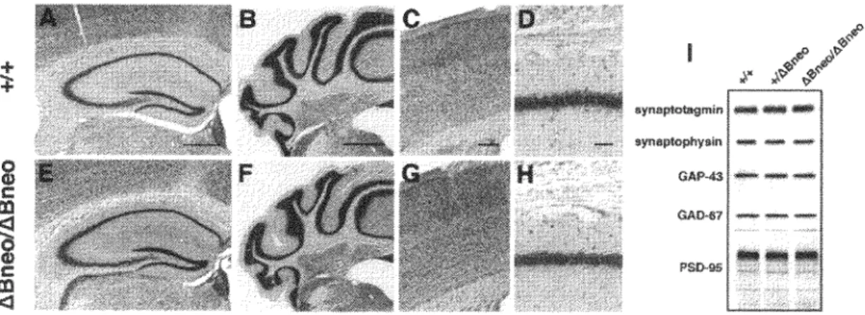
No perinatal lethality was observed and Pcdh-ahBneolhBneo mice were born at the expected Mendelian frequency at the time of weaning. Pcdh-ahBneo/hBneo mice were healthy and fertile. We observed no visible abnormalities in Pcdh-aABneo/hBneo mice during development or adulthood. The brains of Pcdh-aABneo/hBneo mice were similar in size and shape to those of wild-type mice. No obvious impairments were detected in the organization in the neocortex, hippocampus, or cerebellum cortex by Nissl staining of the brain sections (Fig. 4). We also could not detect apparent changes of synaptic proteins such as synaptophysin, synaptotagmin, growth-associated protein 43 (GAP-43), glutamic acid decarboxylase 67 (GAD-67) and postsynaptic density 95 (PSD-95) in the hippocampus between genotypes (Fig. 41).
2
C m1
C m <l aynaplotagmin - ... -GAP-"3 ---OAo.-1 PS0-115Figure 4. Nissl staining of coronal sections and the expression level of synaptic protein of the brain from wild-type (A, B, C, D) and Pcdh-a-1Bncoi!iBneo mice (E, F, G, H). No obviously impairments were detected. (A, E) Niss) staining of the hippocampus at low magnification. (B, F) Cerebellum cortex. (C, G) Layers 1 through 6 in the cerebral cortex. (D, H) Pyramidal cell layer in the hippocampus. (I) lmmunoblot analysis of synaptic-marker proteins in wild-type ( +/+ ), heterozygous (+/ABneo) and homozygous (ABneo/ABneo) target mice. Scale bars, (A) 500 µm;
Emotional behavior in Pcdh-a. .1Bneo /.1Bneo mice
We next analysed behavioral function of Pcdh-a. usmg these mutant mice. All behavioral experiments were performed with male mice that were 10 weeks old at the beginning of the behavior tests. Pcdh-a..1Bneoi.1Bneo mice had no significant defects of physical characteristics such as body weight, whiskers and fur. No disparity was observed in reaching, reflex, the wire hanging test or rotarod test for motor coordination. As shown in Table 1, they did not show any significant abnormalities in the light/dark transition test, home cage activity; however, we detected a tendency toward anxious or fearful phenotypes with time spent in the center area in the open field test (P
=
0.11) and the stay time in open arms in the elevated plus maze (P=
0.13) in Pcdh-a..1Bneo/.1Bneo mice, but there was no significant difference from wild-type mice. Pcdh-a B isoform-specific mutants of Pcdh-a.1Bi.1B mice appeared normal in the open field test and the elevated plus maze test. Pcdh-a.1Bneoi.1Bneo mice also showed the tendency of changes in the porsolt forced swim test which was used to evaluate depression (P = 0.05) and in the hot plate test for pain sensitivity (P = 0.07), but not significant.Table. I Sensor~ anJ motor ab1htics m Pc<lh-c1. genc-rnutatcJ mice.
Pcdh-o:ABneo/ABneo mice +f+ Motor Tests
Wire hang (latency to fall: ,e.:onJs) .'i2..1 (±1.01 Rmamd (latency to fall; seconds I
Day! 59.8 (±6..4 I
Da ·2 s7.o (±7.::n
Open Piel<!
Center time (sewnds) l 1.7 (±2.5) Distance (crnJ 7.188 (±562) Lighl/Jark transition lest
Numher of transitions 24.()(±151 Total time in light (seconds) 172.2(±11.4) Home Cage
Activiti-Distance :\Jight (cm) 100.11± 180) Distance Day (cm) 204(±21)
Elevated Plu, Maze
Time on 01?".n Arms 11.7,-1 14.8 (±4 .. 'i)
l'orsolt Forced Swim
Immobility('!(,) 1-4 minutes 29J (±3.5 I Immobility (ct) l-10 minutes 42.2 (±4J I
Pain Te,t
Ho! Plate (lalenn: se,omb) 11.5(±1.1) Dalt represent Ilic mean (±SEtl--!) (+!+. n,-,15-17: r\Bncn'1\Bne,1. 11=16-17).
Pcdh-«AB/AB mice
Open Field
Center lime (seconds) Distance (cm)
Elevated Plu, Maze
2.l.6 (±2 8) 3028 (±154)
TimeonOpenr\rm,('frl 7,1 (+].7) D.ita represent the mean (±SEtl,1) (+:+, n=l6-17; t\Bit5.B. n=l5-!6J.
18 mutants :\2.0(±.Hll 55.5 (±6..l) 89.1 (±7.7) 6.91±16) 6772 (±653) 23.6(±2.11 160.8 (±11 .01 8241±177) 174 (±17) 74(±16) ]83 (±2 6) 46. l (±.,.2 I 9.0 J,+0.6) 24.:l (±2.6) ,9x:, 1+981 6.(l(+l.2) p-valuc 0.95 0.64 0.84 0.11 0.48 0.89 0.-IX 0.-19 0.2X O.IJ 0.05 0.46 om 0.86 0.81 0.81
Enhanced freezing of Pcdh-a. ABneoiABneo mice in the contextual fear conditioning test
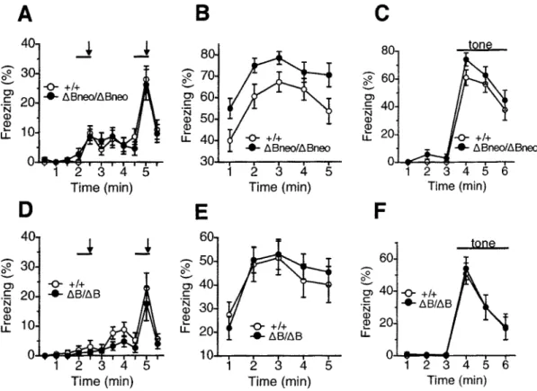
The cognitive functions of Pcdh-aABneoJABneo mice and their sibling were analysed in a contextual and cued fear conditioning test. During the conditioning period, the animals were presented a paired tone and footshock twice as training. In this period, Pcdh-aABneoJABneo mice showed no difference in freezing (genotype effect, F 1,31 = 0.002, P = 0.967) (Fig. 5A). However, when we performed contextual and cued tests 24 h after training, Pcdh-aaBneoiABneo mice showed significant enhancement of freezing in the contextual testing relative to wild-type mice (genotype effect, F 1,31
=
5.48, P=
0.0258)(Fig. SB). On the other hand, Pcdh-aABneotaBneo mice and their wild-type littermates displayed similar levels of freezing during cued testing (genotype effect, F 1,31 = 1.987,
P = 0.169) (Fig. SC). To avoid possible effects of the test sequence in the test battery, we performed the contextual and cued fear conditioning test using another group of mice that had experienced no other behavioral tests. When the littermates received the paired tone and footshock twice, Pcdh-aABneoiABneo mice also showed greater freezing in the contextual test, but not in the cued test than wild-type mice. These results indicated that Pcdh-aaBneotABneo mutants had enhanced contextual learning.
Next, to confirm whether abnormalities in the contextual fear conditioning test in Pcdh-at.Bneolt.Bneo mice were caused by an decrease of Pcdh-a A isoform proteins or loss of Pcdh-a B isoform proteins, we conducted the contextual and cued fear conditioning test for Pcdh-at.BIAB mice that specifically lacked Pcdh-a B isoform proteins. Pcdh-aaB/t.B mice and their wild-type littermates were presented with a pair of tone and footshock twice in the conditioning phase. After 24 h, contextual and cued tests were conducted. Interestingly, Pcdh-aas,aB mice showed no significant difference in the percentage of freezing in the contextual test compared to wild-type littermates (genotype effect, F 1,31
=
0.054, P=
0.818) (Fig. SE). Also, no differences wereobserved in the training (genotype effect, F 1,31 = 1.511, P = 0.228) and cued tests (genotype effect, F 1,31
=
0.027, P = 0.870) (Fig. 5D and F). These findings indicated that enhanced freezing in the altered contextual fear conditioning test displayed in Pcdh-at.Bneoit.Bneo mice was due to the down-regulation of Pcdh-a A isoform proteins.A
B
C
_J
_J
8 8 ~6 -0- +/+ ~ Ol ..._ t.Bneo/t.Bneo Ol C 20 .N C 40 .N (]) (]) (]) ~...
10 20 LL -0- +/+ u. ..._ LlBneo/LlBneo 0 0 1 2 3 4 5 1 2 3 4 5 1 2 3 4 5 6Time (min) Time (min) Time (min)
D
E
F
4_J
_J
60 60 ~ 30 ~ ~ 0) C .N (]) Q)...
LL -0- +/+ 0 ';; 40 20 ..._ LlB/LlB -0- +/+ C ..._LlB/LlB 3 .N Q) 20 10 -0- +/+ ~ ..._ LlB/LlB u. 0 10 0 1 2 3 4 5 1 2 3 4 5 1 2 3 4 5 6Time (min) Time (min) Time (min)
Figure 5. Contextual and cued-fear conditioning in Pcdh-aABneo/ABneo mice (A, B,
C) and Pcdh-aABIAB mice (D, E, F) against wild-type mice (+/+). (A) Percentage of freezing during the conditioning phase. A tone was sounded for 30 sec (bars) followed by a 2-sec footshock (arrows). (B) Pcdh-aABneotABneo mice showed enhanced freezing during contextual testing conducted 24 h after conditioning (P = 0.026). (C) Freezing during the tone-cued testing in wild-type ( +/+) and Pcdh-aA Bneo/ABneo mice. There was no significant difference between wild-type ( +/+) and Pcdh-(1.ABneott.Bneo mice. (D) Percentage of freezing during the conditioning phase in wild-type ( +/+) and Pcdh-aABIAB mice. (E) Freezing during context testing. No significant difference was observed between wild-type ( +/+) and Pcdh-aABneo/ABneo mice. (F) Freezing during the tone-cued testing in wild-type ( +/+) and Pcdh-aAB/AB mice. There was no significant difference between genotypes. Data are given as the means (±SEM) (Pcdh-aABneo/ABneo mice, n
=
16; Pcdh-aAB/AB mice, n = 16; wild-type, n = 17 respectively).Pcdh-ct'\Bneo/ABneo mice in the Morris water maze
To examine spatial learning defects of Pcdh-aABneo/ABneo mice, we tested littermates of Pcdh-aABneo/ABneo and wild-type mice in the Morris water maze (hidden platform version). Mice were given four trials per day for nine consecutive days of the training and probe tests one day after training. In this test, Pcdh-aABneo/ABneo mice generally showed no significant cognitive impairment. Escape latency, which was the time to reach the platform, was similar between genotypes during training (genotype effect, F 1,29 = 0.174, P = 0.679) (Fig. 6A). There was also no abnormality either in swimming speed or in time spent at the perimeter of the pool (Fig. 6B and C) (P > 0.05).
During the probe test, in which the learning platform was removed, Pcdh-aABneofABneo mice spent more time in the training quadrant compared to other quadrants, similar to wild-type littermates (Fig. 6E). In addition, we found no differences in the number of crossings above the place where the platform was located (P = 0.541) (Fig. 6D) and both genotypes spent significantly more time in the training quadrant than in other quadrants in the probe test conducted after reversal training one week after the original training (Fig. 6F). This indicated that Pcdh-aABneo/ABneo mice had no abnormalities in Morris water maze learning.
A
30-
(.)m
20-
(.)>-55
10co
• +/+ ....J • t.Bneo/t.Bneo 0..1..r--r-"T"'""T"""'l--,-"""T"""T"'"",...C-
t:_ 25 ai ci3 20 E -~ 15 a.. C 10 c 123456789 Blocks of 4 trials ~ 5 -0- +/+ (/) • t.Bneo/t.Bneo (l) E o ... ...,.... ... "T"'""..., ... ...,.... ... i= 2 3 4 5 6 7 8 9E
30 25c
Q) 20 a. (/) 15 Q) E 10 i-= 5 0 Blocks of 4 trials D +/+ • t.Bneo/t.Bneo T 0 R LB
u
(l) -!!?. 25 E ~ 20l- _ _
x_A--!
l
15~
(f) E 10 "§: (f) 5 -0- +/+ • t.Bneo/t.Bneo 0 ... ...-...,...,.---,-D
F
2 3 4 5 6 7 8 9 Blocks of 4 trialsg>
15 ·u5 Cl)§
10 0 Q) 5 .0 E ~o..._...__
30 D +t+ D +/+ • t.Bneo/t.Bneo 25c
Q) 20 a. • t.Bneo/t.Bneo (/) 15 Q) E 10 i-= 5 0 T 0 R LFigure 6. Intact performance of Pcdh-at>Bneo/t>Bneo mice in the Morris water maze. (A) Escape latency, (B) swimming speed, (C) time spent at the perimeter of the pool. There were no significant differences between the two genotypes. (D) Both genotypes also crossed the training site more often than the equivalent sites in the other three quadrants in the probe trials performed after training. (E) In probe trials, Pcdh-at>Bneo/t> Bneo mice and their wild-type ( +/+) littermates searched for where the platform had been located. Both genotypes spent more time in the training quadrant T compared with the other quadrants (0, opposite quadrant; R, right quadrant; L, light quadrant) in the probe trials performed after training. (F) Probe trials were conducted again l week after the first probe trials. Both genotypes similarly spent more time in the training quadrant compared with the other quadrants. Data are given as the means (±
SEM) (Pcdh-at>Bneo/t>Bneo mice, n
=
16; wild-type, n=
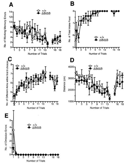
l 5).Disparities of Pcdh-a. ABneo/ABneo mice in the eight-arm radial maze test
Pcdh-a.ABneo/ABneo mice and wild-type littermates were also analysed in the eight-arm radial maze test to examine spatial working memory. In this task, the number of working memory errors of mice revisiting arms that had been visited previously, which is generally interpreted as a measure of working memory performance, was measured (Olton et al., 1979a, b). The number of working memory errors in Pcdh-a.ABneo/ABneo mice was significantly low during first six trials (genotype effect, F 1•23
=
5.015, P=
0.035; interaction between trials and genotype, F 5• 115=
0.045, P=
0.999). No significantgenotype effect of working memory errors during the delay period, 30 sec in the 15th trial, and 2 min in the 16th to 18th trials, was observed (genotype effect, F 1,23 = 2.789, P = 0.109) (Fig. 7 A). The amount of total food intake during a trial was significantly less in Pcdh-aABneo/ABneo mice than in wild-type mice during the first six trials (genotype effect, F 1,23
=
4.547, P=
0.044; interaction between trials and genotype, F 5,115= 0.837,P = 0.526) and delay period (15th to 18th trials) (genotype effect, F 1.z3
=
4.347, P=
0.048; interaction between trials and genotype, F 3,69= 0.486, P = 0.693) (Fig. 7B). However, the number of different arms chosen within the first eight choices was not significantly different (P>
0.05) (Fig. 7C). There were no significant differences in the distance traveled (Fig. 7D) and the latency to take all pellets during the trials, including the delay period (data not shown). In addition, there was no significant genotype effect on the number of omission error trials (genotype effect, F 1,23 = 0.713, P = 0.407;interaction between trials and genotype, F 17•391 = 0.648, P
=
0.853) (Fig. 7E), suggesting that the increased number of revisiting errors displayed during trials without delay was unlikely to be caused by reduced motivation to take the pellets. Thus, Pcdh-aABneo/ABneo mice showed a decreased number of working memory errors in the eight-arm radial maze. Consistent with locomotor activity measured in several tests such as the open field test, light/dark transition and home cage activity (Table 1) there was no distinction in the distance traveled. Taken together, these findings suggested that the hypomorphic Pcdh-a protein level was associated with abnormal memory formation.We also performed the eight-arm radial maze test against Pcdh-aAB/AB mice and their wild-type littermates. There were no significant irregularities between genotypes in the number of working memory errors, the amount of total food intake, the number of different arms chosen within the first eight choices, the distance traveled and the
number of omission error trials (P > 0.05) (Fig. 8). These results indicated that the down-regulation of Pcdh-a A isoform proteins also cause for disparities of spatial working memory.
A
~ 10 g w ~ 8 0 E 6 Q) ~ Cl) C 4 :g 0 3:: 2 0 0 z 0.,C
a, 0 8 ·o {i co 1!! 7 .:: C: ~ 3: "' 61
E l!: 5 "' !!: 0 0 4 ci zE
5 "'e
4 uJ § 3 ·.; Cl) E 2 0 0 o 1 z -0- +/+ • LiBneo/LiBneo~
1 3 5 7 9 11 13 16 18 Number of Trials -0- +I+ • L'>Bneo/LiBneo~
1 3 5 7 9 11 13 16 18 Number of Trials -0- +/+ • LiBneo/L'>Bneo 1 3 5 7 9 11 13 16 18 Number of Trials8
8~
"' 0 0 7 IJ. 5)1 ~ 'iii 6 0 I-0 -0- +/+ ci 5 • LiBneo/LiBneo z 4 7 9 11 13 16 18 Number of TrialsD
3000 -0- +/+ • t.Bneo/t.Bneor
2500 E 3-"' 2000 0 C: ~ 0 1500 1000 1 3 5 7 9 11 13 16 18 Number of TrialsFigure 7. Eight-arm radial maze test of wild-type ( +/+) and Pcdh-al\Bneo II\Bneo mice. Mice were given 14 trials and 4 delay trials (30 sec for the 15th trial and
120 sec for the 16th to 18th trials). In delay trials, mice were confined to the
center platform for each delay time after taking the fourth pellet. (A) The number of working memory errors. A significant genotype effect was observed during the isr to 6h days (P
=
0.035). (B) Amount of total food intake. A significant genotype effect was observed during the first 6 trials (P = 0.044) and delay trials (15th to 18th trials) (P = 0.048). (C) The number of different armschosen within the first eight choices. No significant genotype effects were observed (P > 0.05). (D) Total distance during the trials. There were no significant differences between genotypes. (E) Number of omission errors. Data are given as the means (±SEM) (Pcdh-al\Bneo/tiBneo mice, n = 15; wild-type, n = 9).
A
8
14 -0- +/+ 8 ~ ..._ l\.B/ll.B g 12 'O LU 0 c:' 0 LL 0 3l! 7 E Q) <t! ~ :S g,~
~ :g I-~ 0 -0- +!+ ci ..._ll.B/ll.B 0 z 0 z 5 1 3 5 7 9 11 13 16 18 1 3 5 7 9 11 13 16 18Number of Trials Number of Trials
.,C
D
Q) 0 8 -0- +!+ ·5 -0- +!+ .c (.) ..._l\.8/l\.B 4000 • l\.B/l\.B co '§ 7"
"" C: E 3000 :E ~ ..s. Q) "' (.) E C: "' <( 1n Q 2000 0 1000 ci 3 5 7 9 11 13 16 18 1 3 5 7 9 11 13 16 18 zNumber of Trials Number of Trials
E
5 ~ -0- +/+ ~ ..._l\.B/l\.B C: .2 <I) "' .E 0 0 0 z 1 1 3 5 7 9 11 13 16 18 Number of TrialsFigure 8. Eight-arm radial maze test of wild-type(+/+) and Pcdh-a" 81,rn mice.
(A) The number of working memory errors in Pcdh-aA81" 8 mice. (B) The amount of total food intake in Pcdh-aABIAB mice. (C) The number of different arms chosen within the first eight choices was not significantly different. (D) Total distance during the trials. There were no significant differences between genotypes. (E) The number of omission errors was not different. Data are given as the means (±SEM). (Pcdh-a"801A8 mice, n
=
5; wild-type, n=
5).Prepulse inhibition (PPI) impaired in Pcdh-a ABneo/ABneo mice
Prepulse inhibition (PPI) of the acoustic startle response (ASR) is a measure of sensorimotor gating and concerns an attenuation of the startle response by a week stimulus presented a short time before the startle stimulus. ASR is an unconditioned reflexive response to a sudden acoustic stimulus and the primary circuits mediating PPI is a relatively simple pathway located in the lower brainstem (Davis et al., 1982; Fendt et al., 2001). To investigate a possible role of sensorimotor gating, PPI was preformed in Pcdh-aABneo/ABneo mice and their wild-type littermates. The ASR induced by 110-, 120- dB acoustic stimuli were indistinguishable between mutants and wild-type mice (110-dB, P
=
0.39; 120-dB, P=
0.29) (Fig. 9A). Next, PPI of ASR was analysed. Non-startling week stimuli were presented 100 ms before the 110- or 120-dB startle stimuli. The percentage of PPI, represented the degree of suppression of ASR by 74-, 78- and 82-dB prepulse stimuli, were significant decreased in Pcdh-aaBneo/ABneo mice than wild-type littermates (Fig. 9B). The difference of the percentage of PPI between genotypes was greater when the startle amplitude was 110-dB than 120-dB. This distinction of degree of response between 110-dB and 120-dB probably because of a ceiling effect was induced by massive intensity of the startle stimulus. These results indicated decrease of the sensorimoter gating in Pcdh-aABneo/ABneo mice.A
5 Q) 'U ::,4
;t= 0..3
E <(2
Q) t::cc
1
-(J)o..._.__
110d8B
-
~ 0 .._80
70
*
C 060
-:.0
50
Q) Cl) ::J c.. Q)-§
40
30
20
10
L..*
*
a.. 0
~ ~ ~ ~..._ ~..._ n~ ~ .,i;;: q,v 120d8*
D +t+ • liBneo/liBneoFigure 9. Prepulse inhibition of acoustic startle response of wild-type ( +/+)
and Pcdh-aliBneo ii\Bneo mice. (A) Startle responses to stimuli I l 0- and 120-dB sound. There was no significant difference between genotypes. (B) Prepulse inhibition of acoustic startle response. Three different prepulse stimuli (74-, 78-, 82-dB) were presented I 00 ms before the 110- or 120-dB startle stimuli. Data are given as the means (±SEM) (*, P < 0.05, Pcdh-a 68neo/!iBneo mice, n =
16; wild-type, n = 17).
5-HT level in Pcdh-a. ABneo/ABneo mice
Anxiety or behavioral performance 1s probably associated with alternate monoaminergic transmission in the forebrain such as the frontal cortex or hippocampus. Previous studies have shown that abnormal freezing in the contextual fear conditioning test was related to the amount of monoaminergic transmission (Inoue et al., 1994; Kim
et al., 1997). To evaluate abnormal monoaminergic transmission in Pcdh-a.ABneolABneo mice, detailed neurochemical analysis was performed. In the frontal cortex, no obvious differences were found in noradrenaline (NA), 5-hydroxyindoleacetic acid (5-HIAA, serotonergic metabolites), 5-HT, dopamine (DA), 3,4-dihydroxyphenylacetic acid (DOPAC, dopamine metabolites), and homovanillic acid (HVA, dopamine metabolites) amounts in Pcdh-a.ABneo/ABneo mice (Fig. 10B). In the hippocampus, however, 5-HT level was significantly increased in Pcdh-a.ABneo/ABneo mice (P = 0.0001) (Fig. IOA).
On the other hand, significant increase of 5-HT levels could not be found in Pcdh-a B-specific mutants of Pcdh-aABtAB mice in the hippocampus (Fig. 1 0C) and in the frontal cortex (Fig. 10D). NA and 5-HIAA levels were also not different in Pcdh-aABneo/ABneo mice.These results indicated that the 5-HT level in the hippocampus was associated with the hypomorphic Pcdh-a A isoform.
A
B
•
+I+**
60 80•
+/llBneo•
+/+ 70 D llBneo/llBneo C 50•
+/llBneo -~ 60 ·a-; "§40 D llBneo/llBneo eso a. a. Cl30 E'4o E :::a ~30 0 20 E20 E a. a.10 10 0NA 5-HIAA 5-HT 0 NA 5-HIAA 5-HT DA DOPACHVA
C
D
50 70•
+I+ 60•
+/llB c40•
+/+ C 0 llB/llB ·a-; ]i50 §_30•
+/llB 0 0 llB/llB 0..40 Cl Cl :§20 ..§ 30 0 0 E 20 E a. a.10 10 0NA 5-HIAA 5-HT 0 NA 5-HIAA 5-HT DA DOPACHVA
Figure 10. Monoamines in wild-type(+/+, white bar), heterozygous (+/ABneo, +/AB, gray bar) and homozygous (ABneo/ABneo, AB/AB black bar) mice. The amount of monoamines was measured by HPLC assay. (A) Monoamine level in the hippocampus. 5-HT was significantly increased in Pcdh-aABneo/ABneo mice (* *, P = 0.0001 ). (B) There were no significant differences in the monoamine level in the frontal cortex for genotypes. (C) Monoamine level in the hippocampus in Pcdh-a,.,81118 mice. (D) In the frontal cortex in Pcdh-aABIAB
mice. NA, noradrenaline; 5-HIAA, 5-hydroxyindoleacetic acid; HT, 5-hydroxytryptamine; DA, dopamine; DOPAC, 3,4-dihydroxyphenylacetic acid; HVA, homovanillic acid.
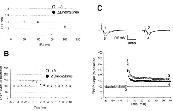
Reduced hippocampal synaptic plasticity in Pcdh-a. ABneo/ABneo mice
Because alternations in the strength of synaptic connection between hippocampal neurons are assumed to underlie contextual and spatial memories, we investigate synaptic plasticity in the hippocampal Schaffer collateral synapses (SC-CAI). There was no significant difference on paired-pulse facilitation (PPF), which is sensitive to presynaptic release probability, between the wild-type and mutant mice (P > 0.05) (Fig. 1 lA). Post-tetanic potentiation (Pf P) was also indistinguishable in magnitude between genotypes (P > 0.05) (Fig. 11B).
However, LTP induced by a strong tetanic stimulation (lO0Hz, ls) was significantly smaller in Pcdh-aABneoiABneo mice (143.8±1.8 % of baseline at 50-60 min after application of tetanus, n
=
9) than in wild-type mice (163.4±3.3 % of baseline, n=
10, t-test, P < 0.01) (Fig. 11C). Initial EPSP slopes were normalized in each experiment to the averaged slope value during the control period (-30 to 0 min). Representative traces (average of 10 consecutive responses) in the inset were EPSPs obtained at the times indicated by the numbers in the graph. Thus, down-regulation of Pcdh-a proteins significantly altered the synaptic plasticity in the SC-CAI pathway.
A
1.8 1.6 1.4 1.2 .. -~- Q·· . Q-1:J+ • D.Bneo/D.BneoC
,~-~--~-~--~-~
~~
3 0.2mVL
4B
f
250j
200 t:, 150 ~ 100 0 ~ 50 0 50 100 150 200 LP.I. (ms) 0 +/+ • D.Bneo/D.Bneo ··· o·o ___ ll •.ll .. ll .. o•-ll o o O 250 400 G> 350 C: $ 300 (/) 1oms i 250 .L # 200 ~~ 3 ~\...:
~
150 1 ---0.. 100 ~ 4 [l 50 2 LlJ ~ o~~~~~~~~~~~~ 0 LlJ -0 ~ ~ ~ ~ ~ 0 1 2 3 4 5 6 7 8 9 10 Time(min) ·30 -20 -10 0 10 20 30 40 50 60 Time(min)Figure 11. Functional properties of hippocampal CAI synapses. (A) Paired-pulse facilitation induced by stimulating afferent fibers twice at intervals of 50, 100, 200 ms was not indistinguishable between wild-type and mutants mice (Pcdh-ahBneottiBneo mice, n = 9; wild-type, n = 10). (B) Post-tetanic potentiation was not different between genotypes (Pcdh-aaBneoltiBneo mice, n
= 13; wild-type, n = 11). (C) The averaged time course of LTP for 60 min after tetanic stimulation in wild-type and mutant mice (Pcdh-ahBneoltiBneo mice, n = 9; wild-type, n = IO). At time 0, tetanic stimulation (lOOHz, ls) was delivered to the Schaffer collateral-commissural pathway. LTP in Pcdh-atiBneolaBneo mice is significantly smaller than that in wild-type mice (P < 0.01).
Discussion
In this study, we reported abnormal phenotypes, which were fear-related learning, working memory, sensorimotor gating and 5-HT level in the hippocampus in Pcdh-a gene-converting mice. However, Pcdh-aAB/AB mutants, which recovered the protein level of Pcdh-a A, but still lacked Pcdh-a B isoforms, normalized these learning abnormalities and the hippocampal 5-HT level. These results suggested that Pcdh-a A isoform proteins could regulate normal contextual learning, working memory and the hippocampal 5-HT level.
In fear conditioning learning tests, hypomorphic Pcdh-a A mutants had significant enhanced abnormalities only in contextual fear learning but not in tone cued fear learning. Pavlovian fear conditioning of contextual cues requires the normal functioning of both the hippocampus and the amygdala, whereas conditioning to tones requires normal functioning of the amygdala, but not the hippocampus (Kim & Fanselow, 1992). These results suggested that down-regulation of Pcdh-a A proteins to approximately 20% influenced hippocampus-dependent normal functions. Moreover, it is known that working memory in the radial maze task and spatial learning of the Morris water maze task are also largely dependent on hippocampal function. Here we also found abnormalities in the working memory of Pcdh-a A hypomorphic mutants, suggesting that their differences in working memory are derived from their hippocampal defects. However, Pcdh-aABneo/ABneo mice showed normal in spatial learning in the Morris water maze task, which is also dependent on hippocampal function. It is possible that enhanced learning ability might hardly be detectable in the Morris water maze task compared to the radial maze task and fear conditioning test. Indeed, a previous study presented that Telencephalin-deficient mice showed enhanced performance in the radial maze task but not the Morris water maze task (Nakamura et al., 2001).
The Pcdh-aABneolMBeo mice also showed significantly reduced PPI of ASR, which is an operational sensorimotor gating. Abnormal sensory inhibition may reflect a deficit in processing incoming sensory information. The prosposed primary circuit of PPI is the pathway through inferior colliculus, superior colliculus and pedunculopontine tegmental nucleus (Fendt et al., 2001). Thus, Pcdh-a proteins would affect the regulation of sensorimotor gating through these neuronal processes. Impairment of