10 Down syndrome: Outcome after surgical revascularization. Pedia trics, 116: e694-e701, 2005 10) Scott RM et a l.: Long-term outcome in children with
moyamoya syndrome after cranial revascularization by pial synangiosis. J Neurosurg, 100(2 Suppl Pediatrics): 142-149, 2004
11) Kelly ME et al.: Progression of unilateral moyamoya disease: A clinical series. Cerebrova sc Dis, 22: 109- 115, 2006
12) Kestle JR et a l.: Moyamoya phenomenon after radiation for optic glioma. J Neurosurg, 79: 32-35, 1993
2. Unilater al cases 1) Definition
Unilateral moyamoya disease is also referred to as probable moyamoya disease and refers to the presence of unilateral stenosis or occlusion of the terminal portion of the internal carotid arteries accompanied by the formation of moyamoya vessels around that region. These unilateral changes may occur concurrently with other underlying diseases, such as hyperthyroidism, intracranial arteriovenous malformation, Down‟s syndrome, Apert‟s syndrome, von Recklinghausen‟s disease, postirradiation of the head, SLE and Sjögren‟s syndrome; when these underlying diseases are present, the condition is classified as quasi-moyamoya disease and not as unilateral moyamoya disease1). In children, when there is a stenosis of the terminal portion of the internal carotid arteries on the other side also, it should be included as definitive moyamoya disease and not as unilateral moyamoya disease2).
2) Epidemiology
In a primary survey conducted in 2,998 Japanese institutions in 2006, the frequency of unilateral moyamoya disease was 10.6% among 2,635 patients with moyamoya disease, including initially diagnosed and re- diagnosed patients3). A family history is occasionally present for patients with unilateral moyamoya disease 4). An analysis of 15 families having a family history of the disease in 3 or more generations revealed 5 patients with concurrent unilateral moyamoya disease in addition to 43 patients with definitive moyamoya disease , and suggested the possibility that the disease was inherited by the same autosomal dominant inheritance pattern. Because of this, unilateral moyamoya disease with a family history is also viewed as a subtype of moyamoya disease 5). In addition, unilateral moyamoya disease is also distinguished from definitive moyamoya disease without a positive family history or increased bFGF levels in the cerebrospinal fluid6).
3) Symptoms and diagnostic methods
The symptoms of unilateral moyamoya disease are basically the same as those of the patients with definitive moyamoya disease. In addition to cerebral ischemic symptoms7), cerebral hemorrhage8), concurrent cerebral
aneurysm9), involuntary movement10), etc., may be noted.
A definitive diagnosis is made based on cerebral angiography, and the severity of cerebral ischemia is determined by brain perfusion scintigraphy11) .
4) Pr ogr ession fr om unilater al to bilater al moyamoya disease
The reported frequency of progression from unilateral to bilateral moyamoya disease varies from 10 to 39% among reports. In a study where 10 patients with unilateral moyamoya disease were followed up for 10 years, the condition progressed to bilateral disease in only 1 (10%) pediatric patient; thus, progression to bilateral disease appears to be rare6). In another study conducted on children, however, a unilateral condition progressed to bilateral disease in 2 of 6 patients (33%) 12) and in a study of 64 patients with unilateral moyamoya disease followed up for 1 to 7 years, progression to bilateral disease was noted in 17 (27%) patients, and such progression to bilateral disease within 5 years was frequent in children with early-onset of moyamoya disease (age at onset 10 years or less)13). In a follow-up study of 12 pediatric patients and 5 adult patients, progression to bilateral disease was noted in only 6 (39%) pediatric patients during a 20-month follow-up period14).
In contrast, in a recent follow-up study of 28 patients with unilateral moyamoya disease, the condition progressed to bilateral disease in 7 (25%) patients, and 5 of these patients were adults. Thus, progression to bilateral disease may be noted not only in pediatric patients, but also in adult patients. The statistically significant risk factors for progression to bilateral disease have been suggested to be the presence of equivocal or mild stenotic changes in the internal carotid artery, middle cerebral artery or anterior cerebral artery o f the other side15).
Refer ences
1) Kitagawa K.: “Guidelines for diagnosis of quasi- moyamoya disease.” Subdivided study report. Health Labour Sciences Research Grant for Research on Measures for Intractable Diseases. 2006 Comprehensive/subdivided study report. p.68
2) Fukui M et a l.: Guidelines for the diagnosis and treatment of spontaneous occlusion of the circle of Willis ('moyamoya' disease). Research Committee on Spontaneous Occlusion of the Circle of Willis (Moyamoya Disease) of the Ministry of Health and Welfare, Japan. Clin Neurol Neurosurg. Suppl 2:
S238-240, 1997
3) Nagata I.: “Pathology and treatment of unilateral moyamoya disease.” Subdivided study report. Health Labour Sciences Research Grant for Research on Measures for Intractable Diseases. 2006 Comprehensive/subdivided study report. P.38
4) Kusaka N et a l.: Adult unilateral moyamoya disease with familial occurrence in two definite cases: a case report and review of the literature. Neurosurg Rev, 29: 82-87, 2006
5) Mineharu Y et a l.: Autosomal dominant moyamoya disease maps to chromosome 17q25.3. Neurology, in press, 2007
6) Houkin K et a l.: Is "unilateral" moyamoya disease different from moyamoya disease? J Neurosurg, 85:
772-776, 1996
7) Nagata S et a l.: Unilaterally symptomatic moyamoya disease in children: Long-term follow-up of 20 patients. Neurosurgery, 59:830-836, 2006
8) Cultrera F et a l.: Hemorrhagic unilateral moyamoya:
Report of one case. Neurologia, 19: 277-279, 2004 9) Kasamo S et a l.: Unilateral moyamoya disease
associated with multiple aneurysms. A case report and review of the literature. Neurol Med Chir (Tokyo), 24: 30-34, 1984
10) Nijdam JR et a l.: Cerebral haemorrhage associated with unilateral Moyamoya syndrome. Clin Neurol Neurosurg, 88: 49-51, 1986
11) Honda N et al.: Iodine-123 IMP SPECT before and after bypass surgery in a patient with occlusion of left anterior and middle cerebral arteries with basal abnormal telangiectasis (unilateral Moyamoya disease). Ann Nucl Med, 1: 43-46, 1987
12) Matsushima T et a l.: Children with unilateral occlusion or stenosis of the ICA associated with surrounding moyamoya vessels - "unilateral"
moyamoya disease. Acta Neurochir (Wien), 131: 196- 202, 1994
13) Kawano T et a l.: Follow-up study of patients with
"unilateral" moyamoya disease. Neurol Med Chir (Tokyo) 34: 744-747, 1994
14) Hirotsune N et a l.: Long-term follow-up study of patients with unilateral moyamoya disease. Clin Neurol Neurosurg, 99 Suppl 2: S178-81, 1997
15) Kelly ME et a l.: Progression of unilateral moyamoya disease: A clinical series. Cerebrova sc Dis, 22: 109- 115, 2006
12
CHAPTER VI. DIAGNOSIS
1. Cerebral angiography, MRI, etc.
1) Recommendation s
Cerebral angiography is essential for a definitive diagnosis of moyamoya disease (Diagnostic criteria, P.1)1).
In MRI, a definitive diagnosis can be made when the following findings are fulfilled on Time of Flight (TOF) imaging conducted using a device with a magnetostatic intensity of 1.5T (especially 3.0 tesla)1-4):
(1) On MRA, stenosis or occlusion of the terminal portion of the intracranial internal carotid artery or proximal portion of the anterior and/or middle cerebral arteries.
(2) On MRA, abnormal vascular networks in the basal ganglia.
(Note) When 2 or more visible flow voids in the basal ganglia are present at least unilaterally on MRI, they can be deemed as representing an abnormal vascular network.
(3) Bilaterality of findings (1) and (2).
Stage classification can be also made based on the MR findings in some cases; it can be performed in consideration of the safety of examination (C1)1).
2) Explanation
In principle, the diagnosis of moyamoya disease is made based on
(1) stenosis or occlusion of the terminal portion of the intracranial internal carotid artery or the proximal portion of the anterior and/or middle cerebral arteries;
(2) abnormal vascular networks in the vicinity of the occlusive or stenotic lesions in the arterial phase; and
(3) bilaterality of findings (1) and (2).
Cerebral angiography is essential for the diagnosis, but when the above MR findings are present, they are, as an exception, recognized as diagnostic criteria (III). However, the above diagnostic criteria should be considered as the standard for designation as an intractable disease by the Ministry of Health, Labour and Welfare. When assuming surgical treatment, conventional cerebral angiography should be performed as far as possible (III)1).
Stage classification based on the cerebral angiographic
Table 1 Stage classification Stage I Narrowing of the carotid fork
Stage II Initiation of the moyamoya (dilated major cerebral artery and a slight moyamoya vessel network)
Stage III Intensification of the moyamoya (disappearance of the middle and anterior cerebral arteries, and thick and distinct moyamoya vessels)
Stage IV Minimization of the moyamoya (disappearance of the posterior cerebral artery, and narrowing of individual moyamoya vessels)
Stage V Reduction of the moyamoya (disappearance of all the main cerebral arteries arising from the internal carotid arterial system, further minimization of the moyamoya vessels, and an increase in the collateral pathways from the external carotid arterial system)
Stage VI Disappearance of the moyamoya (disappearance of the moyamoya vessels, with cerebral blood flow derived only from the external carotid artery and the vertebral-basilar arterial system)
Table 2 Classification and scoring based on the MRA findings 1) Internal carotid artery
Normal 0
Stenosis of C1 1
Discontinuity of the C1 signal 2
Invisible 3
2) Middle cerebral artery
Normal 0
Stenosis of M1 1
Discontinuity of the M1 signal 2
Invisible 3
3) Anterior cerebral artery
Normal A2 and its distal 0
A2 and its distal signal decrease 1
Invisible 2
4) Posterior cerebral artery
Normal P2 and its distal 0
P2 and its distal signal decrease 1
Invisible 2
findings is well known (Table 1)5, 6).
On the other hand, a classification based on the MRA findings has been proposed (Table 2)7). In this system, the stage is determined by simply assigning scores to the MRA findings and then totaling the scores. The stage classification using this method corresponds well to the conventional classification based on angiography, and has been reported to have a high sensitivity and specificity (III)7).
MRA stage 1 identified using the above approach corresponds to stages I and II of the angiographic classification, stage 2 corresponds to stage III, stage 3 corresponds to stage IV, and stage 4 corresponds to stages V and VI; the classification is
A total score of 1) to 4). Calculate individually for the right and left sides.
MRA score MRA stage
0-1 1
2-4 2
5-7 3
8-10 4
MRA is effective for assessing the effects of surgical intervention and observing the angiographic changes after 8) thus practical (III)7). treatment (III) . MRI perfusion imaging is also a useful and
simple tool for cerebral blood flow evaluation (III)9).
References
1) Fukui M: Guidelines for the diagnosis and treatment of spontaneous occlusion of the circle of Willis ('moyamoya' disease). Research Committee on Spontaneous Occlusion of the Circle of Willis (Moyamoya Disease) of the Ministry of Health and Welfare, Japan. Clin Neurol Neurosurg, Suppl 2: S238-40, 1997
2) Houkin K et al.: Diagnosis of moyamoya disease with magnetic resonance angiography. Stroke, 25: 2159-64, 1994
3) Yamada I et al.: Moyamoya disease: Comparison of assessment with MR angiography and MR imaging versus conventional angiography. Radiology, 196: 211-8, 1995 4) Fushimi Y et al.: Comparison of 3.0- and 1.5-T three-
dimensional time-of-flight MR angiography in moyamoya disease: preliminary experience. Radiology, 239: 232-7, 2006
5) Suzuki J et al.: Cerebrovascular "moyamoya" disease:
Disease showing abnormal net-like vessels in base of brain.
Arch Neurol, 20: 288-99, 1969 Mar.
6) Suzuki J et al.: Cerebrovascular "Moyamoya" disease: 2.
Collateral routes to forebrain via ethmoid sinus and superior nasal meatus. Angiology, 22: 223-36, 1971 Apr.
7) Houkin K et al.: Novel magnetic resonance angiography stage grading for moyamoya disease. Cerebrovasc Dis, 20: 347-54, 2005
8) Houkin K et al.: How does angiogenesis develop in pediatric moyamoya disease after surgery? A prospective study with MR angiography. Childs Nerv Syst, 20:734-41, 2004
9) Fujimura M, et al.: Diagnostic value of perfusion-weighted MRI for evaluating postoperative alteration of cerebral hemodynamics following STA-MCA anastomosis in patients with moyamoya disease. No Shinkei Geka (Neurological Surgery), 34:801-809, 2006
2. Cerebral Blood Flow SPECT and PET, etc.
1) Recommendatio n
Evaluation of the cerebral hemodynamics by SPECT and PET is useful for diagnosis and assessment of the severity of cerebral ischemia in patients with ischemic-type moyamoya disease (B).
2) Explanation
1. Clinical significance of the examinations
Cerebral blood flow (CBF)-SPECT and PET have been applied for evaluating the cerebral hemodynamics in patients with moyamoya disease. Assessment of the hemodynamic severity of cerebral ischemia using these diagnostic tools is clinically meaningful for determining the indications of cerebral revascularization and assessment of the therapeutic effects and prognosis, mainly in patients with moyamoya disease.
2. Cerebr al hemodynamics in patients with moyamoya disease
Evaluation of the cerebral hemodynamics using PET has been
14 reported to reveal hemodynamically-induced cerebral ischemia and typical misery perfusion in both pediatric and adult patients with moyamoya disease1-3) (III). This clinicopathological condition is characterized by cerebral ischemia, which induces a series of compensatory responses to maintain the cerebral metabolic rate of oxygen (CMRO2), including increase of the oxygen extraction fraction (OEF) (decrease of the cerebral metabolic reserve), because cerebral blood flow (CBF) cannot be maintained by the cerebral vasodilatory response alone (increase of cerebral blood volume [CBV], decrease of cerebravascular reserve) owing to the marked decrease in the cerebral perfusion pressure (CPP). In regard to CBF-SPECT, with the development of the CBF tracers (123I-IMP, 99mTc- HMPAO and 99mTc-ECD) and advances in the quantitative analysis procedures since the mid-1990‟s, both the CBF at rest and the CBF under acetazolamide-activation can be measured quantitatively;
these advances in the techniques of SPECT have also enabled assessment of the hemodynamic severity of cerebral ischemia in patients with moyamoya disease4) (III). In atherothrombotic stroke, Stage 2 hemodynamic cerebral ischemia, defined as 80% of the normal resting CBF and
10% of cerebrovascular reserve [(CBF under acetazolamide- activation / resting CBF -1) x 100%] as measured by quantitative SPECT is considered to be equivalent to the misery
perfusion demonstrated by PET. Severity assessment using the
same index has also been found to be useful in moyamoya disease patients, but no direct comparisons of the indices obtained using the two examinations have been made.
It should be remembered that acetazolamide-activated SPECT should be performed carefully in pediatric patients with moyamoya disease assumed to have severe ischemia, because cerebral ischemia may deteriorate during the examination.
3. Cerebr al hemodynamics and outcome
A high recurrence rate in patients with misery perfusion demonstrated by PET, or Stage 2 hemodynamic cerebral ischemia demonstrated by CBF-SPECT after a cerebral ischemic attack, has already been identified among patients with atherothrombotic stroke. For pediatric patients with moyamoya disease, a high recurrence rate of cerebral ischemic attacks has also been reported when the cerebrovascular reserve is markedly decreased5) (IIb). In pediatric patients, the outcome is poor in the group showing inadequate improvement of the cerebrovascular reserve after revascularization, and a high probability of residual neurological deficit and recurrent ischemic attacks during the course have been reported6) (IIa).
4. Indications of cerebr al revascular ization based on evaluation of the cerebr al hemodynamics
In general, cerebral revascularization (EC-IC Bypass) is
indicated in patients with misery perfusion (on PET) or Stage 2 hemodynamic cerebral ischemia (on CBF- SPECT), as it can be expected to improve the CPP. For moyamoya disease, because the clinical condition of cerebral ischemia progresses in not only in children but also in adults7) (IIa), cerebral revascularization is considered when CBF-SPECT demonstrates decreased cerebrovascular reserve in patients with moyamoya disease manifesting as cerebral ischemia4)
(III). Nonetheless, cerebral revascularization can be delayed until the development of ischemic symptoms in pediatric patients with
only unilateral symptoms, as long as the cerebral ischemia on the asymptomatic side is not severe8) (III). In contrast, even if the cerebrovascular reserve is not decreased, cerebral revascularization has been performed for preventing rebleeding in patients with moyamoya disease manifesting as cerebral hemorrhage. However, there are practically no studies that can be used as evidence. Currently, an investigation of the cerebral hemodynamics is ongoing in the JAM trial in adult patients with moyamoya disease manifesting as cerebral hemorrhage in Japan9) (III).
5. Cerebr al homodynamics after cerebr al revascular ization
Long-term improvement of the cerebral chemodynamics after cerebral revascularization has been commonly reported, however, the number of reports on the improved postoperative outcome is limited6) (IIb). A hyperperfusion phenomenon, with transient deterioration of the neurological symptoms, has been reported to be seen sometimes immediately after cerebral revascularization in adult patients with moyamoya disease10) (III).
6. Cerebr al angiographic findings and cerebr al hemodynamics
Evaluation of the cerebral angiographic findings and cerebral hemodynamics in adult patients with moyamoya disease manifesting as cerebral ischemia has revealed that cerebral ischemia is more severe in patients with a marked increase over a wide extent of basal moyamoya vessels than in those with less pronounced formation of moyamoya vessels; thus, the degree of development of moyamoya vessels as visualized on cerebral angiograms may well be an index for assessing the severity of cerebral ischemia11) (III).
7. Re-build-up phenomenon on electroencephalogr am and cerebr al hemodynamics
Electroencephalography performed in pediatric patients with moyamoya disease manifesting as cerebral ischemia has revealed the characteristic finding of the re-build-up phenomenon, assumed to be associated with a delay in recovery of the cortical CBF after hyperventilation loading12). Examination by CBF-SPECT has revealed a marked decrease in the cerebrovascular reserve in the region showing the re- build-up phenomenon and also evident improvement of the cerebral hemodynamics in the region where the re-build-up phenomenon disappeared after cerebral revascularization13) (III).
References
1) Ikezaki K et al.: Cerebral circulation and oxygen metabolism in childhood moyamoya disease: A perioperative positron emission tomography study. J Neurosurg, 81: 843-50, 1994
2) Kuwabara Y et al.: Cerebral hemodynamics and metabolism in moyamoya disease - A positron emission tomography study. Clin Neurol Neurosurg, 99 Suppl 2:
S74-8, 1997
3) Morimoto M et al.: Efficacy of direct revascularization in adult Moyamoya disease: Haemodynamic evaluation by positron emission tomography. Acta Neurochir (Wien),
141: 377-84, 1999
4) Saito N et al.: Assessment of cerebral hemodynamics in childhood moyamoya disease using a quantitative and a semiquantitative IMP-SPECT study. Ann Nucl Med, 18:
323-31 2004
5) Touho H et al.: Preoperative and postoperative evaluation of cerebral perfusion and vasodilatory capacity with 99mTc-HMPAO SPECT and acetazolamide in childhood Moyamoya disease. Stroke, 27: 282-9, 1996
6) So Y et al.: Prediction of the clinical outcome of pediatric moyamoya disease with postoperative basal/acetazolamide stress brain perfusion SPECT after revascularization surgery. Stroke, 36: 1485-9, 2005
7) Kuroda S, et al.: Incidence and clinical features of disease progression in adult moyamoya disease. Stroke, 36: 2148- 53, 2005
8) Nagata S et al. : Unilaterally symptomatic moyamoya disease in children: long-term follow-up of 20 patients.
Neurosurgery , 59: 830-6; discussion 6-7, 2006
9) Miyamoto S: Study design for a prospective randomized trial of extracranial-intracranial bypass surgery for adults with moyamoya disease and hemorrhagic onset. The Japan Adult Moyamoya Trial Group. Neurol Med Chir (Tokyo) 2004; 44:218-9.
10) Fujimura M et al.: Temporary neurologic deterioration due to cerebral hyperperfusion after superficial temporal artery-middle cerebral artery anastomosis in patients with adult-onset moyamoya disease. Surg Neurol, 67: 273-82, 2007
11) Piao R et al.; Cerebral hemodynamics and metabolism in adult moyamoya disease: Comparison of angiographic collateral circulation. Ann Nucl Med, 18: 115-21, 2004 12) Kodama N et al.: Electroencephalographic findings in
children with moyamoya disease. Arch Neurol 36:16-19, 1979
13) Kuroda S et al.; Cerebral hemodynamics and "re-build-up"
phenomenon on electroencephalogram in children with moyamoya disease. Childs Nerv Syst, 11: 214-9, 1995
16
CHAPTER VII. TREATMENT
1. Surgical Treatment 1) Recommendation
s
Surgical revascularization is effective for moyamoya disease manifesting with cerebral ischemic symptoms (B).
2) Explanation
1. Candidates for sur gery
Surgical revascularization for moyamoya disease patients with cerebral ischemic attacks has been reported to reduce the frequency of transient ischemic attacks and the risk of cerebral infarction, and improve the postoperative ADL and long-term prognosis of higher brain functions1-8) (IIb). Improvement of the cerebral hempdynamics and metabolism has been reported following revascularization surgery in patients with hemodynamic compromise, noted on preoperative evaluation, by SPECT or PET1,8,9) (IIb).
2. Sur gical procedures
In regard to the revascularization procedures for moyamoya disease, direct revascularizations such as superficial temporal artery–middle cerebral artery (STA-MCA) anastomosis, and indirect pial synangiosis such as encephalo-myo-synangiosis (EMS), encephalo-arterio-synangiosis (EAS), encephalo-duro- synangiosis (EDS) and multiple burr hole surgery have been employed. Both direct and indirect revascularizations alone or a combination of the two types of procedures have been reported to improve cerebral hemodynamics, ameliorating the severity/frequency of ischemic attacks, reducing the risk of cerebral infarction, and improving the postoperative ADL and long-term prognosis of the higher brain functions in the patients1-10) (IIb). The effect of indirect procedure alone is not very significant in adult patients, but direct revascularization is often effective11). In pediatric patients, surgical revascularization, regardless of whether direct or indirect revascularization has been performed, has been reported to improve the prognosis12,13) (IIb).
3. Per ioper ative management
During the perioperative period, the blood pressure should be maintained, normocapnea should be ensured, and adequate body fluid balance should be maintained, while paying attention to ischemic complications, including on the non- surgical side14) (III). When neurological symptoms may appear during the acute phase after revascularization, it has been reported to be useful to bear in mind clinical conditions such as cerebral hyperperfusion syndrome while evaluating the cerebral hemodynamics 15) (III).
4. Postoper ative evaluation
Postoperative assessment of improvement of the cerebral blood flow and of the cerebrovascular reserve capacity by PET and/or SPECT is considered to be useful for evaluating the effect of revascularization1,8,9). Not only cerebral angiography, but also MRA has been reported to be useful for evaluation of
development of the bypass flow 16,17) (III).
References
1) Morimoto M et al.: Efficacy of direct revascularization in adult Moyamoya disease: Haemodynamic evaluation by positron emission tomography. Acta Neurochir (Wien), 141: 377-84, 1999
2) Miyamoto S, et al.: Long-term prognosis after direct bypass in moyamoya disease. No socchu no geka (Surgery for Cerebral Stroke),28: 111-114, 2000
3) Choi JU et al.: Natural history of moyamoya disease:
Comparison of activity of daily living in surgery and non surgery groups. Clin Neurol Neurosurg, 99 Suppl 2: S11-8, 1997
4) Scott RM et al.: Long-term outcome in children with moyamoya syndrome after cranial revascularization by pial synangiosis. J Neurosurg, 100(2 Suppl Pediatrics): 142-9, 2004
5) Matsushima Y, et al.: Long-term prognosis of intelligence in childhood moyamoya patients evaluated by Wechsler tests: II. Childhood Moyamoya Patients who Underwent Encephalo-duro-arterio-synangiosis (EDAS) more than 10 Years Previously. Shoni No Noshinkei (Nervous Dystem In Children), 21: 232-238, 1996
6) Kawaguchi T et al.: Multiple burr-hole operation for adult moyamoya disease. J Neurosurg, 84: 468-76, 2006
7) Houkin K et al. Cerebral revascularization for moyamoya disease in children. Neurosurg Clin N Am, 12: 575-84, 2001
8) Kuroda S et al.: Regional cerebral hemodynamics in childhood moyamoya disease. Childs Nerv Syst, 11: 584-90, 1995
9) Ikezaki K et al.: Cerebral circulation and oxygen metabolism in childhood moyamoya disease: A perioperative positron emission tomography study. J Neurosurg, 81: 843-50, 1994
10) Kawaguchi T et al.: Multiple burr-hole operation for adult moyamoya disease. J Neurosurg, 84: 468-76, 1996
11) Mizoi K et al.: Indirect revascularization for moyamoya disease: Is there a beneficial effect for adult patients? Surg Neurol, 45: 541-9, 1996
12) Matsushima T et al.: Surgical treatment of moyamoya disease in pediatric patients - Comparison between the results of indirect and direct revascularization procedures.
Neurosurgery, 31: 401-5, 1992
13) Ishikawa T et al.: Effects of surgical revascularization on outcome of patients with pediatric moyamoya disease.
Stroke, 28: 1170-3, 1997
14) Iwama T et al.: The relevance of hemodynamic factors to perioperative ischemic complications in childhood moyamoya disease. Neurosurgery, 38: 1120-6, 1996 15) Fujimura M et al.: Temporary neurologic deterioration due
to cerebral hyperperfusion after superficial temporal artery - Middle cerebral artery anastomosis in patients with adult- onset moyamoya disease. Surg Neurol, 67: 273-82, 2007
16) Houkin K et al.: How does angiogenesis develop in pediatric moyamoya disease after surgery? A prospective study with MR angiography. Childs Nerv Syst, 20: 734-41, 2004
17) Honda M et al.: Magnetic resonance angiography evaluation of external carotid artery tributaries in moyamoya disease. Surg Neurol, 64: 325-30, 2005
2. Medical Treatment 1) Recommendation
s
Oral administration of antiplatelet agents is recommended as a medical treatment for moyamoya disease, however, adequate scientific evidence for this recommendation is still lacking (C1).
2) Explanation
The medical treatment of moyamoya disease is roughly classified into treatment for the acute phase of stroke, treatment for preventing recurrence in the chronic phase of stroke, and treatment of asymptomatic moyamoya disease.
1. Acute phase
Intravenous tPA (alteplase) therapy is contraindicated in moyamoya disease manifesting as cerebral ischemia (“Guidelines for Proper Treatment with Intravenous tPA (Alteplase) Therapy” by the Japan Stroke Society)1). In adult patients with moyamoya disease manifesting as cerebral infarction, the use of edaravone, a cerebroprotective agent, and of antithrombotic drugs such as ozagrel, argatroban, aspirin and heparin has been recommended, as specified for the treatment of atherothrombotic cerebral infarction2). Although there is only insufficient evidence, these drugs are considered to be effective in patients with cerebral infarction caused by moyamoya disease (III). For patients with large infarcts causing cerebral edema and intracranial hypertension, glycerol is reportedly effective (III). Furthermore supportive treatment, such as antipyretics for fever, anticonvulsants for convulsions, proper control of blood sugar, oxygen supplementation for maintenance of the arterial oxygen saturation, and prophylactic administration of antiulcer agents for severe case, is considered to be important in patients in the acute phase of cerebral infarction in general (III). When mechanical ventilatory support is necessary, the partial pressure of carbon dioxide in the arterial blood should be kept above 40 mmHg. In regard to blood pressure control, as in the treatment of other cerebral infarction, the blood pressure should not be lowered during the acute phase, as a rule (III).
Treatment of moyamoya disease manifesting as cerebral infarction in children has rarely been reported. Antiplatelet therapy with aspirin (1 to 5 mg/kg) has been reported to be effective (III). Similar to the case in adult patients with moyamoya disease manifesting as ischemia, administration of edaravone, a cerebroprotective agent, and of ozagrel and argatroban, antithrombotic drugs, can be considered for pediatric patients. Anticonvulsants should be used for the treatment of convulsions. The use of aspirin can be considered while keeping in mind that it may increase the risk of development of Reye‟s syndrome in pediatric patients.
For adult patients with moyamoya disease manifesting as
bleeding, antihypertensive therapy is likely to be effective, in
17 accordance with the treatment of cerebral hemorrhage, when the systolic blood pressure is 180 mmHg, diastolic blood pressure is 105 mmHg, or the mean blood pressure is 130 mmHg. Any antiplatelets in use should be discontinued, any anticoagulant therapy should be immediately stopped, and the use of vitamin K and blood products (fresh frozen plasma and factor IX complex) should be considered (III).
2. Prevention of recur rence in the chronic phase
The indications of surgical treatment for the prevention of recurrence should be examined first in patients with moyamoya disease manifesting with a cerebral ischemic attack. Medically, oral administration of aspirin is recommended, but attention is required, because long-term aspirin treatment may convert the
disease type from ischemic to the hemorrhagic type (III).
Whether or not a regular follow-up for the development of microbleeds using MRI T2* might be effective for the prevention of bleeding is a topic that needs to be examined in the future5). When patients cannot tolerate aspirin, or aspirin does not appear to be beneficial for the ischemic attack, use of clopidogrel, a thienopyridine drug, is recommended. Clopidogrel also has good tolerability and safety profiles in children6). However, long-term combination of aspirin and clopidogrel is believed to increase the risk of bleeding complications. Especially, in patients with severe moyamoya disease showing marked cerebral atrophy, or the moyamoya vessels with weakened walls are present in abundance, combined use of two or more antiplatelet agents has been reported to elevate the risk of cerebral hemorrhage (III)6).
Risk factors for stroke should be managed in accordance with that in general: antihypertensive therapy for hypertension, lipid-lowering therapy for dyslipidemia, adequate blood sugar control for diabetes mellitus, and smoking cessation, and weight reduction advice for obese people. In terms of lifestyle guidance, hyperventilation often induces the symptoms of moyamoya disease; therefore, pediatric patients should avoid hot meals (noodles, soup, etc.), strenuous exercise, playing wind instruments such as a flute, and blowing balloons (III). In infants, crying also induces symptoms, therefore crying should be avoided.
3. Medical management of asymptomatic moyamoya disease
Even asymptomatic patients diagnosed as having moyamoya disease are at an elevated risk of developing cerebrovascular events during follow-up, regardless of whether the disease is the ischemic type or the hemorrhagic type7). Unlike in quasi- moyamoya disease with underlying disease (e.g. atherosclerosis and angiitis), there are no effective procedures for preventing vascular lesions in patients with moyamoya disease of unknown cause;
therefore, surgical treatment for the prevention of future stroke can be considered even in asymptomatic
patients. Medically, the management of risk factors and lifestyle guidance should be implemented in accordance with the prevention of recurrence in the chronic phase (III). In adults, the use of antiplatelet agents should not be considered for asymptomatic patients, because nearly a half of the patients with moyamoya disease manifest bleeding.
Referenc
es
1) Members of Subcommittee for Guidelines for Intravenous tPA (Alteplase) Therapy, Committee for Medical Improvement and Social Insurance Japan Stroke Society (Subcommittee leader: Yamaguchi T.): Guidelines for Proper Treatment with Intravenous tPA (Alteplase) Therapy. Oct. 2005; Japanese Journal of Stroke, 27: 327- 354, 2005
2) Committee for Japanese Guidelines for the Management of Stroke (Shinohara Y, Yoshimoto T, Fukuuchi Y. and Ishigami S, editors.): 2004 Guidelines for Stroke Treatment.
3) deVeber G: In pursui of evidence-based treatnments for pediatric stroke: The UK abd Chest guidelines. Lancet Neurol, 4: 432-436, 2005
4) Toriyama T et al.: Post-marketing surveillance on edaravone (Radicut Inj. 30 mg) for cerebral infarction in children: Japanese Journal of Pediatrics (under submission)
5) Kikuta K et al.: Asymptomatic microbleeds in moyamoya disease: T2*-weighted gradient-echo magnetic resonance imaging study. J Neurosurg, 102: 470-475, 2005
6) Soman T et al.: The risks and safety of clopidogrel in pediatric arterial ischemic stroke. Stroke, 37: 1120-1122, 2006
7) Kuroda S et al.: Radiological findings, clinical course, and outcome in asymptomatic moyamoya disease. Results of multicenter survey in Japan. Stroke, 38: 1430-1435, 2007 3. Treatment for Patients with Moyamoya Disease
Manifested as Hemor rhage 1) Recommendation
s
While revascularization can be considered for patients with hemorrhagic-type moyamoya disease, adequate scientific evidence is still lacking (C1).
2) Explanation
Intracranial bleeding in patients with moyamoya disease is the most significant factor worsening the survival and functional prognosis of the patients1) (III). Hemorrhage is assumed to be caused by collapse of the dilated vessels of the collateral circulation (moyamoya vessels), due to hemodynamic loading and rupture of the peripheral aneurysms often formed on moyamoya vessels. The rebleeding rate in patients with hemorrhagic-type moyamoya disease is reportedly 7.09%/year2) (III).
No treatment policy for the prevention of rebleeding has yet been established. In cerebral angiography performed after direct revascularization in patients with moyamoya disease, reduction in the number of moyamoya vessels and/or disappearance of the peripheral aneurysm has been reported3,4) (III). Based on the assumption that the hemodynamic loading on these collateral vessels is reduced, it has been hypothesized that direct revascularization may prevent or reduce the incidence of rebleeding. In patients with ischemic-type moyamoya disease undergoing direct revascularization, the frequency of conversion to the hemorrhagic type of the disease on long-term follow-up has been reported to be reduced as
compared with that in patients treated conservatively5) (III).
It has been reported that the rebleeding rate is significantly lower in patients with hemorrhagic-type moyamoya disease undergoing revascularization procedures as compared with that in patients receiving only conservative medical treatment6) (III);
there are other reports suggesting that the frequency of rebleeding and ischemic attacks is significantly decreased after direct revascularization in patients with hemorrhagic-type moyamoya disease7,8) (III). On the other hand, there are also a number of reports denying the beneficial effect of revascularization on the prevention of rebleeding9-11) (III). It has been reported that the effect of indirect revascularization on hemorrhagic-type moyamoya disease is inferior to that on the ischemic type of the disease, and neovascularization or a decrease in the number of moyamoya vessels cannot be achieved in many cases12) (III). However, a beneficial effect of revascularization on the prevention of cerebrovascular events, including ischemic attacks, has been reported in patients with hemorrhagic-type moyamoya disease 7); thus, it appears that revascularization may be more effective for hemorrhagic-type moyamoya disease patients with ischemic attacks.
A randomized, controlled trial (RCT) to demonstrate the effect of direct revascularization on the prevention of rebleeding in patients with moyamoya disease was initiated in Japan in 2001, and is currently ongoing (Japanese Adult Moyamoya (JAM) Trial)13) (Ib). The JAM Trial is a multicenter study in which patients with hemorrhagic-type moyamoya disease are randomly assigned to a group undergoing direct bilateral revascularization of the cerebral hemispheres or a group administered conservative medical treatment only, and then the patients of both groups are followed up for at least 5 years.
References
1) Han DH et al.: A co-operative study: Clinical characteristics of 334 Korean patients with moyamoya disease treated at neurosurgical institutes (1976-1994). The Korean Society for Cerebrovascular Disease. Acta Neurochir (Wien), 142: 1263-1273, 2000
2) Kobayashi E et al.: Long-term natural history of hemorrhagic moyamoya disease in 42 patients. J Neurosurg, 93: 976-980, 2000
3) Kuroda S et al.: Effects of surgical revascularization on peripheral artery aneurysms in moyamoya disease: Report of three cases. Neurosurgery, 49: 463-467, 2001
4) Houkin K et al.: Surgical therapy for adult moyamoya disease. Can surgical revascularization prevent the recurrence of intracerebral hemorrhage? Stroke, 27: 1342- 1346, 1996
5) Miyamoto S et al.: Long-term outcome after STA-MCA anastomosis for moyamoya disease. Neurosurgery Focus 5(5): article5, 1998
6) Karasawa A, et al.: Revascularization in adult patients with hemorrhagic moyamoya disease. Research committee on the etiology and pathology of spontaneous occlusion of the circle of Willis, Specified Disease by the Ministry of
Health, Labour and Welfare. 2000
comprehensive/subdivided study report. p.55-58, 2001 7) Kawaguchi S et al.: Effect of direct arterial bypass on the
19 prevention of future stroke in patients with the hemorrhagic variety of moyamoya disease. J Neurosurg , 93: 397-401, 2000
8) Nakagawa I, et al.: Direct bypass of STA-MCA anastomosis prevents future stroke in patients with the hemorrhagic type of moyamoya disease. No socchu no geka (Surgery for Cerebral Stroke), 32: 416-420, 2004 9) Suzuki S, et al.: Surgical treatment of adult moyamoya
disease: Mainly hemorrhagic-type. No socchu no geka (Surgery for Cerebral Stroke), 20: 463-467, 1992
10) Yoshida Y et al.: Clinical course, surgical management, and long-term outcome of moyamoya patients with rebleeding after an episode of intracerebral hemorrhage:
An extensive follow-Up study. Stroke, 30: 2272-2276, 1999
11) Fujii K et al.: The efficacy of bypass surgery for the patients with hemorrhagic moyamoya disease. Clin Neurol Neurosurg, 99 Suppl 2: S194-195, 1997
12) Aoki N.: Cerebrovascular bypass surgery for the treatment of moyamoya disease: unsatisfactory outcome in the patients presenting with intracranial hemorrhage. Surg Neurol, 40: 372-377, 1993
13) Miyamoto S.: Study design for a prospective randomized trial of extracranial-intracranial bypass surgery for adults with moyamoya disease and hemorrhagic onset - The Japan Adult Moyamoya Trial Group. Neurol Med Chir (Tokyo), 44: 218-219, 2004
CHAPTER VIII PROGNOSIS (NATURAL HISTORY )
1. Pediatric Moyamoya Disease
Episodes of transient cerebral ischemia occur most frequently a few years after the onset of moyamoya disease, thereafter the frequency usually decreases. However, the frequency increases with the passage of time after the disease onset in patients with intellectual disturbance and dysfunction, and the severity also deteriorates1). In younger infants, cerebral infarction, especially cortical infarction, occurs often, and the presence/absence of cerebral infarction is assumed to be the most important factor associated with the functional prognosis2-4). In children, the disease stage progresses in many patients, but the speed of progression becomes gradual during puberty5,6). It has been reported that during long-term follow-up, unilateral lesions often change to bilateral lesions, and that TIA arising from the cerebral hemispheres occurs in 65% of patients who were originally asymptomatic7). When the disease persists until adulthood, the ADL is favorable in only a small number of patients8), and intracranial hemorrhage may result in death in a few patients5,9).
There are no reported RCTs conducted to examine the effect of cerebral revascularization. However, following cerebral revascularization, it is assumed that the TIAs would decrease in frequency or disappear altogether, that recurrent cerebral infarction would be quite rare regardless of the surgical procedure employed, and that the functional prognosis would be better as compared with that in untreated patients4,10-19). Cerebral revascularization has been shown to result in a reduced frequency/severity of headache, but it has also been reported that even after surgery, irrespective of the improvement of the cerebral circulatory dynamics, headache may persist or even appear anew20,21). Higher brain functions are also an important factor influencing the prognosis, and a decrease of the IQ often becomes evident 5 years or more after the disease onset22). Cerebral revascularization is believed to improve the intellectual prognosis23).
2. Adult Moyamoya Disease
A higher recurrence rate of cerebrovascular events and a poorer prognosis have been reported in adult patients with untreated moyamoya disease than in those undergoing surgical treatment, regardless of the disease type manifested at the initial attack24,
25). As for the case of pediatric patients, cerebral revascularization should be considered.
In recent years, disease progression has been found to be more frequent than previously assumed26-29). Irrespective of the symptomatic/asymptomatic status of the patients or of a definitive/probable diagnosis, disease progression has been reported to occur in approximately 20% of the cases in the non- surgically treated hemisphere, and TIA/cerebral infarction or intracranial hemorrhage has been reported to occur in about half of the cases. Disease progression is known to be more likely to occur in women30). With regard to the complications during pregnancy and delivery in women with moyamoya disease, serious stroke events, such as intracranial hemorrhage,
have been reported to occur occasionally. Evidence-based management policies have not yet been established. Rigorous management during pregnancy, delivery and puerperium in an environment of active collaboration between the obstetrician and neurosurgeon is recommended31,32).
1) Adult ischemic-type moyamoya disease
As for the case of children with the disease, there are no RCTs conducted to examine the efficacy of cerebral revascularization in adult patients with moyamoya disease. A marked decrease of the frequency of TIA and cerebral infarction has been reported after cerebral revascularization. Nonetheless, intracranial hemorrhage and cerebral infarction attributable to disease progression in the non-surgically treated hemisphere may occur in few patients during the follow-up period; therefore, long- term follow-up is believed to be important after surgery for ensuring that a good prognosis is maintained17,33-36).
2) Adult hemor r hagic-type moyamoya disease
The estimated mortality of patients presenting with intracranial hemorrhage at the initial attack ranges from 6.8 to 20%.
Rebleeding worsens the functional prognosis and increases the mortality37,38). Rebleeding may occur at the same site as that in the initial episode, or at a different site39).
It has been reported that following conservative treatment, rebleeding may occur 2 to 20 years after the initial bleeding in 30 to 65% of the patients, and that the incidence tends to increase with increasing duration of the follow-up period37,38,40-
42). The risk of rebleeding is reportedly higher in patients with abnormal dilation of the anterior choroidal artery or posterior communicating artery branches42,43). The disappearance of aneurysms formed in moyamoya vessels after cerebral revascularization has also been reported44).
The effect of revascularization on the prevention of rebleeding is unknown at present. However, long-term follow- up is considered to be essential, regardless of whether or not a patient has been treated by cerebral revascularization.
3. Asymptomatic Moyamoya Disease
In recent years, with the advances and spread of non-invasive diagnostic imaging, the number of patients diagnosed as having moyamoya disease even before the onset of symptoms has been growing. A recent follow-up investigation by the Research Committee revealed that the disease progresses with age, and that as many as 20% and 40% of patients with cerebral infarction and cerebral circulatory disturbance, respectively, are at a high risk for cerebral ischemia45).
The prognosis of asymptomatic moyamoya disease remains unknown for the most part. According to a previous report, 4 out of 33 patients developed TIA, and 2 patients died of intracranial hemorrhage46), and 1 of 10 patients developed cerebral infarction with the progression of the disease47). In a recent follow-up investigation, the disease progressed in 5 of 34 untreated patients, and the risks of cerebral infarction and intracranial hemorrhage were reported to be 3.2%/year. While
20 cerebral infarction occurred more frequently in patients found to have cerebral ischemia on medical examination, no cerebrovascular events reportedly occurred in the 6 patients who underwent cerebral revascularization45). Therefore, patients with asymptomatic moyamoya disease are also considered to be potentially at risk for cerebrovascular events.
When the disease is conservatively followed up, careful long- term observation of the course using MRI/MRA is considered to be necessary.
References
1) Kurokawa T et al, : Prognosis of occlusive disease of the circle of Willis (moyamoya disease) in children. Pediatr Neurol, 1: 274-277, 1985
2) Kim SK et al.: Moyamoya disease among young patients:
Its aggressive clinical course and the role of active surgical treatment. Neurosurgery, 54: 840-846, 2004
3) Kuroda T, et al.: Clinical picture of infants with moyamoya disease. No Shinkei Geka (Neurological Surgery), 31: 1073-1078, 2003
4) Karasawa J et al.: Long-term follow-up study after extracranial-intracranial bypass surgery for anterior circulation ischemia in childhood moyamoya disease. J Neurosurg, 77: 84-89, 1992
5) Ezura M, et al.: Examination in patients with moyamoya disease who were followed up for a long time: Patients who developed moyamoya disease in childhood that continued in adulthood. Shoni no Noshinkei (Nervous System in Children), 18: 461-465, 1993
6) Ezura M et al.: Clinical and angiographic follow-up of childhood - onset moyamoya disease. Childs Nerv Syst, 11: 591-594, 1995
7) Nagata S et al, : Unilaterally symptomatic moyamoya disease in children: Long-term follow-up of 20 patients.
Neurosurgery, 59: 830-837, 2006
8) Imaizumi T et al,: Long-term outcomes of pediatric moyamoya disease monitored to adulthood. Pediatr Neurol, 18: 321-325, 1998
9) Suzuki S, et al.: Surgical treatment of adult moyamoya disease: Mainly hemorrhagic-type. No socchu no geka (Surgery for Cerebral Stroke), 20(6): 463-467, 1992 10) Houkin K et al, : Direct and indirect revascularization for
moyamoya disease surgical techniques and peri- operative complications. Clin Neurol Neurosurg, 99 Suppl 2: S142- 145, 1997
11) Ishikawa T et al, : Effects of surgical revascularization on outcome of patients with pediatric moyamoya disease.
Stroke, 28: 1170-1173, 1997
12) Miyamoto S et al, : Long-term outcome after STA-MCA anastomosis for moyamoya disease. Neurosurg Focus, 5:
e5, 1998
13) Chiu D et al.: Clinical features of moyamoya disease in the United States. Stroke, 29: 1347-1151, 1998
14) Golby AJ et al.: Direct and combined revascularization in pediatric moyamoya disease. Neurosurgery, 45: 50-60, 1999
15) Miyamoto S, et al.: Long-term prognosis after direct bypass in moyamoya disease. No socchu no geka (Surgery for Cerebral Stroke), 28: 111-114, 2000
16) Kuroda S, et al.: Long-term outcome after surgical revascularization in childhood moyamoya disease. No socchu no geka (Surgery for Cerebral Stroke), 28: 421-426, 2000
17) Kuroda S, et al.: Overall outcome after surgical revascularization in childhood and adult moyamoya disease. No socchu no geka (Surgery for Cerebral Stroke), 30: 369-374, 2002
18) Scott RM et al.: Long-term outcome in children with moyamoya syndrome after cranial revascularization by pial synangiosis. J Neurosurg, 100(Suppl Pediatrics): 142- 149, 2004
19) Sainte-Rose C et al.: Multiple bur hole surgery for the treatment of moyamoya disease in children. J Neurosurg, 105: 437-443, 2006
20) Seol HJ et al, : Headache in pediatric moyamoya disease:
Review of 204 consecutive cases. J Neurosurg, 103: 439- 442, 2005
21) Matsushima Y, et al.: Headache in pediatric moyamoya patients: pre- and postoperative changes. Shoni no Noshinkei (Nervous System in Children), 25: 442-447, 2000
22) Imaizumi C et al.: Serial intelligence test scores in pediatric moyamoya disease. Neuropediatrics, 30: 294-299, 1999
23) Matsushima Y et al.: Long-term intelligence outcome of post-encephalo-duro-arterio-synangiosis childhood moyamoya patients. Clin Neurol Neurosurg, 99 Suppl 2:
S147-150, 1997
24) Han DH et al.: A co-operative study: Clinical characteristics of 334 Korean patients with moyamoya disease treated at neurosurgical institutes (1976-1994). The Korean Society for Cerebrovascular Disease. Acta Neurochir (Wien) , 142: 1263-1274, 2000
25) Hallemeier CL et al.: Clinical features and outcome in North American adults with moyamoya phenomenon.
Stroke, 37: 1490-1496, 2006
26) Kawano T et al.: Follow-up study of patients with
“unilateral” moyamoya disease. Neurol Med Chir (Tokyo) , 34: 744-747, 1994
27) Ikezaki K et al.: Clinical features of probable moyamoya disease in Japan. Clin Neurol Neurosurg, 99 Suppl 2:
S173-177, 1997
28) Houkin K et al.: Is "unilateral" moyamoya disease different from moyamoya disease? J Neurosurg, 85(5):
772-6, 1996
29) Kelly ME et al.: Progression of unilateral moyamoya disease: A clinical series. Cerebrovasc Dis, 22(2-3): 109- 15, 2006.
30) Kuroda S et al.: Incidence and clinical features of disease progression in adult moyamoya disease. Stroke, 36: 2148- 2153, 2005
31) Komiyama M, Yasui, T, Kitano S, Sakamoto H, Fujitani K, Matsuo S: Moyamoya Disease and Pregnancy: Case Report and Review of the Literature. Neurosurgery 43:
360-368, 1998
32) Takahashi A, Ikeda T, Iihara K, Miyamaoto S. A questionnaire survey in obstetric institutions throughout Japan and female patients concerning pregnancy and delivery in women affected by moyamoya disease.
Japanese Journal of Neurosurgery, 18: 2009 (in press).
33) Kim DS et al.: Combined direct anastomosis and encephaloduroarteriogaleosynangiosis using inverted superficial temporal artery-galeal flap and superficial temporal artery-galeal pedicle in adult moyamoya disease.
Surg Neurol, 66: 389-395, 2006
34) Kohno K et al.: Cerebral blood flow measurement as an indicator for an indirect revascularization procedure for adult patients with moyamoya disease. Neurosurgery, 42:
752-758, 1998
35) Okada Y et al: Effectiveness of superficial temporal artery- middle cerebral artery anastomosis in adult moyamoya disease: Cerebral hemodynamics and clinical course in ischemic and hemorrhagic varieties. Stroke, 29: 625-630, 1998
36) Wanifuchi H et al: Management of adult moyamoya disease. Neurol Med Chir (Tokyo) , 33: 300-305, 1993 37) Yoshida Y et al.: Clinical course, surgical management,
and long-term outcome of moyamoya patients with rebleeding after an episode of intracerebral hemorrhage:
An extensive follow-up study. Stroke, 30: 2272-2276, 1999 38) Kobayashi E et al.: Long-term natural history of
hemorrhagic moyamoya disease in 42 patients. J Neurosurg, 93: 976-980, 2000
39) Iwama T et al.: Mechanism of intracranial rebleeding in moyamoya disease. Clin Neurol Neurosurg, 99 Suppl 2:
S187-190, 1997
40) Kohno K, et al.: Evaluation of long-term prognosis and cerebral blood flow in adult patients with moyamoya disease with and without vascular reconstructive surgery.
Japanese Journal of Neurosurgery, 6: 456-463, 1997 41) Fujii K et al.: The efficacy of bypass surgery for the
patients with hemorrhagic moyamoya disease. Clin Neurol Neurosurg, 99 Suppl 2: S194-195, 1997
42) Morioka M, et al.: High-risk age for rebleeding in patients with hemorrhagic moyamoya disease: Long-term follow- up study. Neurosurgery, 52(5): 1049-54; discussion 54-5, 2003
43) Irikura K et al.: A source of haemorrhage in adult patients with moyamoya disease: The significance of tributaries from the choroidal artery. Acta Neurochir (Wien), 138:
1282-1286, 1996
44) Kuroda S et al.: Effects of surgical revascularization on peripheral artery aneurysms in moyamoya disease: Report of three cases. Neurosurgery, 49: 463-468, 2001
45) Kuroda S et al.: Radiological findings, clinical course, and outcome in asymptomatic moyamoya disease: results of multicenter survey in Japan. Stroke, 38: 1430-1435, 2007 46) Yamada M, et al.: Clinical picture and prognosis of
asymptomatic moyamoya disease: Based on the results of national questionnaire survey. No Shinkei Geka (Neurological Surgery), 33: 337-342, 2005
47) Nanba R, et al.: Clinical features and outcomes of 10 asymptomatic adult patients with moyamoya disease. No Shinkei Geka (Neurological Surgery), 31: 1291-1295, 2003
22
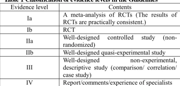
Evidence level Contents
Ia A meta-analysis of RCTs (The results of RCTs are practically consistent.)
Ib RCT
IIa Well-designed controlled study (non- randomized)
IIb Well-designed quasi-experimental study
III Well-designed non-experimental,
descriptive study (comparison/ correlation/
case study)
IV Report/comments/experience of specialists
Recommendation grade Contents
A Strongly recommended
B Recommended
C1 Can be considered, but adequate scientific rationale lacking
C2 Not recommendable because of
absence of scientific rationale
D Not recommended
Classification of evidence levels and recommendation grades in the Guidelines.
Table 1 Classification of evidence levels in the Guidelines
This classification is according to that adopted in the “2004 Guidelines for Stroke Treatment” by the Japan Stroke Society.
Table 2 Classification of recommendation gr ades in the Guidelines
Precautions for the use of the Guidelines
(1) The clinical condition needs to be assessed in individual patients, and the Guidelines are not uniformly applicable to all individual patients.
Therefore, the judgment of the treating physician who most accurately understands his/her patient‟s clinical condition should be afforded priority in the management of patients with moyamoya disease.
(2) The Guidelines should not be referred to without careful consideration, used as data for evaluation of medical examinations, or for medical accidents or lawsuits. Attention should be paid to the fact that the Guidelines include drugs not approved or therapies not authorized in Japan and drugs used for purposes other than the original intent.
(3) The number of patients is small and the cause of moyamoya disease is still unknown. Therefore, there are many aspects of the disease that still remain unresolved and for which adequate amount of evidence has not yet been collected. Therefore, it must be borne in mind while using the Guidelines that there may be many inaccuracies and that the contents may not always necessarily be up to date or the best for the time.
2005 to 2008 Health Labour Sciences Research Grant for Research on Measures for Intractable Diseases
Research Committee on the Pathology and Treatment of Spontaneous Occlusion of the Circle of Willis
Chief Researcher:
Nobuo Hashimoto, Department of Neurosurgery, Kyoto University Graduate School of Medicine
(Current) National Cardiovascular Center Researchers:
Teiji Tominaga, Department of Neurosurgery, Tohoku University Graduate School of Medicine
Susumu Miyamoto, Department of Neurosurgery, Kyoto University Graduate School of Medicine
Izumi Nagata, Department of Neurosurgery, Nagasaki University Graduate School of Biomedical Sciences
Kiyohiro Houkin, Department of Neurosurgery, Hokkaido University Graduate School of Medicine
Norihiro Suzuki, Department of Neurology, Keio University School of Medicine
Akio Koizumi, Department of Health and Environmental Sciences, Kyoto University, Graduate School of Medicine, School of Public Health
Shigeru Nogawa, Department of Internal Medicine, Tokyo Dental College Ichikawa General Hospital
Jyoji Nakagawara, Department of Neurosurgery, Nakamura Memorial Hospital
Kazuo Kitagawa, Department of Neurology, Osaka University, Graduate School of Medicine
Satoshi Kuroda, Department of Neurosurgery, Hokkaido University Graduate School of Medicine
Kenichiro Kikuta, Department of Neurosurgery, Fukui University Graduate School of Medicine
Research collaborators:
Miki Fujimura, Department of Neurosurgery, Sendai Medical Center
Jun Takahashi, Department of Neurosurgery, Kyoto University, Graduate School of Medicine
Kentaro Hayashi, Department of Neurosurgery, Nagasaki University Graduate School of Biomedical Sciences
Koichi Oki, Department of Neurology, Keio University School of Medicine
Haruhiko Hoshino, Department of Neurology, Tokyo Saiseikai Central Hospital
Secretariat:
Yasushi Takagi, Department of Neurosurgery, Kyoto University Graduate School of Medicine
『無症候性もやもや病の予後と治療法の確 立
をめざした多施設共同研究』
Asymptomatic Moyamoya Registry (AMORE) プロトコール
厚生科学研究費特定疾患対策研究事業
〜ウィリス動脈輪閉塞症の病因・病態に関する研究〜
Ver 2.1.0 2011 年 10 月 26
日1
0.概 要
0.1. フローチャート
2
1.2 . 目 的
無症候性ウィリス動脈輪閉塞症(もやもや病)の疫学・病態・予後を明ら かとす る。
1.3 . 登録基準
新たにウィリス動脈輪閉塞症と診断された 20〜70 歳の患者のうち、それま でに一 過性脳虚血発作、脳梗塞、頭蓋内出血(脳出血、脳室内出血あるいはク モ膜下出血) のエピソードを有していない症例。
0.4. 目標登録症例数
200 症例
0.5. 研究期間
登録期間:3 年、観察期間:5 年(合計 8 年)
0.6. 研 究 デ ザ イ
ン
多施設共同観察研究
0.7. 連絡先 研究事務局
黒田 敏、浜田秀雄
富山大学附属病院 脳神経外科
〒076-0194 富山市杉谷 2630
Tel 076-43407348 Fax 076-434-5034 E-mail [email protected] 竹内佐和子、高戸賀子(担当秘書)
E-mail [email protected]
本研究実施計画書は、本研究に直接かかわる者および倫理審査委員会以外の者に情報を開示してはならな い。また、本状法は事前の書面による主任研究者の承諾なしに本件の実施あるいは評価以外の目的
3
に利用 してはならない。
4
本研究に関与する全ての者は「世界医師会ヘルシンキ宣言」および「臨床研究に関する倫理指針」に従 う。