• 検索結果がありません。

 Gastric cancer is the second most common cause of cancer related mortality worldwide

N/A
N/A
Protected

Academic year: 2021

シェア " Gastric cancer is the second most common cause of cancer related mortality worldwide"

Copied!
7
0
0

読み込み中.... (全文を見る)

全文

(1)

Introduction

 Gastric cancer is the second most common cause of cancer related mortality worldwide

1)

. Complete surgical resection, the only potentially curative treatment for advanced gastric cancer, can only be performed on a small subgroup of patients. In 30% to 50% of patients undergoing surgical exploration, the intent of the surgery is to cure the patient. However, almost 60% of patients who undergo an R0 curative resection relapse and die of their disease

2, 3)

.

 Clinical staging is determined using TNM classification : T indicating depth of invasion of the primary tumor, N nodal metastasis, and M distant metastasis. This classification system is useful to estimate patient prognosis. Currently, endoscopic ultrasound(EUS)and com- puted tomography(CT)are widely used for preoperative tumor staging

4)

. The accuracy of

Expression of Standard CD44 in Advanced Gastric Cancer : Relationship with Metastasis to Lymph Nodes

Shuei A

RIMA

, Toshiaki K

UNIMURA

, Hiromi D

ATE

, Kai M

ATSUO

, Tomoaki M

ORI

, Yasuo O

CHIAI

, Takeyoshi K

ITAYAMA

and Toshio M

OROHOSHI

Abstract : Standard CD44(CD44s)is reported to play an important role in determining the malignant potential of various carcinomas. The aim of the present study was to evaluate CD44s expression in T2-T3 gastric cancer(Japa- nese Classification of Gastric Cancer stages MP, SS, SE)and the relationship between CD44s expression and clinicopathological parameters. CD44s expres- sion was measured using immunohistochemistry in tumors from 98 patients with primary gastric cancer. Cases were categorized into two groups based on CD44s staining ; the CD44s positive group had >10% positively stained tumor cells and the CD44s negative group had < 10%. CD44s positivity was demonstrated in 59.1%(58 / 98)of tumors. CD44s expression showed no significant relationship with patient age or gender, or tumor location, size or macroscopic / microscopic classification. However, CD44s expression showed a significantly negative relationship with metastasis to lymph nodes(p <0.0001) . Thus, in T2-T3 gastric cancer, loss of CD44s expression suggests that metastasis of the tumor to lymph nodes is likely.

Key words : CD44s, advanced gastric cancer, lymph node metastasis Original

First Department of Pathology, Showa University School of Medicine, 1-5-8 Hatanodai, Shinagawa-ku, Tokyo 142-

8666, Japan.

(2)

staging tumors by determining the T value has been much improved with EUS having an accuracy of between 78%-92%

5-11)

, and CT between 69%-89%

9, 12-18)

.

 However, the accuracy of establishing the N category is still poor ; 63%-78% using EUS

5-11)

and 51%-78% using CT

9, 12-18)

. The N category estimates 5-year survival rate in patients with N0 gastric cancer as 89.4%, but 68.3% in those with N1 disease

19)

. Thus, improving the accuracy of the N value in patients with gastric cancer, could improve the accuracy of the preoperative prognosis.

 The transmembrane glycoprotein molecule standard CD44(CD44s)is a cell surface hyaluronate receptor, and is expressed in most epithelial and nonepithelial tissues. The clinical significance of CD44s expression in gastric cancer, however, remains unclear. Various studies have reported an association of CD44s expression with an advanced stages of gastric cancer and a poor prognosis

20-23)

, with one study reporting adverse correlation between CD44s expression and tumor progression

24-29)

.

 The aim of the present study was to evaluate CD44s expression in T2-T3 gastric cancer

(Japanese Classification of Gastric Cancer stages ; muscularis propria MP, subserosa SS, serosa exposed SE)and the relationship between CD44s expression and clinicopathologi- cal parameters. We thought that it would be possible to improve preoperative prognostic precision by using CD44s, if a relationship would be present between CD44s and various clinicopathological factors(especially N value)in gastric cancer.

Materials and Methods

 Tissue samples from 98 patients who had T2-T3 primary gastric cancer surgically resected at Showa University Hospital from 2004 through 2009 were collected. Samples were pre- served in 10% formalin for 24 h and the nonnecrotic portion of the tumors was routinely processed and paraffin embedded. Representative sections were stained with hematoxylin and eosin. Various clinicopathological parameters were evaluated based on the Japanese Classification of Gastric Carcinoma(The 13th Edition, web site : http://www.jgca.jp/PDFfiles/

JCGC-2E.PDF) .

Immunohistochemistry

 Three micron sections from representative blocks of each case were stained immunohis-

tochemically using monoclonal antibody clone 2C5(anti-CD44s ; R&D Systems, Minneapolis,

MN, USA)at a dilution of 1 : 500. The labeled streptavidin-biotin-peroxidase labeling

technique was used. Sections were deparaffinized in xylene, dehydrated in descending

grades(100-50%)of ethanol, and subjected to antigen retrieval(microwave, citrate pH 6.0,

25 min) . After cooling to room temperature, slides were incubated to quench endogenous

peroxidase activity with 1% hydrogen peroxide in ethanol for 30 min. Nonspecific immu-

noreactivity was blocked by incubation with normal donkey serum for 30 min each. The

sections were then incubated with primary antibody at 4℃ overnight. After three 5 minute

(3)

washes with phosphate-buffered saline(PBS) , sections were incubated for 60 min with the multilink biotinylated anti-immunoglobulin. Three 5-minute washes with PBS preceded and followed treatment with streptavidin-peroxidase reagent for 30 min. The reactions were visu- alized with diaminobenzidine(DAKO A-S, Copenhagen, Denmark)as a chromogen. Finally, sections were counterstained with hematoxylin, dehydrated and mounted.

 CD44s expression was seen as brown, fine to coarse granular staining on the cytoplasmic membrane. Our study aimed to estimate the averaged CD44s expression in the entire tumor area, and defined CD44s-positivity to be when more than 10% of the tumor cells was stained(Fig. 1) . This definition was chosen as it is the most commonly used in the literature

20, 24, 25)

. And cases were categorized into two groups based on CD44s staining ; the CD44s positive group had > 10% positively stained tumor cells and the CD44s negative group had <10%. All histological slides were examined by two experienced pathologists blinded to clinical data or disease outcome.

Statistical analysis

 Association between CD44s expression and clinicopathological parameters was tested using the chi-square test, with significance set at P < 0.05.

Fig. 1. Sections of gastric tumors stained using CD44s immunohistochemis- try demonstrating the tumor grading system

A : Section showing more than 10% of the tumor cells with positive staining for CD44s, and so recorded as a tumor with positive CD44s expression. 400

B : Section showing lack of CD44s staining of tumor cells, and so recorded

as a tumor with negative CD44s expression. 400

(4)

Results

 The characteristics of the study population are shown in Table 1. The subjects consisted of 70 men(71.4%)and 28 women(28.6%) , with a mean age of 69.4 years(range, 45-91 years)and a mean gastric tumor size of 5.2 cm(range, 1.5-13.8 cm) .

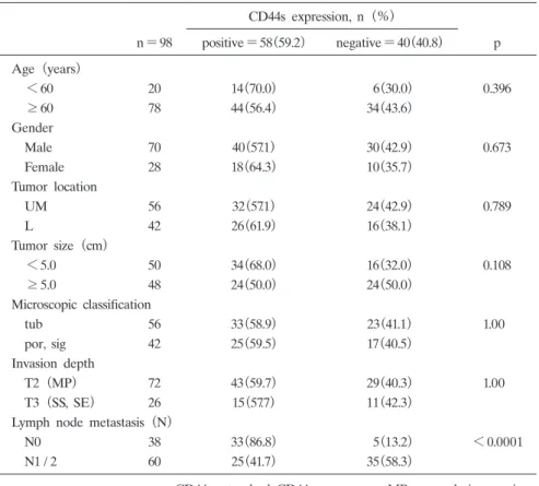
 CD44s expression in the primary tumor was demonstrated in 59.1%(58 / 98)of the tumors. Associations between tumor CD44s expression and the clinicopathological param- eters of patients and gastric tumors are summarized in Table 1. No statistically significant differences were found between positive CD44s expression in gastric tumors in patients aged less than 60 years(70%)compared with those 60 years or older(56.4%) . No statistically significant differences were found between positive CD44s expression in gastric tumors in male(57.1%)or female patients(64.3%) . No statistically significant differences were found between positive CD44s expression in gastric tumors occurring in either the lower stomach

(61.9%)or the middle or upper stomach(57.1%) . Similarly for tumor size, no statistically significant differences were found between positive CD44s expression in gastric tumors less

Table 1 CD44s expression and clinicopathogical parameters CD44s expression, n(%)

n=98 positive=58 (59.2) negative=40 (40.8) p Age(years)

<60 20 14 (70.0) 6 (30.0) 0.396 60 78 44 (56.4) 34 (43.6)

Gender

Male 70 40 (57.1) 30 (42.9) 0.673

Female 28 18 (64.3) 10 (35.7)

Tumor location

UM 56 32 (57.1) 24 (42.9) 0.789 L 42 26 (61.9) 16 (38.1)

Tumor size(cm)

<5.0 50 34 (68.0) 16 (32.0) 0.108 5.0 48 24 (50.0) 24 (50.0)

Microscopic classification

tub 56 33 (58.9) 23 (41.1) 1.00

por, sig 42 25 (59.5) 17 (40.5)

Invasion depth

T2(MP) 72 43 (59.7) 29 (40.3) 1.00

T3(SS, SE) 26 15 (57.7) 11 (42.3)

Lymph node metastasis(N)

N0 38 33 (86.8) 5 (13.2) <0.0001

N1 / 2 60 25 (41.7) 35 (58.3)

CD44s ; standard CD44 tub ; well differentiated type por, sig ; poorly differentiated type

MP ; muscularis propria SS ; subserosa

SE ; serosa exposed

(5)

than 5.0 cm in diameter, compared with tumors 5.0 cm in diameter or greater. For micro- scopic tumor type, no statistically significant differences were found between positive CD44s expression in gastric tumors tub1 / tub2(58.9%)or in por / sig(59.5%) . However, strong statistical significance was found in the association between positive CD44 expression and lymph node metastases ; positive expression was present in 86.8% of gastric tumors from patients with no detectable lymph node metastasis(classification N0) , but in only 41.6%

of patients with lymph node metastasis(classification N1 / 2)(P <0.0001) . Thus, CD44s expression in gastric tumors had a significantly negative relationship with the presence of lymph node metastasis in the patient.

Discussion

 CD44 is a large family of cell surface transmembrane glycoproteins whose members have different extracellular domains as a result of alternative gene splicing

30, 31)

. CD44s is a receptor for hyaluronate and is highly expressed on human lymphocytes. In malignant tumors originating from various organs, there is some controversy as to whether CD44s acts as a growth / invasion-promoting molecule or tumor suppression cofactor. While a number of studies have shown that CD44s expression is associated with metastasis and a poor patient prognosis

21)

, others have shown that down-regulation of CD44s expression is correlated with an adverse patient outcome. For example, reduced CD44s expression has been shown to be associated with a high incidence of lymphatic or vascular infiltration in endometrial carcinoma

26)

and breast carcinoma

24)

, and a poor prognosis in patients with pancreatic carcinoma, thyroid carcinoma, and squamous cell carcinoma of the head and neck

27, 28)

. These differences in study findings may be due to the use of different antibodies, immunohistochemistry methods, patient material, and duration of patient follow-up.

 Similarly, the clinical significance of CD44s expression in the progression and metastasis of malignant gastric tumor is also controversial. However, Montgomery et al reported that loss of CD44s expression in gastric stromal tumors was consistent with tumor aggressiveness

29)

, and Joo et al found a significant relationship between CD44s expression and lymph node metastasis in gastric cancer

22)

. Further, Mayer et al demonstrated expression of CD44s was significantly positively associated with recurrence of gastric adenocarcinoma, distant metastasis and patient mortality

23)

. These findings have been speculated to be due to CD44s activa- tion of p185 tyrosine kinase and c-Src kinase, interactions which are important in tumor progression

29)

.

 Our study found an adverse relationship between CD44s expression and lymph node metastasis in T2-T3 gastric cancer. This finding may be due to the loss of CD44s expres- sion and hence, reduced intercellular adhesion and adhesion between cells and basement membranes. This could facilitate detachment of tumor cells from their primary sites and allow infiltration of lymphatic or vascular spaces

32)

.

 The results of this study support a role for CD44s in the inhibition of metastasis of

(6)

gastric tumor cells to lymph nodes. This possible suppression of gastric tumor metastasis by CD44s may result from its role in binding gastric tumor cells to the extracellular matrix in the gastric muscularis propria and subserosal layers and thus restricting cancer cell spread.

 CD44s expression may thus be a useful prognostic indicator in cases with gastric cancer, and may facilitate patient-specific tailoring of treatment options, such as the use of chemo- therapy.

References

1) Hohenberger P and Gretschel S : Gastric cancer. Lancet 362:305-315(2003)

2) De Vita F, Giuliani F, Galizia G, Belli C, Aurilio G, Santabarbara G, Ciardiello F, Catalano G and Orditura M : Neo-adjuvant and adjuvant chemotherapy of gastric cancer. Ann Oncol 18:120-123(2007)

3) Dicken BJ, Bigam DI, Cass C, Mackey JR, Joy AA and Hamilton SM : Gastric adenocarcinoma : review and consideration for future direction. Ann Surg 241:27-39(2005)

4) Kwee RM and Kwee TC : Imaging in local staging of gastric cancer : a systematic review. J Clin Oncol 25:

2107-2116(2007)

5) Wang JY, Hsieh JS, Huang YS, Huang CJ, Hou MF and Huang TJ : Endoscopic ultrasonography for preopera- tive locoregional staging and assessment of resectability in gastric cancer. Clin Imaging 22:355-359(1998)

6) Mancino G, Bozzeti F, Schicchi A, Schiavo M, Spinelli P and Andreola S : Preoperative endoscopic ultrasonog- raphy in patients with gastric cancer. Tumori 86:139-141(2000)

7) Xi WD, Zhao C and Ren GS : Endoscopic ultrasonography in preoperative staging of gastric cancer : determi- nation of tumor invasion depth, nodal involvement and surgical respectability. World J Gastroenterol 9:254-257

(2003)

8) Willis S, Truong S, Gribnitz S, Fass J and Schumpelick V : Endoscopic ultrasonography in the preoperative stag- ing of gastric cancer : accuracy and impact on surgical therapy. Surg Endosc 14:951-954(2000)

9) Bhandari S, Shim CS, Kim JH, Jung IS, Cho JY, Lee JS, Lee MS and Kim BS : Usefulness of three-dimen- sional, multidetector row CT(virtual gastroscopy and multiplanar reconstruction)in the evaluation of gastric cancer : a comparison with conventional endoscopy, EUS, and histopathology. Gastrointest Endosc 59:619-626

(2004)

10) Shimoyama S, Yasuda H, Hashimoto M, Tatsutomi Y, Aoki F, Mafune K and Kaminishi M : Accuracy of liner- array EUS for preoperative staging of gastric cardia cancer. Gastrointest Endosc 60:50-55(2004)

11) Ganpathi IS, So JB and Ho KY : Endoscopic ultrasonography for gastric cancer : does it influence treatment?

Surg Endosc 20:559-562(2006)

12) DʼElia F, Zingarelli A, Palli D and Grani M : Hydro-dynamic CT preoperative staging of gastric cancer : cor- relation with pathological findings. A prospective study of 107 cases. Eur Radiol 10:1877-1885(2000)

13) Habermann CR, Weiss F, Riecken R, Honarpisheh H, Bohnacker S, Staedtler C, Dieckmann C, Schoder V and Adam G : Preoperative staging of gastric adenocarcinoma : comparison of helical CT and endoscopic US.

Radiology 230:465-471(2004)

14) Takao M, Fukuda T, Iwanaga S, Hayashi K, Kusano H and Okudaira S : Gastric cancer : evaluation of triphasic spiral CT and radiologic-pathologic correlation. J Comput Assist Tomogr 22:288-294(1998)

15) Hundt W, Braunschweig R and Reiser M : Assessment of gastric cancer : value of breathhold technique and two-phase spiral CT. Eur Radiol 9:68-72(1999)

16) Shimizu K, Ito K, Matsunaga N, Shimizu A and Kawakami Y : Diagnosis of gastric cancer with MDCT using the water-filling method and multiplanar reconstruction : CT-histologic correlation. AJR Am J Roentgenol 185:

1152-1158(2005)

17) Chen CY, Hsu JS, Wu DC, Kang WY, Hsieh JS, Jaw TS, Wu MT and Liu GC : Gastric cancer : preoperative

local staging with 3D multi-detector row CT-correlation with surgical and histopathologic results. Radiology

(7)

242:472-482(2007)

18) Hur J, Park MS, Lee JH, Lim JS, Yu JS, Hong YJ and Kim KW : Diagnostic accuracy of multidetector row computed tomography in T and N staging of gastric cancer with histopathologic correlation. J Comput Assist Tomogr 30:372-377(2006)

19) Park SR, Kim MJ, Ryu KW, Lee JH, Lee JS, Nam BH, Choi IJ and Kim YW : Prognostic value of preopera- tive clinical staging assessed by computed tomography in respectable gastric cancer patients. Ann Surg 251:

428-435(2010)

20) Huh JW, Kim HR, Kim YJ, Lee JH, Park YS, Cho SH and Joo JK : Expression of standard CD44 in human colorectal carcinoma : association with prognosis. Pathol Int 59:241-246(2009)

21) Sneath RJ and Mangham DC : The normal structure and function of CD44 and its role in neoplasia. Mol Pathol 51:191-200(1998)

22) Joo M, Lee HK and Kang YK : Expression of E-cadherin, beta-catenin, CD44s and CD44v6 in gastric adeno- carcinoma : relationship with lymph node metastasis. Anticancer Res 23:1581-1588(2003)

23) Mayer B, Jauch KW, Günthert U, Figdor CG, Schildberg FW, Funke I and Johnson JP : De-novo expression of CD44 and survival in gastric cancer. Lancet 342:1019-1022(1993)

24) Gong Y, Sun X, Huo L, Wiley EL and Rao MS : Expression of cell adhesion molecules, CD44s and E-cadher- in, and microvessel density in invasive micropapillary carcinoma of the breast. Histopathology 46:24-30(2005)

25) Kunimura T, Yoshida T, Sugiyama T and Morohoshi T : The relationships between loss of standard CD44 expression and lymph node, liver metastasis in T3 colorectal carcinoma. J Gastrointest Cancer 40:115-118

(2009)

26) Fujita N, Yaegashi N, Ide Y, Sato S, Nakamura M, Ishiwata I and Yajima A : Expression of CD44 in normal human versus tumor endometrial tissues : possible implication of reduced expression of CD44 in lymph-vascular space involvement of cancer cells. Cancer Res 54:3922-3928(1994)

27) Carinci F, Stabellini G, Calvitti M, Pelucchi S, Targa L, Farina A, Pezzetti F and Pastore A : CD44 as prognos- tic factor in oral and oropharyngeal squamous cell carcinoma. J Craniofac Surg 13:85-89(2002)

28) Sato S, Miyauchi M, Takekoshi T, Zhao M, Kudo Y, Ogawa I, Kitagawa S, Fujita M and Takata T : Reduced expression of CD44 variant 9 is related to lymph node metastasis and poor survival in squamous cell carci- noma of tongue. Oral Oncol 36:545-549(2000)

29) Montgomery E, Abraham SC, Fisher C, Deasel MR, Amr SS, Sheikh SS, House M, Lilliemoe K, Choti M, Brock M, Ephron DT, Zahuruk M and Chadburn A : CD44 loss in gastric stromal tumors as a prognostic marker. Am J Surg Pathol 28:168-177(2004)

30) Günthert U, Hofmann M, Rudy W, Reber S, Zöller M, Haussmann I, Matzku S, Wenzel A, Ponta H and Her- rlich P : A new variant of glycoprotein CD44 confers metastatic potential to rat carcinoma cells. Cell 65:13-24

(1991)

31) Stamenkovic I, Amiot M, Pesando JM and Seed B : A lymphocyte molecule implicated in lymph node homing is a member of the cartilage link protein family. Cell 56:1057-1062(1989)

32) Asao T, Nakamura J, Shitara Y, Tsutsumi S, Mochiki E, Shimura T, Takenoshita S and Kuwano H : Loss of standard type of CD44 expression in invaded area as a good indicator of lymph-node metastasis in colorectal carcinoma. Dis Colon Rectum 43:1250-1254(2000)

[Received March 19, 2010 : Accepted April 23, 2010]

Fig. 1. Sections  of  gastric  tumors  stained  using  CD44s  immunohistochemis- immunohistochemis-try  demonstrating  the  tumor  grading  system
Table 1 CD44s  expression  and  clinicopathogical  parameters CD44s  expression,  n(%) n=98 positive=58 (59.2) negative=40 (40.8) p Age(years) <60 20 14 (70.0) 6 (30.0) 0.396 60 78 44 (56.4) 34 (43.6) Gender Male 70 40 (57.1) 30 (42.9) 0.673 Female 28 18 (

参照

関連したドキュメント

In this study, we investigated whether tranilast inhibits the effects of TGF-b-induced EMT in HPMCs by inhibiting the TGF-b/Smad pathway, and whether fibrosis can be attenuated in

reported that gemcitabine-mediated apoptosis is caspase- dependent in pancreatic cancers; Jones et al [14] showed that gemcitabine-induced apoptosis is achieved through the

NELL1 (a) and NELL2 (b) mRNA expression levels in renal cell carcinoma cell lines OS-RC-2, VMRC-RCW, and TUHR14TKB and control HEK293T cells were analyzed using quantitative

Keywords: homology representation, permutation module, Andre permutations, simsun permutation, tangent and Genocchi

Habiro con- siders an abelian group A k (H) dened by unitrivalent graphs with k trivalent vertices and with univalent vertices labelled by elements of H , subject to anti- symmetry,

Physiologic evaluation of the patient with lung cancer being considered for resectional surgery: Diagnosis and management of lung cancer, 3rd ed: American College of Chest

Applications of msets in Logic Programming languages is found to over- come “computational inefficiency” inherent in otherwise situation, especially in solving a sweep of

Shi, “The essential norm of a composition operator on the Bloch space in polydiscs,” Chinese Journal of Contemporary Mathematics, vol. Chen, “Weighted composition operators from Fp,