Title
Expression of macrophage migration inhibitory factor and CD74 in the inner ear and
middle ear in lipopolysaccharide-induced otitis media
Running Head
MIF and CD74 in otitis media
Authors
Hisashi Ishihara, Shin Kariya, Mitsuhiro Okano, Pengfei Zhao, Yukihide Maeda,
Kazunori Nishizaki
Author’s affiliations
Department of Otolaryngology-Head and Neck Surgery, Okayama University Graduate
School of Medicine, Dentistry and Pharmaceutical Sciences, Okayama, Japan
Abstract:
Conclusion: Significant expression of macrophage migration inhibitory factor and its
receptor (CD74) was observed in both the middle ear and inner ear in experimental
otitis media in mice. Modulation of macrophage migration inhibitory factor and its
signaling pathway might be useful in the management of inner ear inflammation due to
otitis media.
Objectives: The inner ear dysfunction secondary to otitis media has been reported.
However, the specific mechanisms involved are not clearly understood. The aim of this
study is to investigate the expression of macrophage migration inhibitory factor and
CD74 in the middle ear and inner ear in lipopolysaccharide-induced otitis media.
Method: BALB/c mice received a transtympanic injection of either lipopolysaccharide
or phosphate-buffered saline (PBS). The mice were sacrificed 24 h after injection, and
temporal bones were processed for polymerase chain reaction (PCR) analysis, histologic
examination, and immunohistochemistry.
Results: PCR examination revealed that the lipopolysaccharide-injected mice showed a
significant up-regulation of macrophage migration inhibitory factor in both middle ear
and inner ear as compared with the PBS-injected control mice. Immunohistochemical
study showed positive reactions for macrophage migration inhibitory factor and CD74
in infiltrating inflammatory cells, middle ear mucosa, and inner ear in the
lipopolysaccharide-injected mice.
Keywords
hearing loss; tinnitus; cochlea; hair cell; spiral ganglion cells; stria vascularis; spiral
ligament; cytokine; inflammation; labyrinthitis
Introduction
Otitis media is a common childhood disease and is the major cause of hearing
loss in children. Multiple factors including infection, Eustachian tube dysfunction, and
inflammatory cytokines are involved in the pathogenesis of otitis media [1].
Inflammatory mediators in the middle ear cavity can spread from the middle ear into the
inner ear through the round window during the course of otitis media [2, 3]. This
condition can lead to cochlear pathology including the loss of hair cells in the cochlea,
resulting in sensorineural hearing loss [4-6].
Macrophage migration inhibitory factor is an inflammatory cytokine, and is
also an essential factor for neural development [7]. The expression of macrophage
migration inhibitory factor in middle ear effusion in patients with otitis media and the
effect of macrophage migration inhibitory factor in cochlear function have been
reported [8-10]. However, the role of macrophage migration inhibitory factor in the
inner ear is still under debate.
Up-regulation of macrophage migration inhibitory factor in the middle ear in
experimental otitis media has been reported [11]. In addition, the blocking macrophage
migration inhibitory factor activity relieves middle ear inflammation in
lipopolysaccharide-induced otitis media [12]. However, to the best of our knowledge,
no previous study reported the finding of macrophage migration inhibitory factor and its
receptor (CD74) in the inner ear during the course of otitis media. The purpose of this
study is to determine the expressions of macrophage migration inhibitory factor and
CD74 in the middle ear and inner ear in experimental otitis media in mice.
Material and Methods
Animals
BALB/c mice were used in this study. The mice were deeply anesthetized
with an intraperitoneal injection of a mixture of ketamine (100 mg/kg body weight) and
xylazine (10 mg/kg body weight). An otoscopic examination was performed on all mice
prior to treatment in order to ensure that the tympanic membranes were normal and that
no middle ear effusion was present.
The mice were randomly divided into two groups. The experimental group
(n=10) received lipopolysaccharide (1.0 mg/mL; Sigma-Aldrich, St Louis, Missouri,
USA) via transtympanic injection using a 30-gauge needle. Phosphate-buffered saline
(PBS) at 0.01 M was injected into the middle ear of the animals in the control group
(n=10). The mice were sacrificed 24 hours after injection of the lipopolysaccharide or
PBS. The temporal bones were removed immediately after sacrifice and processed for
polymerase chain reaction (PCR) analysis, histologic examination, and
immunohistochemistry.
This study conformed to the current laws of Japan and was performed in
accordance with the relevant animal protection rules. The Animal Research Control
Committee of Okayama University approved the study (Approval number,
OKU-2013121).
Polymerase chain reaction
Inner ear (cochlea and vestibular end organ) was dissected from the tympanic
bulla. Tympanic bulla harvested for analysis included middle ear mucosa, bulla wall,
and ossicles. Inner and middle ear RNA collected separately from the BALB/c mice
(experimental group, n=6, 12 ears; control group, n=6, 12 ears) were subjected to
quantitative PCR. Total RNA was extracted using an RNeasy Mini Kit (Qiagen,
Valencia, California, USA) according to the manufacturer's protocol. PCR was
performed with a TaqMan® gene expression assay, using a specific pre-made TaqMan®
probe for macrophage migration inhibitory factor gene (Mm01611157_gH;
CATCAGCCCGGACCGGGTCTACATC; Applied Biosystems, Foster City, California, USA), TaqMan® One-step RT-PCR Master Mix Reagents (Applied Biosystems), and
Applied Biosystems 7500 Real-Time PCR System (Applied Biosystems).
Glyceraldehyde 3-phosphate dehydrogenase (GAPDH) (Mm99999915_g1,
GAACGGATTTGGCCGTATTGGGCGC, Applied Biosystems) was used as an
endogenous control in all analyses.
Histologic examination
The temporal bone specimens (experimental group, n=2, 4 ears; control group,
n=2, 4 ears) were placed in 4% paraformaldehyde for 72 hours and decalcified in 10%
ethylenediaminetetraacetic acid for 3 weeks at 4 ℃. After dehydration, the specimens
were embedded in paraffin and sectioned at a thickness of 10 μm, then mounted on
glass slides, processed using hematoxylin and eosin staining, and evaluated under light
microscopy.
Immunohistochemistry
The paraffin-embedded tissues (experimental group, n=2, 4 ears; control
group, n=2, 4 ears) were sectioned at a thickness of 4 μm and mounted on glass slides.
The sections were deparaffinized and rehydrated. Endogenous peroxidase activity was
quenched with 0.3% hydrogen peroxide in methanol for 30 minutes at room temperature.
Antigen retrieval was performed by microwave heating. Non-specific protein binding
was blocked with goat serum albumin (Vector Laboratories, Inc., Burlingame,
California, USA) for 1 hour at room temperature. Immunohistochemical staining was
performed using rabbit anti-macrophage migration inhibitory factor antibody (sc-20121;
Santa Cruz Biotechnology, Inc., Santa Cruz, California, USA) and rabbit anti-CD74
antibody (NBP1-33109; Novus Biologicals, Littleton, Colorado, USA) overnight at 4 ℃.
Rabbit Immunoglobulin Fraction (X0903, Dako, Glostrup, Denmark) was used as a
negative control. For visualization, a VECTASTAIN Elite ABC Kit (Vector
Laboratories, Inc.) and 3, 3’-diaminobenzidine (DAB) reagent (K3467, Dako) were
used according to the manufacturer’s instructions.
Statistical analysis
Data are presented as means ± standard deviation. For statistical analysis, the
non-parametric Mann-Whitney U test was used for comparison between the two groups.
Significant differences were established at a level of P < 0.05 (IBM SPSS Statistics;
IBM, New York, USA).
Results
Histologic examination
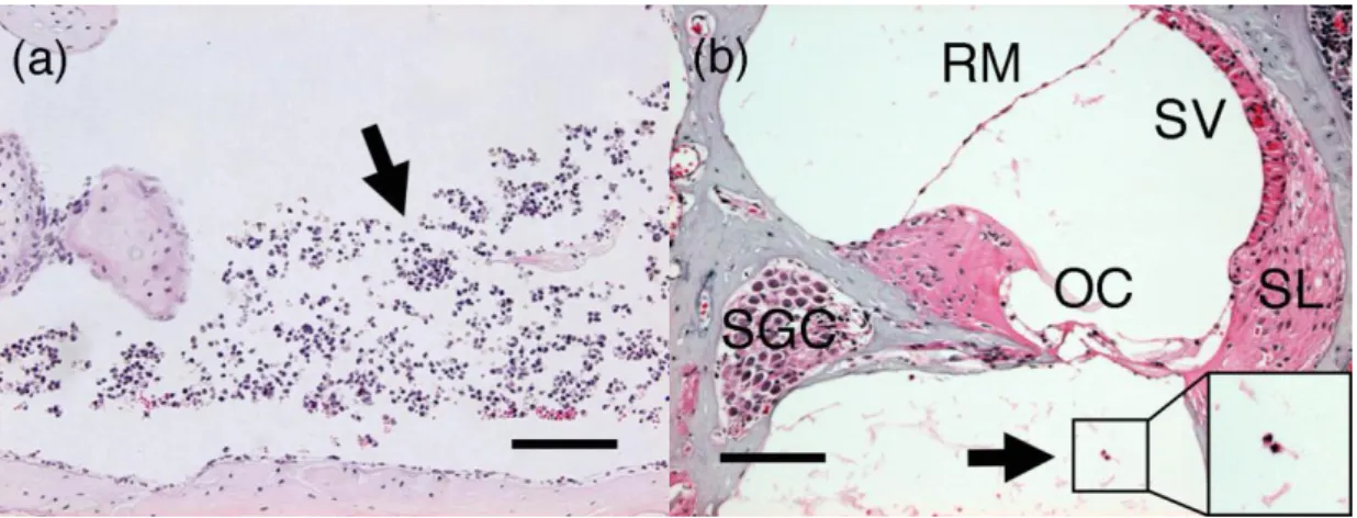
The lipopolysaccharide-injected mice showed a remarkable infiltration of
inflammatory cells (polymorphonuclear leukocyte and monocyte) in the middle ear
cavity (Figure 1a). Labyrinthitis with inflammatory cell infiltration was observed in the
cochlea of the lipopolysaccharide-injected mice (Figure 1b). No significant
inflammatory finding was detected in the middle ear and inner ear of the PBS-injected
mice.
Quantitative PCR
The expressions of macrophage migration inhibitory factor gene relative to
GAPDH in the middle ear and inner ear tissues are shown in Figure 2. The
lipopolysaccharide-injected mice showed a significant increase in the gene expression
of macrophage migration inhibitory factor in both the middle ear (P < 0.05) and inner
ear (P < 0.05) as compared with the PBS-injected control mice.
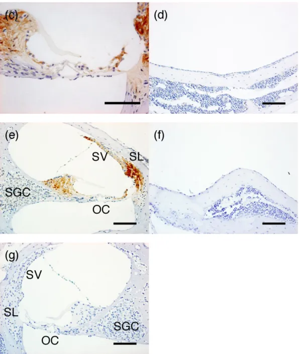
Expression of macrophage migration inhibitory factor
The PCR findings suggest the possibility of a role played by macrophage
migration inhibitory factor in the middle ear and inner ear in
lipopolysaccharide-induced otitis media. Next, we examined the localization of the
macrophage migration inhibitory factor. Strong positive immunostaining was found for
macrophage migration inhibitory factor in the infiltrating inflammatory cells as well as
mucosal epithelium in the middle ear of the lipopolysaccharide-injected mice (Figure
3a). Positive immunostaining for macrophage migration inhibitory factor was observed
in the spiral ligament, stria vascularis, spiral limbus, spiral ganglion cells, organ of Corti,
and infiltrating inflammatory cells in the cochlea of the lipopolysaccharide-treated mice
(Figure 3b, 3c). Macrophage migration inhibitory factor was not detected in the middle
ear mucosa of the PBS-treated mice (Figure 3d). Positive immunostaining for
macrophage migration inhibitory factor was observed in the spiral ligament, stria
vascularis, spiral limbus, spiral ganglion cells, and organ of Corti in the PBS-treated
mice (Figure 3e). Macrophage migration inhibitory factor was strongly expressed in the
saccular macula, utricular macula, crista ampullaris, and cells lining the membranous
labyrinth, but not in the facial nerve in the PBS-treated mice. There was no significant
immunostaining in the middle ear and inner ear in the negative controls using Rabbit
Immunoglobulin Fraction in the PBS-treated mice (Figure 3f, 3g).
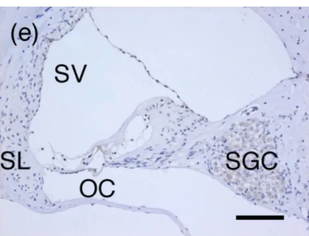
Expression of CD74
Up-regulation of macrophage migration inhibitory factor was observed in
lipopolysaccharide-induced otitis media in both the mRNA level and protein level.
Therefore, we next examined the presence of the receptor for macrophage migration
inhibitory factor (CD74) in lipopolysaccharide-induced otitis media. CD74 was
expressed in the infiltrating inflammatory cells in the middle ear cavity and middle ear
mucosa (Figure 4a). CD74 was also detected in fibrocytes of the spiral ligament, stria
vascularis, spiral limbus, spiral ganglion cells, and organ of Corti in
lipopolysaccharide-induced otitis media (Figure 4b, 4c). Unlike macrophage migration
inhibitory factor, CD74 was detected in middle ear mucosa in PBS-treated mice (Figure
4d). CD74-positive cells were also detected in inner ear (spiral ligament, stria vascularis,
spiral limbus, spiral ganglion cells, and organ of Corti) in PBS-treated mice (Figure 4e).
Discussion
Gram-negative bacteria are the major pathogens in otitis media.
Lipopolysaccharide, also known as endotoxin, is the structural component of the outer
membrane of gram-negative bacteria, and is a critical pathogenic mediator of
inflammatory diseases. A previous study reported that lipopolysaccharide was detected
in the middle ear in 96% of patients with otitis media [8]. The injection of bacterial
lipopolysaccharide into the middle ear can cause labyrinthitis, and induces cochlear
damage [13]. We showed here for the first time that macrophage migration inhibitory
factor and CD74 were significantly expressed in inner ear in
lipopolysaccharide-induced otitis media.
The significant role of macrophage migration inhibitory factor in acute
infections and chronic inflammatory diseases has been reported. For example, the role
of macrophage migration inhibitory factor in sepsis has been extensively examined. The
plasma level of macrophage migration inhibitory factor in patients with sepsis
correlated positively with the severity of sepsis, and was significantly higher in septic
patients who died than in those who survived [14]. In the middle ear, down-regulation
of macrophage migration inhibitory factor activity reduced middle ear inflammation in
experimental otitis media [12]. Macrophage migration inhibitory factor is also an
important factor in maintaining normal cochlear function [7]. We showed that
macrophage migration inhibitory factor was expressed in inner ear in the PBS-treated
control mice. Macrophage migration inhibitory factor-positive inflammatory cell
infiltration might be related to the significant expression of macrophage migration
inhibitory factor shown by PCR in inner ear in lipopolysaccharide-treated mice.
CD74, also known as a MHC class II invariant chain, is a type II
transmembrane protein and is a major component of the macrophage migration
inhibitory factor receptor complex. Macrophage migration inhibitory factor binds to cell
surface CD74, and induces p44/p42 MAPK phosphorylation and cell proliferation [15].
Macrophage migration inhibitory factor induces neutrophilic inflammation in rodent
lung, and administration of anti-CD74 antibody inhibits the infiltration of neutrophils
and the production of inflammatory cytokine and chemokine [16]. On the other hand,
CD74 is required for the maintenance of normal alveolar structure in mice [17]. We
showed here that CD74 was expressed in both the middle ear and the inner ear in
lipopolysaccharide-induced otitis media.
In conclusion, middle ear injection of lipopolysaccharide induced significant
expression of macrophage migration inhibitory factor, and the receptor (CD74) for
macrophage migration inhibitory factor was observed in the middle ear and inner ear of
the lipopolysaccharide-injected mice. Macrophage migration inhibitory factor and
CD74 might play some roles in both the middle ear and inner ear in
lipopolysaccharide-induced otitis media in mice. Our findings suggest that macrophage
migration inhibitory factor and its signaling pathway serve as a therapeutic target in the
management of inner ear damage induced by otitis media.
Acknowledgments
This work was supported by JSPS KAKENHI (Grants-in-Aid for Scientific Research
from The Ministry of Education, Culture, Sports, Science and Technology of Japan;
Grant Number 25462642). The authors have no other funding, financial relationships, or
conflicts of interest to disclose.
References
[1] Juhn SK, Jung MK, Hoffman MD, Drew BR, Preciado DA, Sausen NJ, et al. The
role of inflammatory mediators in the pathogenesis of otitis media and sequelae. Clin
Exp Otorhinolaryngol 2008;1:117-38.
[2] Schachern PA, Tsuprun V, Cureoglu S, Ferrieri P, Briles D, Paparella M, et al. The
round window membrane in otitis media: effect of pneumococcal proteins. Arch
Otolaryngol Head Neck Surg 2008;134:658-62.
[3] Tsuprun V, Cureoglu S, Schachern PA, Ferrieri P, Briles DE, Paparella MM, et al.
Role of pneumococcal proteins in sensorineural hearing loss due to otitis media. Otol
Neurotol 2008;29:1056-60.
[4] Joglekar S, Morita N, Cureoglu S, Schachern PA, Deroee AF, Tsuprun V, et al.
Cochlear pathology in human temporal bones with otitis media. Acta Otolaryngol
2010;130:472-6.
[5] MacArthur CJ, Hausman F, Kempton JB, Sautter N, Trune DR. Inner ear tissue
remodeling and ion homeostasis gene alteration in murine chronic otitis media. Otol
Neurotol 2013;34:338-46.
[6] MacArthur CJ, Hausman F, Kempton JB, Choi D, Trune DR. Otitis media impacts
hundreds of mouse middle and inner ear genes. PLoS One 2013;8:e75213.
[7] Bank LM, Bianchi LM, Ebisu F, Lerman-Sinkoff D, Smiley EC, Shen YC, et al.
Macrophage migration inhibitory factor acts as a neurotrophin in the developing inner
ear. Development 2012;139:4666-74.
[8] Kariya S, Okano M, Aoji K, Kosaka M, Chikumoto E, Hattori H, et al. Role of
macrophage migration inhibitory factor in otitis media with effusion in adults. Clin
Diagn Lab Immunol 2003;10:417-22.
[9] Kariya S, Okano M, Maeda Y, Hirai H, Higaki T, Noyama Y, et al. Role of
macrophage migration inhibitory factor in age-related hearing loss. Neuroscience
2014;279:132-8.
[10] Kariya S, Okano M, Maeda Y, Hirai H, Higaki T, Noyama Y, et al. Macrophage
migration inhibitory factor deficiency causes prolonged hearing loss after acoustic
overstimulation. Otol Neurotol 2015;36:1103-8.
[11] Kariya S, Schachern PA, Cureoglu S, Tsuprun V, Okano M, Nishizaki K, et al.
Up-regulation of macrophage migration inhibitory factor induced by endotoxin in
experimental otitis media with effusion in mice. Acta Otolaryngol 2008;128:750-5.
[12] Zhang J, Xu M, Zheng Q, Zhang Y, Ma W, Zhang Z. Blocking macrophage
migration inhibitory factor activity alleviates mouse acute otitis media in vivo. Immunol
Lett 2014;162(1 Pt A):101-8.
[13] Choi CH, Jang CH, Cho YB, Jo SY, Kim MY, Park BY. Matrix metalloproteinase
inhibitor attenuates cochlear lateral wall damage induced by intratympanic instillation
of endotoxin. Int J Pediatr Otorhinolaryngol 2012;76:544-8.
[14] Kerschbaumer RJ, Rieger M, Völkel D, Le Roy D, Roger T, Garbaraviciene J, et al.
Neutralization of macrophage migration inhibitory factor (MIF) by fully human
antibodies correlates with their specificity for the β-sheet structure of MIF. J Biol Chem
2012;287:7446-55.
[15] Leng L, Metz CN, Fang Y, Xu J, Donnelly S, Baugh J, et al. MIF signal
transduction initiated by binding to CD74. J Exp Med 2003;197:1467-76.
[16] Takahashi K, Koga K, Linge HM, Zhang Y, Lin X, Metz CN, et al. Macrophage
CD74 contributes to MIF-induced pulmonary inflammation. Respir Res 2009;10:33.
[17] Sauler M, Leng L, Trentalange M, Haslip M, Shan P, Piecychna M, et al.
Macrophage migration inhibitory factor deficiency in chronic obstructive pulmonary
disease. Am J Physiol Lung Cell Mol Physiol 2014;306:L487-96.
Figure captions
Figure 1: Histopathological findings of the (a) middle ear and (b) inner ear in
lipopolysaccharide (LPS)-injected mice. Infiltration of inflammatory cells (arrow) was
observed. (hematoxylin and eosin staining; OC, organ of Corti; RM, Reissner’s
membrane; SV, stria vascularis; SL, spiral ligament; SGC, spiral ganglion cell; Scale bar,
100 μm).
Figure 2: Mean expression of macrophage migration inhibitory factor mRNA by
real-time reverse transcription polymerase chain reaction (RT-PCR) assays. Quantitative
analysis of the real-time RT-PCR results revealed that macrophage migration inhibitory
factor mRNA expression was significantly up-regulated in the lipopolysaccharide
(LPS)-injected mice both in the middle ear and inner ear as compared with normal
control ear tissues from mice with middle ear injection of phosphate-buffered saline
(PBS). (LPS, lipopolysaccharide; PBS, phosphate-buffered saline; MIF, macrophage
migration inhibitory factor; GAPDH, glyceraldehyde-3-phosphate dehydrogenase; *,
P<0.05).
Figure 3: Immunohistochemical staining for macrophage migration inhibitory factor
expression in (a) middle ear cavity in lipopolysaccharide (LPS)-treated mice, (b) inner
ear in LPS-treated mice, (c) organ of Corti in LPS-treated mice, (d) middle ear cavity in
phosphate-buffered saline (PBS)-treated mice, and (e) inner ear in PBS-treated mice.
Negative control immunohistochemical staining using Rabbit Immunoglobulin Fraction
in (f) middle ear cavity and (g) inner ear of PBS-treated mice. Positive immunostaining
for macrophage migration inhibitory factor was observed in infiltrating inflammatory
cells (arrow) of LPS-treated mice (a, b). Macrophage migration inhibitory
factor-positive staining was also observed in the inner ear of PBS-treated mice (e). (OC,
organ of Corti; SV, stria vascularis; SL, spiral ligament; SGC, spiral ganglion cell; Scale
bar, 100 μm)
Figure 4: Immunohistochemical staining for CD74 expression in (a) middle ear cavity
in lipopolysaccharide (LPS)-treated mice, (b) inner ear in LPS-treated mice, (c) organ of
Corti in LPS-treated mice, (d) middle ear mucosa in phosphate-buffered saline
(PBS)-treated mice, and (e) inner ear in PBS-treated mice. Positive immunostaining for
CD74 was observed in infiltrating inflammatory cells (arrow). CD74-positive staining
was also observed in the inner ear of LPS-treated mice (b, c). (OC, organ of Corti; SV,
stria vascularis; SL, spiral ligament; SGC, spiral ganglion cell; Scale bar, 100 μm)