1
PKR induces the expression of NLRP3 by regulating the NF-B pathway in Porphyromonas
gingivalis-infected osteoblasts
Kaya Yoshida1*, Hirohiko Okamura2, 7, Yuka Hiroshima3, Kaori Abe4, Jun-ichi Kido5, Yasuo Shinohara3, Kazumi Ozaki6
1
Department of Oral Healthcare Education and 2Department of Histology and Oral Histology, Institute of Biomedical Sciences, Tokushima University Graduate School,
3-18-15
Kuramoto, Tokushima 770-8504, Japan3
Institute for Genome Research, Tokushima University, 3-18-15 Kuramoto, Tokushima 770-8503,
Japan
4
SHIBASAKI, Inc., 507 Horikiri, Chichibu 368-0066, Japan
5
Department of Periodontology and Endodontology and 6 Oral Healthcare Promotion, Institute of Biomedical Sciences, Tokushima University Graduate School,
3-18-15
Kuramoto, Tokushima 770-8504, Japan7
Department of Oral Morphology, Graduate School of Medicine, Dentistry and Pharmaceutical
Sciences, Okayama University, 2-5-1 Shikata, Kita-ku, Okayama 770-8525, Japan
Abbreviated title: PKR is required for P.gingivalis-induced NLRP3 expression in osteoblasts.
*Address correspondence to:
Kaya Yoshida, DDS, PhD,
3-18-15, Kuramoto, Tokushima 770-8504, Japan.
TEL: +81-88-633-7898, FAX: +81-88-633-7898
2
AbstractThe double-stranded RNA-dependent kinase (PKR), which is activated by double stranded RNA,
induces inflammation by regulating NF-B signaling. The NLR family pyrin domain-containing 3 (NLRP3) inflammasome also modulates inflammation in response to infection. Porphyromonas
gingivalis (P.gingivalis) is an oral bacterium which is implicated in the pathogenesis of periodontal
diseases. We previously reported that PKR is a key modulator of bone metabolism and inflammation
in the periodontal tissue. PKR was also reported to induce inflammation in response to microbes by
regulating the NLRP3 inflammasome, suggesting that PKR could affect inflammation along with
NLRP3 in periodontal diseases. In this study, we investigated the effects of PKR on NLRP3
expression and NF-B activity in P. gingivalis infected osteoblasts. We first constructed a SNAP26b-tagged P.gingivalis (SNAP-P. g.) and traced its internalization into the cell. SNAP-P. g.
increased the activity of PKR and NF-B and also induced NLRP3 expression in osteoblasts. Inhibition of NF-B attenuated SNAP-P. g.-induced NLRP3 expression. The knockdown of PKR using shRNA decreased both the activity of NF-B and the expression of NLRP3 induced by SNAP-P.g.. We therefore concluded that in osteoblasts, P. gingivalis activated PKR, which in turn
increased NLRP3 expression by activating NF-B. Our results suggest that PKR modulates inflammation by regulating the expression of the NLRP3 inflammasome through the NF-B pathway in periodontal diseases.
Keywords: Double-stranded RNA-dependent kinase; inflammasomes; NF-kappa B; osteoblasts; Porphyromonas gingivalis
3
1. IntroductionThe serine-threonine kinase PKR is ubiquitously expressed in mammalian cells and was originally
reported to be activated by binding to the double-stranded RNA derived from viruses. In
virus-infected cells, activated PKR inhibits protein synthesis and promotes apoptosis through
phosphorylation of the subunit of eukaryotic initiation factor 2 (eIF2), resulting in an induction of the immune response [1]. PKR is now known to mediate inflammation, cancer [2] and obesity [3, 4]
in response to a variety of factors, including cytokines [5], growth factors, lipopolysaccharide (LPS)
[6] and oxidative stresses [7].
The diverse functions of PKR are considered to be dependent on its ability to act as a signal
transducer. For example, activated PKR affects multiple pathways mediated by several kind of
transcriptional factors such as interferon regulatory factor 3 (IRF3), nuclear factor B (NF-B), c-Jun, and activating transcription factor 2 (ATF2), leading to regulation of inflammation and immunity [8].
Among these pathways, the mechanisms by which PKR regulate NF-B signaling has been well studied. PKR is autophosphorylated and activated by the inflammation-induced stimuli, and directly
interacts with the IB kinase (IKK) subunit of IKK complex to phosphorylate it [9, 10]. The activated IKK then phosphorylates the inhibitory of B (IB) which retain NF-B in the cytoplasm, while the IB is degraded by the 26S proteasome in an ubiquitin-dependent manner. This degradation allows NF-B to translocate to the nucleus where it promotes the transcription of genes that regulate inflammation. Thus, PKR is considered to be a crucial factor in modulating inflammation by
regulating NF-B signaling.
The NLR family pyrin domain-containing 3 (NLRP3) inflammasome- a multiprotein complex
comprising of NLRP3, the adaptor apoptosis-associated speck-like protein (ASC) and pro-caspase-1,
is also reported to modulate inflammation. In viral and bacterial infected cells, NLRP3
4
and p10 subunits. The subunits assemble to form active caspase-1 hetero-dimers that catalytically
cleave pro-IL-1 into the biologically active form. IL-1 is one of most potent proinflammatory cytokines and the increase of IL-1secretion by the inflammasomes is related to the pathogenesis of several diseases [11, 12].
NLRP3 inflammasome is known to be activated by two steps—an initial priming step and an
activation step. Recently, NF-B has been found to be required for the priming step [11]. In response to stimuli, the priming step is mediated through the Toll-like receptors (TLRs), which activates
NF-B and increases the expression of NLRP3, which in turn convert the pro-IL-1 to its functional form. In the activation step, the induced NLRP3 is subsequently able to assemble and form the
complex of NLRP3, ASC and pro-caspase-1, resulting in an increase in cleavage of pro-caspase-1
and IL-1 secretion. Indeed, the inhibition of either TLRs or NF-B resulted in the reduction of NLRP3 expression and caspase-1 cleavage in macrophages [13], suggesting that the priming step is
important to activate inflammasomes. However the molecular mechanism regulating this remains
poorly understood.
Porphyromonas gingivalis (P.gingivalis) is an oral microorganism which is implicated in the
pathogenesis of periodontal disease, a chronic inflammatory disorder of the periodontal tissue.
Progress in periodontal disease induces inflammation and resorption of alveolar bone, resulting in
tooth loss. An understanding of how bacterial infection affects bone metabolism and induces bone
resorption is therefore necessary to prevent periodontal disease.
Little is known about the relationship between PKR and periodontal disease. We previously reported
that PKR positively regulates the differentiation of osteoblasts by inducing GSK-3 activity using mouse osteoblastic cell line MC3T3-E1 cells, which have the capacity to undergo osteoblastic
differentiation and mineralization in vitro [14, 15]. We also showed that LPS isolated from E. coli
phosphorylates PKR to increase the expression of pro-inflammatory cytokines through NF-B signaling in human gingival cells [6]. Our results suggested that PKR might be a key modulator for
5
bone metabolism and inflammation in periodontal tissue. However, the effects of the periodontal
bacteria on PKR activity and PKR-regulated inflammation in osteoblasts still remain unknown.
Several studies have also indicated that NLRP3 is involved in the pathogenesis of periodontal
disease. Bostanci et al. reported that NLRP3 is expressed at significantly higher levels in gingival
tissue from patients with periodontal disease [16]. They also showed that P. gingivalis regulates the
NLRP3 inflammasome complex by increasing NLRP3 and down-regulating NLRP2 and ASC
expression in human myelomonocytic cell line Mono-Mac-6 [16]. It was found that in human
monocytic cell line THP-1, P. gingivalis activates the NLRP3 inflammasome via TLR2 and TLR4
[17] pathways. Infection with another periodontal bacteria Aggregatibacter actinomycetemcomitans
(A. actinomycetemcomitans) also promotes apoptosis of human osteoblastic MG63 cells by activating
the NLRP3 inflammasome [18]. Moreover, A. actinomycetemcomitans enhances NLRP3 and reduces
NLRP6 expression leading to the release of IL-1 in human mononuclear leukocytes [19].
Recently, PKR has been reported to induce the immune response and inflammation in response to
microbes by regulating multiple inflammasomes [20]. PKR directly interacts with inflammasome
components such as NLRP3, NLRP4, and AIM2 to mediate inflammasome activity [21]. Inhibition of
PKR by p58IPK suppressed the activity of NLRP3 inflammasomes in macrophages [22].
These observations suggest that PKR, along with NLRP3 affects inflammation in osteoblasts during
periodontal diseases. We therefore hypothesized that PKR could be activated by P. gingivalis and
contribute to inflammation by regulating the NLRP3 in osteoblasts. In this study, we therefore
investigated the effects of PKR on NLRP3 expression and NF-B activity in P. gingivalis-infected osteoblasts.
6
2. Materials and Methods2.1. Materials
Caffeic acid phenethyl ester (CAPE) was purchased from Calbiochem (San Diego, USA). Antibodies
against phosphorylated eIF2a (Ser51), NLRP3 (D4D8T), IB-and GAPDH (14C10) were obtained from Cell Signaling Technology (Danvers, MA, USA). Antibodies against PKR (M-515),
phospho-PKR (Thy 446, sc-101783), NF-B p65 (F-6), caspase-1 p10 (M-20), IL-1 (H-153) and
-actin were obtained from Santa Cruz Biotechnology (Santa Cruz, CA, USA) and the SNAP-tag® rabbit polyclonal antibody was obtained from New England Biolabs.
2.2. Establishment of SNAP-P.g.
SNAP-P.g. was constructed by transforming P. gingivalis ATCC33277 as described previously [23].
Briefly, SNAP26b was amplified from the bacterial expression plasmid pSNAP-tag® (T7). The
promoter (protrxB) and terminator (termtrxB) regions of trxB (Gene ID 6330860) were also amplified.
The three PCR fragments were ligated into pBlueScript®II Phagemid Vector (pSK), and the resulting
recombinant plasmids (pSK- protrxB_SNAP26b_termtrxB) were transformed into E. coli Single Step
(KRX) competent cells. The protrxB_SNAP26b_termtrxB (pst) fragment was cloned into a shuttle
vector, pT-COW and the resulting plasmid (pT-COW_pst) was introduced into P. gingivalis strain
ATCC33277 by electroporation.
2.3. Bacterial cultures
SNAP-P.g. was cultured in Brain Heart Infusion (BD Bioscience, Franklin Lakes, NJ) containing
0.5% yeast extract (BD Bioscience), 10 g/mL hemin (Wako Chemicals, Osaka, Japan), 1 g/mL 2-methyl-1, 4-naphthoquinone (vitamin K3) (Tokyokasei, Tokyo, Japan) and 5g/mL tetracycline in an anaerobic condition at 37°C.
7
2.4. Cell culture and treatment with SNAP-P.g. preparations
The mouse osteoblastic cell line MC3T3-E1 cells were plated at a density of 3 × 10³ cells/ mL and cultured in plastic dishes in -MEM supplemented with 10% FBS for 5 days as previously described [24]. For experiments, the medium was changed to -MEM supplemented with 10% FBS containing 50 g/mL ascorbic acid and 10 mM -glycerophosphate, and the cells were infected with SNAP-P.g. at a multiplicity of infection (MOI) of 100 for the desired time period.
2.5. SDS-PAGE and western blot analysis
Cells were washed with PBS and were scraped into TN lysis buffer (50 mM Tris [pH 8.0], 150 mM
NaCl, 0.1% NP-40) in the presence of protease inhibitors (4 g/mL aprotinin, 1g/mL leupeptin, 0.2 mM PMSF). The protein extracts obtained were subjected to immunoblotting according to the
protocol described previously [25].
2.6. Real-time PCR
Total RNA was isolated from MC3T3-E1 cells using ISOGEN (Nippon Gene, Tokyo, Japan),
followed by phenol extraction and ethanol precipitation. The cDNA was synthesized using Prime
ScriptTM RT (Takara Bio, Kyoto, Japan) as per manufacturer’s protocol. The levels of mRNA were measured by the real-time reverse transcription-PCR method using a 7300 Real-Time PCR system
(Applied Biosystems, Carlsbad, CA, USA) and SYBR Premix Ex TaqTM (Takara Bio) for detection. The primer sequences were as follows: mouse GAPDH (NM_001001303): forward,
5'-TGTGTCCGTCGTGGATCTGA-3', reverse, 5'-TTGCTGTTGAAGTCGCAGGAG-3'; mouse
NLRP3 (NM_145827.3): forward, 5'-ACTGAAGCACCTGCTCTGCAAC-3', reverse,
5'-AACCAATGCGAGATCCTGACAAC-3'.
8
The NF-B promoter reporter assay was performed by transfection of the luciferase plasmid pNF-B-Luc containing 5
×
binding sites (TGGGGACTTTCCGC) (Agilent Technologies, La Jolla, CA). One g of pNF-B-Luc and 0.05 g of pRL-TK Renilla luciferase vector (Promega) expressingRenilla luciferase (as control of transfection efficiency) were used for electroporation into MC3T3-E1
cell using an Amaxa® Nucleofector II -program T-20 (Lonza). The luciferase activity of NF-B was measured by Dual-Luciferase® Reporter Assay System (Promega) according to the manufacturer’s
directions and normalized to the control Renilla luciferase activity.
2.8. Immunocytochemistry
MC3T3-E1 cells were cultured on sterile 18-mm round coverslips and immunocytochemistry was
performed as described previously [6]. Briefly, the cells were fixed with 3% formalin for 30 min and
permeabilized with 0.1% Triton X-100 in PBS for 2 min on ice. After blocking with 4% BSA in PBS
for 1 h, the cells were incubated with anti-NF-B p65 antibody overnight at 4°C, followed by Alexa Fluor 488-conjugated anti-mouse IgG. The samples were mounted and observed using an inverted
fluorescence microscope (ECLIPSE Ti-U, Nikon, Tokyo). Images were acquired using ECLIPSE
Ti-U microscope and analyzed with its NIS-Elements software (Nikon, Tokyo, Japan).
2. 9. shRNA
MC3T3-E1 cells were plated at a density of 0.5
×
104 cells in 96-well plates and infected with MISSION® shRNA lentiviral transduction particles specific for mouse PKR (NM_011163,Sigma-Aldrich) according to the manufacturer’s directions. The infected cells were selected as stable
clones by treatment with 5 g/mL of puromycin. MISSION® Non-Target shRNA lentiviral transduction particles were used as negative control.
9
All data are expressed as means ± SD with a minimum of 3 independent experiments performed for
10
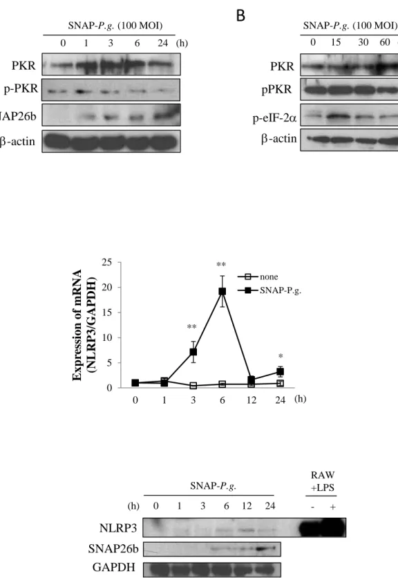
3. Results3.1. SNAP-P.g. activates PKR and induces NLRP3 expression in osteoblasts
We first established a SNAP26b-tagged P.gingivalis (SNAP-P. g.) in order to specifically trace the
internalization of P. gingivalis into osteoblastic cells. MC3T3-E1 cells were infected with SNAP-P.g.
at a MOI of 100 for 0 to 24 h, and SNAP-P.g. was detected by western blotting using antibody against
the SNAP26b protein. As shown in Fig. 1A, the intensity of the band corresponding to SNAP26b
increased at 1 h and remained increased up to 24 h post infection, indicating that SNAP-P.g.
internalized into MC3T3-E1 cells. We then examined whether the infection of SNAP-P. g. induced
PKR activation in these cells. The level of total PKR was increased at 1 h and persisted up to 6 h post
infection. The phosphorylation of PKR at threonin 446, however, did not change upon SNAP-P.g.
infection (Fig. 1A). We therefore examined the effect of SNAP-P.g. on PKR phosphorylation at
earlier time points. As shown in Fig. 1B, SNAP-P.g. induced the phosphorylation of PKR at threonin
446, 15 min after infection. eIF-2, the substrate phosphorylated by PKR, was also phosphorylated after 15 min of infection (Fig. 1B). -actin, used as an internal control, remained unchanged for the samples (Fig. 1A, 1B).
Next, the cells were incubated with or without SNAP-P.g. (at a concentration of 100 MOI) for 0 to
24 h and the effect of SNAP-P.g. on NLRP3 expression was analyzed by real-time PCR. The mRNA
levels of NLRP3 increased and reached maximum expression 6 h post infection, after which it
reduced. No increase in the mRNA expression was observed at the 12 h time-point and there was only
a slight increase in its level 24 h post-infection (Fig. 1C). Furthermore, the expression of NLRP3
protein in SNAP-P.g. treated MC3T3-E1 cells was examined by western blotting. The intensity of the
band corresponding to SNAP26b increased in a time-dependent manner up to 24 h (Fig. 1D, middle
11
Consistent with the results obtained by real-time PCR, the increase of NLRP3 was very little in
SNAP-P.g. treated MC3T3-E1 cells 24 h post infection (Fig. 1D, upper panel).
3.2. SNAP-P.g. activates NF-B by inducing IB degradation in osteoblasts
It is reported that NLRP3 expression is increased through the NF-B pathway in the priming step. We therefore investigated whether NF-B is implicated in the up-regulation of NLRP3 expression induced by SNAP-P.g. in MC3T3-E1 cells. We infected cells with SNAP-P.g. for 60 min and then
detected the phosphorylation of p65, a component of NF-B, at serine 536 by western blotting. As shown in figure 2A, the level of phosphorylated p65 was increased upon infection (Fig. 2A, upper
panel). Because NF-B phosphorylates in the cytoplasm and translocates to the nucleus only after the degradation of inhibitor IB (which bind to NF-B), we examined the effect of SNAP-P.g. on the level of IB protein. As expected, SNAP-P.g. infection reduced the IB levels in MC3T3-E1 cells (Fig. 2A, middle panel). We then analyzed the localization of p65 in cells with or without infection by
immunocytochemistry using an antibody for p65. In control cells, p65 was observed in cytoplasm (Fig.
2B, a, c), while in cells infected with SNAP-P.g., the major part of p65 translocated from cytoplasm
to nucleus (Fig. 2B, b, d, e). We then assessed the transcriptional activity of p65 using a dual
luciferase reporter assay. The luciferase plasmid pNF-B-Luc and pRL-TK Renilla luciferase vector were transfected into MC3T3-E1 cells. After transfection, the cells were treated with SNAP-P.g for
up to 3 h, and then luciferase activity was assessed by a luminometer. The transcriptional activity of
p65 was not altered 1 h post infection, however, it was significantly increased at 3 h (p < 0.01) (Fig.
2C).
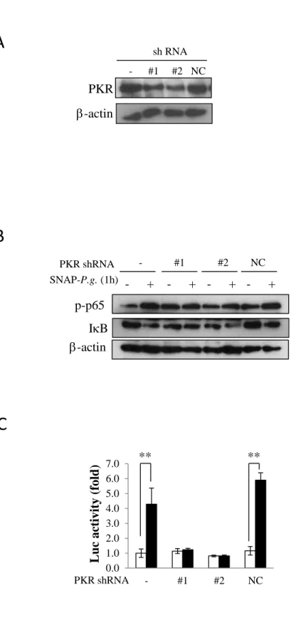
3.3. NF-B regulates SNAP-P.g.-induced NLRP3 expression in osteoblasts
To clarify whether the NF-B activation induced by SNAP-P.g. was involved in the increase of NLRP3 expression, we pre-treated cells with BAY11-7085, an inhibitor of IB phosphorylation, and
12
then infected the cells with SNAP-P.g. for 1 h. As shown in figure 3A, BAY11-7085 inhibited the
SNAP-P.g.-induced IB degradation and p65 phosphorylation. These inhibitions also partly decreased the mRNA expression of NLRP3 (Fig. 3B). Consistent with the results obtained by real-time PCR,
BAY 11-7085 also inhibited the protein level of NLRP3 induced by SNAP-P.g. (Fig. 3C).
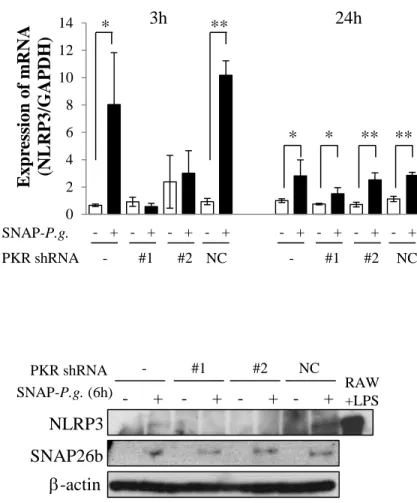
3.4. Reduction of PKR alters NF-B activation and NLRP3 expression induced by SNAP-P. g. in
osteoblasts
To clarify whether PKR is implicated in the molecular mechanism by which SNAP-P.g. increased
NLRP3 expression, PKR knockdown cells were constructed by infecting MC3T3-E1 cells with a
lentivirus expressing a shRNA specific for PKR. We established two clones of the PKR knockdown
cells (#1, #2), and confirmed the efficiency of shRNA knockdown in them. Figure 4A shows that
PKR knockdown cells (#1, #2) expressed lower levels of PKR protein when compared to the
untransfected wild type cells (-). The lower level of PKR protein was not observed in the cells
infected with non-target shRNA as negative control (NC) (Fig. 4A). Next, we examined the effect of
PKR on the IB degradation and NF-B phosphorylation in SNAP-P.g.-infected cells. The infection with SNAP-P.g. for 1 h induced IB degradation and NF-B phosphorylation in (-) and NC cells, however the effect was not observed in PKR knockdown cells (#1, #2) (Fig. 4B). The transcriptional
activity of NF-B was also significantly increased by SNAP-P.g. infection in wild type (-) and NC cells. However, SNAP-P.g. did not affect the transcriptional activity of NF-B in PKR knockdown cells (#1, #2) (Fig. 4C).
We later examined if the NF-B inactivation by shRNA specific for PKR affects the expression of NLRP3. In wild type and NC cells, infection with SNAP-P.g. for 3 h significantly increased the
mRNA expression of NLRP3. This increase was not observed in PKR knockdown cells (#1, #2) (Fig.
4D, left). 24 h post infection, SNAP-P.g. slightly increased the mRNA expression of NLRP3 in(-) and
13
(-) and NC cells (Fig. 4D, right). Figure 4E shows that SNAP-P.g. increased the level of NLRP3
protein in (-) and NC cells 6 h post infection, while the expression of the protein was repressed by the
14
4. DiscussionIn this study, SNAP-P. g. increased the activity of PKR and NF-B, and induced NLRP3 expression in osteoblasts. The inhibition of NF-B attenuated SNAP-P. g.-induced NLRP3 expression, suggesting that SNAP-P. g. increases NLRP3 expression through the NF-B signaling pathway. Finally, the knockdown of PKR by shRNA decreased both the activity of NF-B and expression of NLRP3 induced by SNAP-P. g.. Based on these observations, we concluded that PKR was activated
by SNAP-P. g. and then increased the expression of NLRP3 by activating NF-B in osteoblasts.
Bacteria and its individual components such as LPS are known to induce PKR activation and
inflammation in different kinds of cells [26]. We also observed that LPS from E. coli induces PKR
activation, resulting in the production of pro-inflammatory cytokines in human gingival cells [6].
However, it was not known whether the infection of oral microorganisms implicated in periodontal
diseases can activate PKR in osteoblasts. In this study, PKR was phosphorylated 15 min after P.
gingivalis infection followed by the phosphorylation of eIF-2, (Fig. 1B), suggesting that P.
gingivalis activates PKR in osteoblasts.
In contrast, the mechanism by which P. gingivalis activates PKR remains unknown. Since P.
gingivalis has been known to internalize into cells and cause chronic inflammation [27], it was
initially expected that P.gingivalis affect PKR after its invasion of osteoblasts. We therefore used P.
gingivalis labeled with SNAP26b tag-protein (SNAP-P.g.) in order to trace its internalization. In
SNAP-P.g.-treated osteoblasts, SNAP26b was detected starting from 1 h up to 24 h post infection (Fig.
1A). In contrast, the phosphorylation of PKR and eIF-2 was observed as early as at 15 min (Fig. 1B) but not 1 h after the internalization of P.gingivalis (Fig. 1A). These results indicated that P. gingivalis
activates PKR without invading the osteoblasts, and therefore other mechanisms are implicated in P.
15
We hypothesized that TLRs might mediate PKR activation in osteoblasts infected with P. gingivalis.
TLRs have been reported to recognize the extracellular milieu and the pathogen-associated molecular
pattern (PAMP) such as flagellin and LPS. LPS from P. gingivalis is known to be an agonist of TLR2,
and it enhances the production of receptor activator of NF-B ligand (RANKL) in a TLR2/NF-B dependent manner in mouse parietal osteoblasts [28]. Moreover, the stimulation of TLRs promotes the
priming signal of NLRP3 and increases the release of IL-1 release in human monocytes [29]. Thus, it is likely that TLR2 could be involved in the activation of PKR induced by P. gingivalis, resulting in
the increase of NLRP3 expression. Further studies are needed to address this possibility.
Our results showed that P. gingivalis activated PKR almost immediately after infection leading to an
increase in both NF-B phosphorylation and activity within 3 h (Fig. 2). Following NF-B activation, the expression of NLRP3 mRNA increased at 3 h and reached maximum expression at 6 h, and then
decreased and exhibited only a modest level of increase after 24 h of P. gingivalis infection (Fig. 1C).
In agreement with the report that the priming of NLRP3 is regulated by NF-B [11], the induction of NLRP3 mRNA by P. gingivalis at 3 h was partially dependent on NF-B in these osteoblasts (Fig. 3B, C). PKR is well known to be essential for NF-B activation in inflammatory processes [30]. In consistent with those reports, we also observed the knockdown of PKR by shRNA markedly reduced
the NF-B activity (Fig. 4D, left), showing that PKR might increase NLRP3 expression by regulating NF-B activity. Interestingly, the regulation of PKR on NLRP3 mRNA expression was observed only at 3 h (Fig. 4D, left), but not 24 h after P. gingivalis infection (Fig. 4D, right). These results suggested
that PKR might play a critical role in NLRP3 expression, especially in early stages of infection.
We concluded that PKR is required for the priming step for NLRP3 in P. gingivalis-infected
16
inflammasome activation remains unanswered in the present study. In agreement with the report
showing that IL-1 production is tightly controlled [31], the level of IL-1 expression in osteoblasts was very low and infection with P. gingivalis did not increase its extracellular release (data not
shown). Moreover, we observed that P. gingivalis induced cleavage of pro-caspase-1 into p10 subunit
up to 6 h post infection and the PKR shRNA treatment did not affect it (data not shown). Therefore, it
is likely that PKR is necessary for the P. gingivalis-induced NLRP3 expression in the priming step,
but not sufficient for the subsequent activation of NLRP3 in osteoblasts. These results agree with the
previous report that PKR was dispensable for caspase-1 activation, processing of IL-1 and secretion of IL-1 induced by LPS in macrophages [32]. In contrast, other studies demonstrated that PKR activates inflammasomes in macrophages treated with LPS [21, 22]. Due to these conflicting data, the
role of PKR in the activation of NLRP3 inflammasome still needs further clarification.
The danger signals, such as ATP, released from infected cells are considered to be one of the factors
that regulate NLRP3 activation. It has been reported that the activation of inflammasomes requires a
second signal such as exogenous ATP in macrophages [13, 33]. In gingival epithelial cells, LPS from
P. gingivalis induced IL-1 gene, but the cytokine was not secreted unless co-stimulated with LPS
and ATP [34]. Recently, the reactive oxygen species (ROS) induced by ATP-P2X7 receptor has been
reported to positively regulate the inflammasome activation in gingival epithelial cells infected with P.
gingivalis [35], and the expression of P2X7 receptor, IL-1and NLRP3 was also modulated in human
chronic periodontitis [36]. Moreover, it was reported that periodontal pathogens such as P. gingivalis,
Treponema denticola and Tannerella forsythia induces release of ATP in macrophages [37]. These
observations suggest that ATP and ROS produced by oral bacterial infections may contribute to the
inflammasome activation, thereby promoting periodontal diseases [38, 39]. To validate the roles of
PKR in NLRP3 activation in periodontal diseases, further experiments must be conducted using PKR
specific shRNA to show the effects of PKR on caspase-1 cleavage and IL-1 release and the difference in inflammasome activation in the presence of ATP.
17
Moreover, to clarify the role of PKR in the pathogenesis of periodontal diseases, the effect of
biofilms on PKR needs to be further investigated; in this study, P. gingivalis was specially evaluated.
The formation of biofilms, which contain multiple species of oral bacteria, result in periodontal
diseases. Contrary to our results here, subgingival biofilms have been reported to downregulate the
expression of NLRP3 mRNA and IL-1 in primary human gingival fibroblasts [40]. Notably, the exclusion of P. gingivalis from the biofilm reportedly partially rescues this downregulated expression
of NLRP3 mRNA and IL-1 [41]. In contrast, our results show that P. gingivalis may accelerate inflammation by increasing NLRP3 inflammasomes. These observations suggest that P. gingivalis is
implicated in the host immune system by reducing the expression of NLRP3 inflammasomes in
response to an oral biofilms challenge, and that these distinct roles of P. gingivalis are important in
18
5. ConclusionWe demonstrated that PKR rapidly induces NF-B in response to infection with P. gingivalis, leading to the up-regulation of NLRP3 expression in osteoblasts. The present study suggests that PKR is
responsible for P. gingivalis-induced NLRP3 expression in the priming step, even though its effects
on NLRP3 activation remain unclear. Our findings provide further understanding of the molecular
19
AcknowledgementsWe thank the Support Center for Advanced Medical Sciences, Institute of Biomedical Sciences,
Tokushima University Graduate School, for technical support in performing the experiments. This
study was supported by grant-in-Aid for Scientific Research from the Ministry of Education, Science,
20
References[1]
C.E. Samuel, The eIF-2 alpha protein kinases, regulators of translation in eukaryotes
from yeasts to humans, J Biol Chem 268 (1993) 7603-7606.
[2]
A. Pataer, S.G. Swisher, J.A. Roth, C.J. Logothetis, P.G. Corn, Inhibition of
RNA-dependent protein kinase (PKR) leads to cancer cell death and increases
chemosensitivity, Cancer Biol Ther 8 (2009) 245-252.
[3]
T. Nakamura, M. Furuhashi, P. Li, H. Cao, G. Tuncman, N. Sonenberg, C.Z. Gorgun,
G.S. Hotamisligil, Double-stranded RNA-dependent protein kinase links pathogen
sensing with stress and metabolic homeostasis, Cell 140 (2010) 338-348.
[4]
T. Nakamura, A. Arduini, B. Baccaro, M. Furuhashi, G.S. Hotamisligil,
Small-molecule inhibitors of PKR improve glucose homeostasis in obese diabetic
mice, Diabetes 63 (2014) 526-534.
[5]
H. Shinohara, J. Teramachi, H. Okamura, D. Yang, T. Nagata, T. Haneji, Double
Stranded RNA-Dependent Protein Kinase is Necessary for TNF-alpha-Induced
Osteoclast Formation In Vitro and In Vivo, J Cell Biochem 116 (2015) 1957-1967.
[6]
K. Yoshida, H. Okamura, Y. Hoshino, M. Shono, M. Yoshioka, D. Hinode, H.
Yoshida, Interaction between PKR and PACT mediated by LPS-inducible NF-kappaB
in human gingival cells, J Cell Biochem 113 (2012) 165-173.
[7]
C.W. Pyo, S.H. Lee, S.Y. Choi, Oxidative stress induces PKR-dependent apoptosis
via IFN-gamma activation signaling in Jurkat T cells, Biochem Biophys Res Commun
377 (2008) 1001-1006.
[8]
R. Kang, D. Tang, PKR-dependent inflammatory signals, Sci Signal 5 (2012) pe47.
[9]
M.C. Bonnet, R. Weil, E. Dam, A.G. Hovanessian, E.F. Meurs, PKR stimulates
NF-kappaB irrespective of its kinase function by interacting with the IkappaB kinase
complex, Mol Cell Biol 20 (2000) 4532-4542.
[10]
M. Zamanian-Daryoush, T.H. Mogensen, J.A. DiDonato, B.R. Williams, NF-kappaB
activation by double-stranded-RNA-activated protein kinase (PKR) is mediated
through NF-kappaB-inducing kinase and IkappaB kinase, Mol Cell Biol 20 (2000)
1278-1290.
[11]
D. De Nardo, E. Latz, NLRP3 inflammasomes link inflammation and metabolic
disease, Trends Immunol 32 (2011) 373-379.
[12]
L. Franchi, T. Eigenbrod, R. Munoz-Planillo, G. Nunez, The inflammasome: a
caspase-1-activation platform that regulates immune responses and disease
pathogenesis, Nat Immunol 10 (2009) 241-247.
21
[13]
F.G. Bauernfeind, G. Horvath, A. Stutz, E.S. Alnemri, K. MacDonald, D. Speert, T.
Fernandes-Alnemri, J. Wu, B.G. Monks, K.A. Fitzgerald, V. Hornung, E. Latz,
Cutting edge: NF-kappaB activating pattern recognition and cytokine receptors
license NLRP3 inflammasome activation by regulating NLRP3 expression, J
Immunol 183 (2009) 787-791.
[14]
K. Yoshida, H. Okamura, B.R. Amorim, A. Ozaki, H. Tanaka, H. Morimoto, T.
Haneji, Double-stranded RNA-dependent protein kinase is required for bone
calcification in MC3T3-E1 cells in vitro, Exp Cell Res 311 (2005) 117-125.
[15]
K. Yoshida, H. Okamura, B.R. Amorim, D. Hinode, H. Yoshida, T. Haneji,
PKR-mediated degradation of STAT1 regulates osteoblast differentiation, Exp Cell
Res 315 (2009) 2105-2114.
[16]
N. Bostanci, G. Emingil, B. Saygan, O. Turkoglu, G. Atilla, M.A. Curtis, G.N.
Belibasakis, Expression and regulation of the NALP3 inflammasome complex in
periodontal diseases, Clin Exp Immunol 157 (2009) 415-422.
[17]
E. Park, H.S. Na, Y.R. Song, S.Y. Shin, Y.M. Kim, J. Chung, Activation of NLRP3
and AIM2 inflammasomes by Porphyromonas gingivalis infection, Infect Immun 82
(2014) 112-123.
[18]
P. Zhao, J. Liu, C. Pan, Y. Pan, NLRP3 inflammasome is required for apoptosis of
Aggregatibacter actinomycetemcomitans-infected human osteoblastic MG63 cells,
Acta Histochem 116 (2014) 1119-1124.
[19]
G.N. Belibasakis, A. Johansson, Aggregatibacter actinomycetemcomitans targets
NLRP3 and NLRP6 inflammasome expression in human mononuclear leukocytes,
Cytokine 59 (2012) 124-130.
[20]
H.J. Stunden, E. Latz, PKR stirs up inflammasomes, Cell Res 23 (2013) 168-170.
[21]
B. Lu, T. Nakamura, K. Inouye, J. Li, Y. Tang, P. Lundback, S.I. Valdes-Ferrer, P.S.
Olofsson, T. Kalb, J. Roth, Y. Zou, H. Erlandsson-Harris, H. Yang, J.P. Ting, H.
Wang, U. Andersson, D.J. Antoine, S.S. Chavan, G.S. Hotamisligil, K.J. Tracey,
Novel role of PKR in inflammasome activation and HMGB1 release, Nature 488
(2012) 670-674.
[22]
E. Boriushkin, J.J. Wang, J. Li, M. Bhatta, S.X. Zhang, p58(IPK) suppresses NLRP3
inflammasome activation and IL-1beta production via inhibition of PKR in
macrophages, Sci Rep 6 (2016) 25013.
[23]
M. Ishikawa, K. Yoshida, H. Okamura, K. Ochiai, H. Takamura, N. Fujiwara, K.
Ozaki, Oral Porphyromonas gingivalis translocates to the liver and regulates hepatic
glycogen synthesis through the Akt/GSK-3beta signaling pathway, Biochim Biophys
Acta 1832 (2013) 2035-2043.
22
[24]
K. Yoshida, H. Okamura, K. Ochiai, Y. Hoshino, T. Haneji, M. Yoshioka, D. Hinode,
H. Yoshida, PKR plays a positive role in osteoblast differentiation by regulating
GSK-3beta activity through a beta-catenin-independent pathway, Mol Cell Endocrinol
361 (2012) 99-105.
[25]
H. Okamura, D. Yang, K. Yoshida, J. Teramachi, T. Haneji, Reduction of PP2A
Calpha stimulates adipogenesis by regulating the Wnt/GSK-3beta/beta-catenin
pathway and PPARgamma expression, Biochim Biophys Acta 1843 (2014)
2376-2384.
[26]
J. Teramachi, Y. Inagaki, H. Shinohara, H. Okamura, D. Yang, K. Ochiai, R. Baba, H.
Morimoto, T. Nagata, T. Haneji, PKR regulates LPS-induced osteoclast formation
and bone destruction in vitro and in vivo, Oral Dis (2016).
[27]
I. Olsen, A. Progulske-Fox, Invasion of Porphyromonas gingivalis strains into
vascular cells and tissue, J Oral Microbiol 7 (2015) 28788.
[28]
A. Kassem, P. Henning, P. Lundberg, P.P. Souza, C. Lindholm, U.H. Lerner,
Porphyromonas gingivalis Stimulates Bone Resorption by Enhancing RANKL
(Receptor Activator of NF-kappaB Ligand) through Activation of Toll-like Receptor
2 in Osteoblasts, J Biol Chem 290 (2015) 20147-20158.
[29]
R. Lavieri, P. Piccioli, S. Carta, L. Delfino, P. Castellani, A. Rubartelli, TLR
costimulation causes oxidative stress with unbalance of proinflammatory and
anti-inflammatory cytokine production, J Immunol 192 (2014) 5373-5381.
[30]
J.A. Marchal, G.J. Lopez, M. Peran, A. Comino, J.R. Delgado, J.A. Garcia-Garcia, V.
Conde, F.M. Aranda, C. Rivas, M. Esteban, M.A. Garcia, The impact of PKR
activation: from neurodegeneration to cancer, Faseb j 28 (2014) 1965-1974.
[31]
P.A. Keyel, How is inflammation initiated? Individual influences of IL-1, IL-18 and
HMGB1, Cytokine 69 (2014) 136-145.
[32]
Y. He, L. Franchi, G. Nunez, The protein kinase PKR is critical for LPS-induced
iNOS production but dispensable for inflammasome activation in macrophages, Eur J
Immunol 43 (2013) 1147-1152.
[33] Z. Zhong, A. Umemura, E. Sanchez-Lopez, S. Liang, S. Shalapour, J. Wong, F. He, D.
Boassa, G. Perkins, S.R. Ali, M.D. McGeough, M.H. Ellisman, E. Seki, A.B.
Gustafsson, H.M. Hoffman, M.T. Diaz-Meco, J. Moscat, M. Karin, NF-kappaB
Restricts Inflammasome Activation via Elimination of Damaged Mitochondria, Cell
164 (2016) 896-910.
[34]
O. Yilmaz, A.A. Sater, L. Yao, T. Koutouzis, M. Pettengill, D.M. Ojcius,
ATP-dependent activation of an inflammasome in primary gingival epithelial cells
infected by Porphyromonas gingivalis, Cell Microbiol 12 (2010) 188-198.
23
[35]
S.C. Hung, C.H. Choi, N. Said-Sadier, L. Johnson, K.R. Atanasova, H. Sellami, O.
Yilmaz, D.M. Ojcius, P2X4 assembles with P2X7 and pannexin-1 in gingival
epithelial cells and modulates ATP-induced reactive oxygen species production and
inflammasome activation, PLoS One 8 (2013) e70210.
[36]
E.S. Ramos-Junior, A.C. Morandini, C.L. Almeida-da-Silva, E.J. Franco, J. Potempa,
K.A. Nguyen, A.C. Oliveira, D.S. Zamboni, D.M. Ojcius, J. Scharfstein, R.
Coutinho-Silva, A Dual Role for P2X7 Receptor during Porphyromonas gingivalis
Infection, J Dent Res 94 (2015) 1233-1242.
[37]
H.K. Jun, Y.J. Jung, B.K. Choi, Treponema denticola, Porphyromonas gingivalis, and
Tannerella forsythia induce cell death and release of endogenous danger signals, Arch
Oral Biol 73 (2017) 72-78.
[38]
R. Spooner, O. Yilmaz, The role of reactive-oxygen-species in microbial persistence
and inflammation, Int J Mol Sci 12 (2011) 334-352.
[39]
I. Olsen, O. Yilmaz, Modulation of inflammasome activity by Porphyromonas
gingivalis in periodontitis and associated systemic diseases, J Oral Microbiol 8 (2016)
30385.
[40]
N. Bostanci, A. Meier, B. Guggenheim, G.N. Belibasakis, Regulation of NLRP3 and
AIM2 inflammasome gene expression levels in gingival fibroblasts by oral biofilms,
Cell Immunol 270 (2011) 88-93.
[41]
G.N. Belibasakis, B. Guggenheim, N. Bostanci, Down-regulation of NLRP3
inflammasome in gingival fibroblasts by subgingival biofilms: involvement of
Porphyromonas gingivalis, Innate Immun 19 (2013) 3-9.
24
Figure legendsFig. 1. SNAP-P.g. activates PKR and induces NLRP3 expression in osteoblasts.
(A and B) Western blot analysis of MC3T3-E1 cells infected with SNAP-P.g. (MOI =100) for 0, 1, 3,
6 and 24 h (A), for 15, 30 and 60 min (B). (C) Real-time PCR analysis of NLRP3 mRNA expression
in MC3T3-E1 cells with (closed squares) or without (opened squares) SNAP-P.g. infection for the
indicated time periods. Values represent the mean ± S.E.M. (n = 4). ** p < 0.01 compared to
non-infected MC3T3-E1 cells. (D) The changes in NLRP3 protein levels in the cells infected with
SNAP-P.g. were analyzed by western blotting.
Fig. 2. SNAP-P.g. activates NF-B by degrading IB in osteoblasts.
MC3T3-E1 cells were treated with SNAP-P.g. at MOI of 100 for the indicated time points. (A) The
levels of p65 phosphorylation at serine 536 and IB were analyzed by western blotting. (B) p65 was detected using an anti-p65 antibody and observing under a fluorescence microscope. a, b; Lower
magnification ( 1,000) images of cells non-treated (a) or infected cells (b). The scales were set to 50
m. c, d, e; Higher magnification images of a and b ( 10,000) in SNAP-P.g.-infected (d, e) and control (c) cells. The scales were set to 10 m. (C) MC3T3-E1 cells were infected with SNAP-P.g. at MOI of 100 for 0 to 3 h. The transcriptional activity of NF-B in SNAP-P.g. was assessed by a luminometer. Values represent the mean ± S.E.M. (n = 4). ** p < 0.01 when compared to non-infected
MC3T3-E1 cells.
Fig. 3. NF-B regulates SNAP-P. g.-induced NLRP3 expression in osteoblasts.
MC3T3-E1 cells were pre-treated with BAY11-7085 and then either infected with SNAP-P.g or left
uninfected for (A) 1 h (B), 3 h (C) and 6 h. (A) The levels of phospho-p65 and IB were analyzed by western blotting. (B) The mRNA levels of NLRP3 was measured by real-time PCR. Data were
25
represent the mean ± S.E.M. (n = 4). ** p < 0.01 compared to individual controls. (C) The expression
of NLRP3 protein was analyzed by western blotting.
Fig. 4. Reduction of PKR alters NF-B activation and expression of NLRP3 induced by SNAP-P. g. in osteoblasts.
The two clones of PKR knockdown cells (#1, #2) and non-target shRNA-transfected cells (NC) were
established by infecting MC3T3-E1 cells with a lentivirus producing either a shRNA or non-target
control shRNA, respectively. (A) The expression of PKR was analyzed by western blotting. The wild
type MC3T3-E1 cells (-) were used as a control. (B) The cells were either infected with (+) or left
uninfected (-)for 1 h, and subjected to western blot analysis using specific antibodies for phospho-p65,
IB and -actin. (C) Luciferase activity in MC3T3-E1 cells either infected (closed bar) or uninfected (opened bar) with SNAP-P.g. for 3 h. Values represent the mean ± S.E.M. (n = 4). ** p < 0.01
compared to individual controls. (D) The expression of NLRP3 mRNA in MC3T3-E1 cells either
infected (closed bar) or uninfected (opened bar) for 3 h (left panel) or 24 h (right panel). Values
represent the mean ± S.E.M. (n = 4). * p < 0.05, ** p < 0.01 compared to individual controls. (E) The
cells were either infected with (+) or left uninfected (-). for 6 h, and the expression of NLRP3,
Figure 1. SNAP-P. g. activates PKR and induces NLRP3 expression in osteoblasts.
SNAP-P.g. (100 MOI)PKR
0 1 3 6 24 (h)SNAP26b
b
-actin
b
-actin
PKR
0 15 30 60 (min) SNAP-P.g. (100 MOI)p-PKR
pPKR
p-eIF-2
a
(h) ** * ** 0 5 10 15 20 25 0 1 3 6 12 24E
xp
re
ssi
on
of
m
RN
A
(N
L
RP
3/G
AP
DH)
none SNAP-P.g. 0 SNAP-P.g. 1 3 6 12 24 (h)GAPDH
NLRP3
- + RAW +LPSSNAP26b
A
B
C
D
b
-actin
p-p65
0 15 30 60I
k
B
SNAP-P.g. (min) 0 0.5 1 1.5 2 2.5 3 3.5 4 0 1 3L
u
c ac
tivity (
fold
)
**
Figure 2. SNAP-P. g. activates NF-
k
B by inducing I
k
B degradation in osteoblasts.
0 60 (min)
a
b
c
d
e
(h)A
B
C
b
-actin
p-p65
I
k
B
BAY SNAP-P.g. - - + - + + - + 0 0.5 1 1.5 2 2.5 3 3.5 4 4.5cont P.g. P.g.+BAY BAY
E
xp
re
ssi
on
of
m
RN
A
(N
L
RP
3/G
AP
DH)
**
**
- + RAW +LPSNLRP3
SNAP26b
BAY SNAP-P.g. - - + - + + - +b
-actin
Figure 3. NF-
k
B regulates SNAP-P. g.-induced NLRP3 expression in osteoblasts.
A
B
#1 #2 NC -