E
ndogenous bacterial endophthalmitis (EBE) is a rare but severe intraocular infection with a poor visual prognosis [1], accounting for 2-10% of all cases of endophthalmitis [1-4]. EBE occurs when an organ-ism reaches the eye via the bloodstream and crosses the blood — ocular barrier. A previous study detected an extraocular focus of infection was detected in 64% patients, with the most common sites being the liver, lungs, and endocardium [2]. EBE secondary to an abscess in the skeletal muscle is less common — and to our knowledge, EBE secondary to an iliopsoas abscess (IPA) has only been reported twice [5,6]. Here we present a case involving an elderly woman whose vision significantly improved after drainage of IPA and sys-temic and local antibacterial treatment.Case Presentation
A 79-year old woman presented to our hospital with a 10-day history of gradual reduction in her binocular vision and severe backache but without eye pain. She had been diagnosed with uveitis at a local clinic and treated with steroid and antibiotic eye drops for 3 days. However, her best-corrected visual acuity (BCVA) fell to 20/500 and 20/33 in the right and left eyes, respec-tively, and she was referred to our hospital the follow-ing day.
The patient’s medical history included hypertension well-controlled by oral medication (amlodipine 10 mg/ day) with no other history of systemic or infectious dis-eases. She had undergone right total hip arthroplasty for a traumatic fracture 2 years prior; moreover, she had a history of penicillin allergy that caused a systemic cutaneous drug eruption. Her ophthalmological history CopyrightⒸ 2021 by Okayama University Medical School.
http ://escholarship.lib.okayama-u.ac.jp/amo/
Case Report
Endogenous Bacterial Endophthalmitis Caused by Iliopsoas Abscess
Weiying Sun
a, Yusuke Shiode
a, Kazuki Tokumasu
b, Shuhei Kimura
a,
Mio Hosokawa
a, Shinichiro Doi
a, Kosuke Takahashi
a, Ryo Matoba
a,
Fumio Otsuka
b, and Yuki Morizane
a*Departments of aOphthalmology, bGeneral Medicine, Okayama University Graduate School of Medicine,
Dentistry and Pharmaceutical Sciences, Okayama 700-8558, Japan
A 79-year-old woman presented to our hospital with a 10-day history of gradually worsening binocular vision and severe backache. Further investigations revealed poor bilateral best-corrected visual acuities (BCVA), bilateral vitreous opacities, gray-white lesions scattered throughout the retina, and a left iliopsoas abscess on CT that later grew out methicillin-sensitive S. aureus. The abscess was drained and intravenous antibiotics were initiated, but the left eye additionally required intravitreal vancomycin. BCVA for both eyes normalized within 1 year. Intramuscular abscess should be considered as a possible primary lesion in cases of endogenous bacte-rial endophthalmitis.
Key words: endogenous bacterial endophthalmitis, methicillin-sensitive Staphylococcus aureus, iliopsoas abscess,
intravitreal injection, vancomycin
Received September 29, 2020 ; accepted January 18, 2021.
*Corresponding author. Phone : +81-86-235-7297; Fax : +81-86-222-5059
included the following: photocoagulation therapy for central serous chorioretinopathy in the left eye 15 years prior, vitrectomy combined with cataract surgery for epiretinal membrane in the right eye 3 years prior, and cataract surgery in the left eye 2 years prior. After receiving these treatments, she visited the local clinic regularly and exhibited a BCVA of 20/20 in both eyes.
At the first visit to our hospital, her BCVA of count-ing fcount-ingers (30 cm) in the right eye and hand motion in the left eye, with intraocular pressures of 9 and 6 mmHg, respectively. Slit-lamp examination of both eyes revealed scattered white blood cells in the anterior chamber and slight pigmentation on the surface of the intraocular lens. The fundus of both eyes was obscured by vitreous opacities, and gray-white retinal exudates (Fig.1A and B, arrowheads) were scattered across the retina. In addition, white retinal vessels were seen in the temporal retina of the left eye (Fig.1B, arrows). Optical coherence tomography (OCT) revealed retinal thickening due to the previously diagnosed epiretinal membrane in the right eye (Fig.1C). Details of the ret-inal structure in the left eye were unclear because of the vitreous opacity (Fig.1D).
Moreover, the patient’s vital signs were unstable.
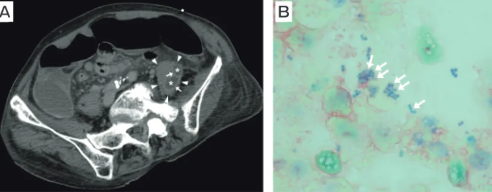
Her blood pressure was 84/56 mmHg, pulse was 112/ min, and body temperature was 35.6°C. She com-plained of mild drowsiness (Japan Coma Scale, I-1), and there was remarkable swelling in bilateral forearms and lower legs. Blood examination revealed severe sys-temic inflammation with a white blood cell count of 18,870/µl and a C-reactive protein (CRP) level of 20.34 mg/dl. Computed tomography (CT) with con-trast revealed enlargement of the left iliopsoas muscle (Fig.2A, arrowheads) with low-density areas (Fig.2A, asterisks) in a ring-enhancing lesion (Fig.2A, arrows) within the muscle. Accordingly, iliopsoas abscesses were suspected.
The patient was diagnosed with septic shock, binoc-ular EBE and left iliopsoas abscess. She was immedi-ately moved to the Emergency Intensive Care Unit, where intravenous meropenem 1 g b.i.d, clindamycin 0.6 g q.i.d, and vancomycin 1 g q.i.d. were initiated. The same day, percutaneous CT-guided pus drainage from the left IPA was performed. Gram stain of the pus showed gram-positive-cocci in clusters (Fig.2B, arrows). Three days after hospitalization, the condition of the right eye remained unchanged, whereas the view of the left fundus was significantly obscured by
worsen-A B
C D
Fig. 1 Fundus photography and optical coherence tomography images at the first visit of a 79-year-old woman with endogenous bacte-rial endophthalmitis secondary to iliopsoas abscess. At the first visit, the fundus in both eyes appears obscured by vitreous opacities, and gray-white retinal exudates are scattered around the retina (arrowheads, A and B). White retinal vessels are also seen in the temporal retina of the left eye (arrows, B). Optical coherence tomography reveals retinal thickening associated with a previous epiretinal membrane in the right eye (C), and the retinal structure in the left eye is unclear because of the vitreous opacity (D).
ing of the vitreous opacity. Thus, intravitreal vancomy-cin 1 mg was administered in the left eye; the usual treatment of ceftazidime could not be used because she had a history of penicillin allergy and we were con-cerned that administration of the β-lactam antibiotics could cause similar systemic side effects. Four days after
hospitalization, culture of pus from the IPA revealed methicillin-sensitive S. aureus (MSSA). MSSA was also detected in the blood culture. One week after hospital-ization, her general condition had improved and EBE in both eyes had improved. Although bedside visual acu-ity measurements were not performed, fundus
exam-A B
Fig. 2 Initial abdominal computed tomography (CT) image and results of Gram staining for a 79-year-old woman with endogenous bac-terial endophthalmitis secondary to iliopsoas abscess. Initial abdominal CT with contrast reveals enlargement of the left iliopsoas muscle (arrowheads, A) with low-density areas (asterisks, A) in a ring-enhancing lesion (arrows, A); iliopsoas abscesses were suspected. Puncture of the abscess and examination of the specimen revealed gram-positive-cocci in clusters (arrows, B), and culture confirmed methicillin-sensitive Staphylococcus aureus.
A B
C D
Fig. 3 Fundus photography and optical coherence tomography images obtained at the final visit of a 79-year-old woman with endoge-nous bacterial endophthalmitis secondary to iliopsoas abscess. A fundus photograph obtained 1 year after the first visit shows that the vitreous opacity has disappeared in both eyes. The retinal exudate has resulted in scarring (arrowheads, A and B). The white retinal blood vessels in the left eye show reperfusion (arrows, B). Optical coherence tomography shows no retinal detachment or retinal edema associ-ated with inflammation, although retinal thickening associassoci-ated with a previous epiretinal membrane can be observed in the right eye (C), while retinal thinning associated with previous central serous chorioretinopathy can be observed in the left eye (D).
ination showed clearing in both eyes with a reduction in the vitreous opacities. One month after hospitalization, her general condition improved and her BCVA had improved to 20/29 and 20/40 in the right and left eyes, respectively. Therefore, she was discharged from our hospital after discontinuing administration of antibiot-ics. One year later, the vitreous opacities in both eyes had almost disappeared, with BCVAs of 20/20 and 20/25 in the right and left eyes, respectively (Fig.3).
Discussion
We report a case of bilateral EBE secondary to IPA, with successful visual outcomes after abscess drainage and intensive antibiotic treatment. Although intraocu-lar fluid culture was not performed in this case, the presumptive diagnosis of bilateral EBE due to IPA was confirmed by the clinical course. EBE due to skeletal muscle abscess is uncommon, and to the best of our knowledge, there are only 2 cases of EBE due to psoas/ iliopsoas abscess whose clinical course were known in the NCBI database, one by Nishida et al. [5] and another by Voros et al. [6] (Table 1). In both cases,
there was severe systemic inflammation with high- sensitivity CRP in the peripheral venous blood, and S.
aureus was detected in the fluid from the abscess.
Systemic administration of antibiotics and vitreous injection were effective, and the visual outcome was relatively good without the need for vitrectomy. These features were consistent with those of the present case.
In the present case, there were no apparent foci of infection or any systemic disease history suggesting a clear source for the IPA. IPA is characterized by pus accumulation within the iliopsoas muscle, and its inci-dence is approximately 0.4 cases for 100,000 population per year [8]. It can occur in patients with diabetes mel-litus, intravenous drug abuse, AIDS, renal failure, and immunosuppression [9]. Therefore, the etiology of the abscess in the present case remains unclear.
The most common agent of IPA is S. aureus [10-12], reported by one study as the causative organism in over 88% of patients with primary IPA [10]. The prognosis of IPA is generally good; however, delayed or inadequate treatment usually results in death. Therefore, early diagnosis is crucial. The rate of mortality from an undrained abscess is close to 100%, with septicemia
Table 1 Previously reported cases and the present case of endogenous bacterial endophthalmitis secondary to iliopsoas abscess Nishida et al. [5] Voros et al. [6] Present case
Eye OS OS OU
Age (years) 60 45 79
Sex F M F
Organism S. aureus S. aureus S. aureus
CRP (mg/dl) 21.4 14.5 20.3
Culture
Blood + - +
Vitreous ND - ND
Iliopsoas abscess + + +
Antibiotics Systemic PAPM/PM, VCM, CLDM, CTRX RFP, CLDM, MEPM MEPM, CLDM, VCM
Intravitreal CAZ VCM, CAZ VCM
BCVA Initial 20/25 20/120 CF (OD) HM (OS)
Outcome 20/17 20/80 20/20 (OD) 20/25 (OS)
Backache at the first visit Unknown + +
OD, right eye; OS, left eye; OU, both eyes; F, female; M, male; CRP, C-reactive protein; ND, not done; PAPM/PM, panipenem; VCM, vancomycin; CLDM, clindamycin; CTRX, ceftriaxone; CAZ, ceftazidime; RFP, rifampicin; BCVA, best-corrected vision acuity; CF, counting fingers; HM, hand motion.
being the most common cause of death [13]. In the present case, MSSA was detected in the blood as well as the IPA, and the initiation of appropriate broad- spectrum antibiotics along with CT-guided drainage of the abscess and intravitreal injection of vancomycin resulted in healing and favorable visual outcomes.
In conclusion, intramuscular abscess should be con-sidered as a possible primary lesion in cases of EBE, complaints of localized pain, together with CT exam, may be useful in identifying the source.
References
1. Okada AA, Johnson RP, Liles WC, DʼAmico DJ and Baker AS: Endogenous bacterial endophthalmitis: report of ten-year retro-spective study. Ophthalmology (1994) 101: 832-838.
2. Jackson TL, Paraskevopoulos T and Georgalas I: Systematic review of 342 cases of endogenous bacterial endophthalmitis. Surv Ophthalmol (2014) 59: 627-635.
3. Jackson TL, Eykyn SJ, Graham EM and Stanford MR: Endogenous bacterial endophthalmitis: a 17-year prospective series and review of 267 reported cases. Surv Ophthalmol (2003) 48: 403-423. 4. Wong JS, Chan TK, Lee HM and Chee SP: Endogenous bacterial
endophthalmitis: an east Asian experience and a reappraisal of a severe ocular affliction. Ophthalmology (2000) 107: 1483-1491. 5. Nishida T, Ishida K, Niwa Y, Kawakami H, Mochizuki K and
Ohkusu K: An eleven-year retrospective study of endogenous bac-terial endophthalmitis. J Ophthalmol (2015) 2015: 261310. 6. Voros GM, Pandit R and Griffiths PG: Metastatic endogenous
endophthalmitis secondary to Staphylococcus aureus iliopsoas abscess. Eye (2004) 18: 97-98.
7. Todokoro D, Mochizuki K, Nishida T, Eguchi H, Miyamoto T,
Hattori T, Suzuki T, Inoue T, Nejima R, Hoshi S and Akiyama H: Isolates and antibiotic susceptibilities of endogenous bacterial endophthalmitis: a retrospective multicenter study in Japan. J Infect Chemother (2018) 24: 458-462.
8. Bartolo DC, Ebbs SR and Cooper MJ: Psoas abscess in Bristol: a 10-year review. Int J Colorectal Dis (1987) 2: 72-76.
9. Walsh TR, Reilly JR, Hanley E, Webster M, Peitzman A and Steed DL: Changing etiology of iliopsoas abscess. Am J Surg (1992) 163: 413-416.
10. Ricci MA, Rose FB and Meyer KK: Pyogenic psoas abscess: worldwide variations in etiology. World J Surg (1986) 10: 834-843. 11. Navarro López V, Ramos JM, Meseguer V, Pérez Arellano JL,
Serrano R, García Ordóñez MA, Peralta G, Boix V, Pardo J, Conde A, Salgado F and Gutiérrez F; GTI-SEMI Group: Microbiology and outcome of iliopsoas abscess in 124 patients. Medicine (Baltimore) (2009) 88: 120-130.
12. Santaella RO, Fishman EK and Lipsett PA: Primary versus sec-ondary psoas abscess. Presentation microbiology and treatment. Arch Surg (1995) 130: 1309-1313.
13. Gruenwald I, Abrahamson J and Cohen O: Psoas abscess: case report and review of the literature. J Urol (1992) 147: 1624-1626. 14. Chen SC, Lee YY, Chen YH, Lin HS, Wu TT and Sheu SJ:
Klebsiella pneumoniae infection leads to a poor visual outcome in endogenous endophthalmitis: a 12-year experience in Southern Taiwan. Ocul Immunol Infl (2017) 25: 870-877.
15. Sridhar J, Flynn Jr HW, Kuriyan AE, Dubovy S and Miller D: Endophthalmitis caused by Klebsiella species. Retina (2014) 34: 1875-1881.
16. Connell PP, OʼNell EC, Fabinyi D, Islam FM, Buttery R,
McCombe M, Essex RW, Roufail E, Clark B, Chiu D, Campbell W and Allen P: Endogenous endophthalmitis: 10-year experience at a tertiary referral centre. Eye (Lond) (2011) 25: 66-72.
17. Ang M, Jap A and Chee SP: Prognostic factors and outcomes in endogenous Klebsiella pneumoniae endophthalmitis. Am J Ophthalmol (2011) 151: 338-344. e2.