鳥取大学研究成果リポジトリ
Tottori University research result repository
タイトル
Title
Therapeutic Value of Lymph Node Dissection Along the
Superior Mesenteric Vein and the Posterior Surface
of the Pancreatic Head in Gastric Cancer Located in
theLower Third of the Stomach
著者
Auther(s)
Saito, Hiroaki; Kono, Yusuke; Murakami, Yuki;
Shishido, Yuji; Kuroda, Hirohiko; Matsunaga, Tomoyuki;
Fukumoto, Yoji; Osaki, Tomohiro; Ashida, Keigo;
Fujiwara, Yoshiyuki
掲載誌・巻号・ページ
Citation
Yonago Acta Medica , 61 (3) : 175 - 181
刊行日
Issue Date
2018
資源タイプ
Resource Type
学術雑誌論文 / Journal Article
版区分
Resource Version
出版社版 / Publisher
権利
Rights
注があるものを除き、この著作物は日本国著作権法により保
護されています。
DOI
Yonago Acta Medica 2018;61:175–181 Original Article
Corresponding author: Hiroaki Saito, MD, PhD sai10@med.tottori-u.ac.jp
Received 2018 March 5 Accepted 2018 August 30
Abbreviations: LN, lymph node; JCGC, Japanese Classification of Gastric Carcinoma; RCT, randomized control trial
Therapeutic Value of Lymph Node Dissection Along the Superior Mesenteric Vein
and the Posterior Surface of the Pancreatic Head in Gastric Cancer Located in the
Lower Third of the Stomach
Hiroaki Saito, Yusuke Kono, Yuki Murakami, Yuji Shishido, Hirohiko Kuroda, Tomoyuki Matsunaga, Yoji Fukumoto, Tomohiro Osaki, Keigo Ashida and Yoshiyuki Fujiwara
Division of Surgical Oncology, Department of Surgery, School of Medicine, Tottori University Faculty of Medicine, Yonago 683-8504, Japan
ABSTRACT
Background Therapeutic value of lymph node
dis-section along the superior mesenteric vein (14v) and the posterior surface of the pancreatic head (13) remains unclear in gastric cancer patients.
Methods We reviewed 355 patients with advanced
gastric cancer in the lower third of the stomach who had undergone gastrectomy at our hospital.
Results The frequency of lymph node (LN) metastasis
was 10.2% and 7.4% in stations 13 and 14v, respectively. The frequency of station 13 metastasis was 26.8% for T3/T4 tumors with group 2 LNs metastasis and 1.4% for all other tumors. The frequency of station 14v metastasis was 22.2% for T3/T4 tumors with group 2 LNs metasta-sis and 1.8% for all other tumors. The therapeutic values for dissecting LN stations 13 and 14v were 1.9 and 0.9, respectively, similar to the therapeutic value for group 2 LN dissection.
Conclusion Because metastasis to stations 13 and 14v
occurs frequently in patients with T3/T4 gastric cancer located in the lower third of the stomach who also have metastasis to group 2 LNs, stations 13 and 14v should be dissected in these patients.
Key words gastric cancer; lymph node dissection;
prognosis
Gastric cancer is one of the most common cancers in Asia, and its mortality still ranks second among all
cancer deaths worldwide.1 In Japan, gastrectomy with
D2 lymph node (LN) dissection is performed safely and is widely accepted as a standard treatment for locally
advanced gastric cancer.2, 3 According to the Gastric
Cancer Treatment Guidelines, the extent of dissection required for D2 LN dissection varies according to the
type of gastrectomy.4 Because distal gastrectomy is
usu-ally performed for lower-third gastric cancer, required LNs stations are 1, 3a, 3b, 4sb, 4d, 5, 6, 7, 8a, 9, 11p, and 12a to achieve D2 LN dissection. However, the suitable extent of LN dissection is still controversial. The LNs along the superior mesenteric vein are referred to as station 14v according to the 2nd English edition of the
Japanese Classification of Gastric Carcinoma (JCGC).5
These nodes receive the majority of lymphatic drainage from the lower third of the stomach and used to be con-ventionally included in the N2 group according to the
previous Japanese classification.5 In the past, the need
for 14v dissection has been assessed based on anatom-ical location in the lymphatic drainage pathway and on the frequency of 14v metastasis as reported in several
Japanese studies.6, 7 However, some papers have
demon-strated that 14v dissection does not contribute to the
prognosis of gastric cancer patients.8 Therefore, the
lat-est version of the Gastric Cancer Treatment Guidelines excludes station 14v from the nodes required for D2 LN dissection in gastric cancer located in the lower third of
the stomach.4
The LNs on the posterior surface of the pancreatic head are referred to as station 13 according to the second English edition of the Japanese Classification of Gastric
Carcinoma.5 Because of its anatomical location, gastric
cancer in the lower third of the stomach preferentially
metastasizes to station 13 LNs.9, 10 However, the clinical
significance and prognostic value of station 13 LN dissection remain controversial as well. The aim of this study was to evaluate therapeutic value of lymph node dissection of 13 and 14v LNs in gastric cancer located in the lower third of the stomach.
MATERIALS AND METHODS
Patients
From 1980 to 2010, 2262 patients underwent gastrec-tomy for gastric cancer at Tottori University Hospital, and 1210 patients were pathologically diagnosed with advanced gastric cancer. Among the patients with advanced gastric cancer, 355 patients had gastric cancer restricted to the lower third of the stomach and the
H. Saito et al.
duodenum; these patients were recruited for analysis in this study. Patients with invasion involving the middle or upper third of the stomach were excluded from the pres-ent study. All patipres-ents underwpres-ent open gastrectomy with regional LN dissection. The clinicopathological findings
were determined according to the JCGC.5 Pathological
diagnosis was made by single pathologist in our hospi-tal. Clinicopathological features were retrospectively collected from the institute’s database. This study was approved by our institutional review board (1608A086), and informed consent requirement was waived for this retrospective study.
Therapeutic value of lymph node dissection
The therapeutic value of LN dissection was evaluated by multiplying the frequency of metastasis to the station by the 5-year survival rate of patients with metastasis in
that station as proposed by Sasako et al.9
Statistical analysis
Associations among factors were evaluated by the chi-squared test. Five-year survival rates were calculated according to the Kaplan–Meier method. Survival data shown in the current study are for cancer-specific surviv-al. To this end, patients who died from causes other than gastric cancer were considered lost to follow-up as of the
time of death. The accepted level of significance was P
< 0.05. Stat View software (Abacus Concepts; Berkeley, CA) was used for all statistical analyses.
RESULTS
Among 355 patients included in this study, station 13 and 14v LN dissection was performed in 157 and 121 patients, respectively. Table 1 shows the relationships among 13 and 14 lymph node dissection and clini-copathological variables in gastric cancer patients. Patients who underwent station 13 LN dissection were significantly younger than those who did not undergo
station 13 LN dissection (P < 0.0001). Tumor size was
Table 1. Relationships among stations 13 and 14 lymph node dissection and clinicopathological variables in pa-tients with gastric cancer
Variable 13 lymph node dissection 14v lymph node dissection Performed
(n = 157) Not performed(n = 198) P value Performed(n = 121) Not performed(n = 234) P value Age 60.6 ± 11.1 68.0 ± 11.2 < 0.0001 61.2 ± 12.0 66.6 ± 11.2 0.0002 Tumor size 7.1 ± 2.6 6.0 ± 2.7 0.0003 6.9 ± 2.6 6.3 ± 2.7 0.026 Sex 0.53 0.072 Male (n = 228) 98 (62.4%) 130 (65.7%) 70 (62.4%) 158 (65.7%) Female (n = 127) 59 (37.6%) 68 (34.3%) 51 (37.6%) 76 (34.3%) Histology a 0.17 0.47 Differentiated (n = 159) 64 (40.8%) 95 (48.0%) 51 (42.1%) 108 (46.2%) Undifferentiated (n = 196) 93 (59.2%) 103 (52.0%) 70 (57.9%) 126 (53.8%) Depth of invasion b 0.09 0.62 T2 / T3 (n = 158) 62 (39.5%) 96 (48.5%) 52 (43.0%) 107 (45.7%) T4 (n = 197) 95 (60.5%) 102 (51.5%) 69 (57.0%) 127 (44.3%)
Lymph node metastasis 0.0023 0.0049
Absent (n = 104) 33 (21.0%) 71 (35.9%) 24 (19.8%) 80 (34.2%) Present (n = 251) 124 (79.0%) 127 (64.1%) 97 (90.2%) 154 (65.8%) Lymphatic invasion 0.35 0.29 Absent (n = 76) 30 (19.1%) 46 (23.2%) 22 (18.2%) 54 (23.1%) Present (n = 279) 127 (80.9%) 152 (76.8%) 99 (81.8%) 180 (76.9%) Venous invasion 0.99 0.021 Absent (n = 138) 61 (38.9%) 77 (38.9%) 37 (30.6%) 101 (43.2%) Present (n = 217) 96 (61.1%) 121 (61.1%) 84 (69.4%) 133 (56.8%) Stage of disease 0.013 0.14 I / II (n = 148) 54 (34.4%) 94 (47.5%) 44 (36.4%) 104 (44.4%) III / IV (n = 207) 103 (65.6%) 104 (52.5%) 77 (63.6%) 130 (55.6%) All results are expressed as the mean ± SD.
a Differentiated, papillary, or tubular adenocarcinoma; undifferentiated, poorly differentiated, mucinous adenocarcinoma, and signet-ring
cell carcinoma.
b Depth of invasion: T2, tumor invasion of the muscularis propria; T3, tumor invasion of the subserosa; T4, tumor penetration of the serosa
Lymph node dissection of gastric cancer
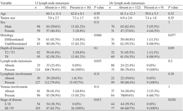
Table 2. Relationship between the presence of stations 13 and 14 lymph node metastasis and clinicopathologi-cal variables in patients with gastric cancer
Variable 13 lymph node metastasis 14v lymph node metastasis
n Absent (n = 141) Present (n = 16) P value n Absent (n = 112) Present (n = 9) P value
Age 60.3 ± 11.4 62.8 ± 8.1 0.59 61.3 ± 12.3 59.4 ± 5.3 0.32 Tumor size 7.0 ± 2.7 7.2 ± 1.7 0.51 6.9 ± 2.6 7.4 ± 1.8 0.35 Sex 0.11 1 Male 98 84 (59.6%) 13 (81.2%) 70 65 (62.4%) 5 (55.5%) Female 59 57 (40.4%) 3 (18.8%) 51 47 (37.6%) 4 (44.5%) Histology 0.066 0.077 Differentiated 74 61 (43.3%) 3 (18.8%) 51 50 (40.8%) 1 (11.1%) Undifferentiated 83 80 (56.7%) 13 (81.2%) 70 62 (59.2%) 8 (88.9%) Depth of invasion 0.1 0.09 T2 / T3 62 59 (41.8%) 3 (18.8%) 52 51 (45.5%) 1 (11.1%) T4 95 82 (58.2%) 13 (81.2%) 69 61 (54.5%) 8 (88.9%)
Lymph node metastasis 0.025 0.2
Absent 33 33 (23.4%) 0 (0%) 88 24 (21.4%) 0 (0%) Present 124 108 (76.6%) 16 (100%) 33 88 (78.6%) 9 (100%) Lymphatic involvement 0.31 0.36 Absent 30 29 (20.6%) 1 (6.3%) 22 22 (19.6%) 0 (0%) Present 127 112 (79.4%) 15 (93.7%) 99 90 (80.4%) 9 (100%) Venous involvement 0.11 1 Absent 61 58 (41.1%) 3 (18.8%) 37 34 (30.4%) 3 (33.3%) Present 96 83 (58.9%) 13 (81.2%) 84 78(69.6%) 6 (66.7%) Stage of disease 0.013 0.026 I / II 54 54 (38.3%) 0 (0%) 44 44 (39.3%) 0 (0%) III / IV 103 87 (61.7%) 16 (100%) 77 68 (60.7%) 9 (100%) All results are expressed as the mean ± SD. See table 1 for the detail of histology and depth of invasion.
significantly larger in patients who underwent station 13 LN dissection than in those who did not undergo
station 13 LN dissection (P = 0.0003). Furthermore, the
presence of LN metastasis was significantly more in patients who underwent station 13 LN dissection than in
those who did not undergo station 13 LN dissection (P =
0.0023). With regard to 14v LN dissection, patients who underwent station 14v LN dissection were significantly younger than those who did not undergo station 14v LN
dissection (P = 0.0002). Tumor size was significantly
larger in patients who underwent station 14v LN dissec-tion than in those who did not undergo stadissec-tion 14v LN
dissection (P = 0.026). Furthermore, the presence of LN
metastasis and venous invasion was significantly more in patients who underwent station 14v LN dissection than in those who did not undergo station 14v LN dissection (P = 0.0049 for LN metastasis and P = 0.021 for venous invasion). We then determined the frequency of LN metastasis associated with gastric cancer located in the lower third of the stomach (Fig. 1). Station 1, 3, 4sb, 4d, 5, 6, and 7 LNs are ones that are required to be dissected to achieve D1 LN dissection in gastric cancer located in the lower third of the stomach. Station 8a, 9, 11p, and 12a LNs are ones that are required to be dissected to
achieve D2 LN dissection in gastric cancer located in the lower third of the stomach. The frequency of LN metastasis was 10.2% and 7.4% for stations 13 and 14v, respectively, similar to that for the group 2 LNs, such as stations 9, 11p, and 12a. We then determined relation-ship between the presence of 13 and 14 LN metastasis and clinicopathological variables in patients with gastric cancer (Table 2). Patients with LN metastasis and those with stage III / IV tumors had significantly more station 13 LN metastasis than did those without LN metastasis (P = 0.025) and those with stage I / II tumors (P = 0.013). Patients with stage III / IV tumors had significantly more station 14v LN metastasis than did those with stage I / II
tumors (P = 0.026). Figure 2 shows correlations between
the depth of invasion and the frequency of metastasis to each LN station. Metastasis in station 1, 3, 4sb, 4d, 5, 6, 7, 8a, 9, and 11p LNs was observed for T2 tumors. However, both station 13 and station 14v LN metastasis were observed for T3 and T4 tumors, but not for T2 tumors.
We then determined the frequency of LN metas-tasis to each station according to the status of station 13 LN metastasis. Tumors with station 13 metastasis metastasized significantly more often to stations 6, 8a,
H. Saito et al.
Lymph node station 0 10 20 30 40 50 60 1 3 4sb 4d 5 6 7 8a 9 11p 12a 13 14v
Saito et al. Figure 1
Fr equ en cy o f l ym ph n ode m etas tas is (%) 0 10 20 30 40 50 60 70 80 90 100 1 3 4sb 4d 5 6 7 8a 9 11 12a 14v 13 negative 13 positive * P < 0.05 ** P < 0.01 *** P < 0.0001 * *** ****** **
Lymph node station
Saito et al. Figure 3
Fr equ en cy o f l ym ph n ode m etas tas is (%)
Saito et al. Figure 5
0 5 10 15 20 25 30 T3/T4 with LNs metastasis to group 2 LNs Fr equ en cy o f l ym ph n ode m etas tas is (%) T3/T4 without LNs metastasis to group 2 LNs T2 with LNs metastasis to group 2 LNs T2 without LNs metastasis to group 2 LNs
Saito et al. Figure 2
Fr equ en cy o f l ym ph n ode m etas tas is (%) 0 10 20 30 40 50 60 70 80 T2 T3 T4 T2 T3 T4 T2 T3 T4 T2 T3 T4 T2 T3 T4 T2 T3 T4 T2 T3 T4 T2 T3 T4 T2 T3 T4 T2 T3 T4 T2 T3 T4 T2 T3 T4 T2 T3 T4 1 3 4sb 4d 5 6 7 8a 9 11p 12a 13 14v 1 3 4sb 4d 5 6 7 8a 9 11p 12a 13 14v T2 T3 T4
Lymph node station
0 10 20 30 40 50 60 70 80 90 100 1 3 4sb 4d 5 6 7 8a 9 11p 12a 13 14v negative 14vpositive * P < 0.05 ** P < 0.01 *** P < 0.0001 * *** ** *** * *
Lymph node station
Saito et al. Figure 4
Fr equ en cy o f l ym ph n ode m etas tas is (%)
Saito et al. Figure 6
0 5 10 15 20 25 T3/T4 with LNs metastasis to group 2 LNs T3/T4 without LNs metastasis to group 2 LNs T2 with LNs metastasis to group 2 LNs T2 without LNs metastasis to group 2 LNs Fr equ en cy o f l ym ph n ode m etas tas is (%)
Fig. 1. The frequency of metastasis to each lymph node station in
gastric cancer located in the lower third of the stomach.
Fig. 3. The frequency of metastasis to each lymph node station
according to the presence of station 13 lymph node metastasis in gastric cancer located in the lower third of the stomach.
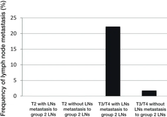
Fig. 5. The frequency of station 13 lymph node metastasis
accord-ing to the depth of invasion and the presence of metastasis to the group 2 lymph nodes in gastric cancer located in the lower third of the stomach. LN, lymph node. See Fig. 2 for the detail of depth of invasion.
Fig. 2. The frequency of metastasis to each lymph node station
according to the depth of invasion in gastric cancer located in the lower third of the stomach. Depth of invasion: T2, tumor invasion of the muscularis propria; T3, tumor invasion of the subserosa; T4, tumor penetration of the serosa or tumor invasion of adjacent organs.
Fig. 4. The frequency of metastasis to each lymph node station
according to the presence of station 14 lymph node metastasis in gastric cancer located in the lower third of the stomach.
Fig. 6. The frequency of station 14v lymph node metastasis
according to the depth of invasion and the presence of metastasis to the group 2 lymph nodes in gastric cancer located in the lower third of the stomach. LN, lymph node. See Fig. 2 for the detail of depth of invasion.
Lymph node dissection of gastric cancer
11, 12a, and 14v. Furthermore, this difference was more strikingly observed in the group 2 LNs, such as stations 8a, 11p, and 12a, compared with the group 1 LN such as station 6 (Fig. 3). Tumors with station 14v metastasis metastasized signifi cantly more often to stations 3, 8a, 9, 11p, 12a, and 13. This difference was also more striking-ly observed in the group 2 LNs, such as stations 8a and 12a, compared with the group 1 LN such as station 3 (Fig. 4). The frequency of station 13 LN metastasis was 26.8% for T3/T4 tumors with group 2 LN metastasis and 1.4% for all other tumors (Fig. 5). Furthermore, the frequency of station 14v LN metastasis was 22.2% for T3/T4 tumors with group 2 LN metastasis and 1.8% for all other tumors (Fig. 6). These results indicate that the presence of 13 and 14v LN metastasis could be predict-ed baspredict-ed on the depth of invasion and the presence of metastasis to the group 2 LNs.
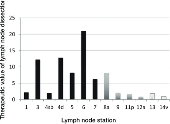
The 5-year disease specifi c survival rates of patients with metastasis in each LN station were as follows: station 1, 21.9%; station 3, 28.2%; station 4sb, 33.3%; station 4d, 32.4%; station 5, 30.7%; station 6, 37.7%; sta-tion 7, 30.6%; stasta-tion 8a, 29.3%, stasta-tion 9, 22.7%; stasta-tion 11p, 21%; station 12a, 10.5%; station 13, 18.8%; station 14v, 12.5%. Finally, we determined the therapeutic value of dissecting each LN station (Fig. 7). The therapeutic values for LN dissection of stations 13 and 14v were 1.9 and 0.9, respectively, similar to the therapeutic value for group 2 LN dissection (i.e., stations 9, 11p, and 12a).
DISCUSSION
There is a general consensus regarding the role of
regional LN dissection in gastric cancer surgery.11–13
The optimal method to evaluate the therapeutic value of dissecting each LN station is randomized control trial (RCT) and some RCTs have been done in gastric cancer thus far. For instance, Songun et al. demonstrated
that D2 lymphadenectomy is associated with lower locoregional recurrence and gastric-cancer-related death rates than D1 surgery, indicating that group 2 lymph node dissection was useful in improving the prognosis
of gastric cancer patients.14 On the other hand, it is now
accepted that prophylactic para-aortic LN dissection
confers no survival advantage,15, 16 However, the optimal
extent of LN dissection is still controversial since it is diffi cult to conduct RCT. Therefore, it is indispensable to develop other methods to evaluate the therapeutic value of each LN dissection. In this regard, Sasako et al. de-veloped new method, known as therapeutic value index, to evaluate the therapeutic value of each LN dissection for gastric cancer. This index includes the prognosis of the patients with metastasis to the station, as well as the metastatic incidence and used in many studies of gastric cancer to evaluate the theoretical therapeutic impact of
dissecting each LN station.10, 17 The high therapeutic
val-ue index of the LN station indicates that the dissection of that LN station is useful in improving the prognosis of gastric cancer patients.
The therapeutic value of station 13 LN dissection remains controversial. Tokunaga et al. showed that the frequency of station 13 LN metastasis in patients with advanced gastric cancer restricted to the lower third of the stomach and the duodenum was 23.9% in cases with duodenal invasion and 7% in cases without duodenal
invasion; this difference was statistically significant.10
In our study, the frequency of station 13 LN metastasis in patients with advanced gastric cancer restricted to the lower third of the stomach and the duodenum was 16.7% in cases with duodenal invasion and 9.7% in cases without duodenal invasion, which is almost identical to the previous report, indicating that metastasis to the 13 and 14v LNs occurs frequently in patients with gastric cancer located in the lower third of the stomach. Sasako et al. reported a 5-year survival rate of 0% for cases of lower-third advanced gastric cancer with station 13 LN metastasis. Therefore, these authors discounted any
therapeutic value in station 13 LN dissection.9 In
con-trast, Tokunaga et al. reported a 5-year survival rate of 17.5% in advanced gastric cancer patients with duodenal invasion and station 13 LN metastasis; the frequency of metastasis to these LNs was 23.9%. As a result, the therapeutic index for station 13 LN dissection in these advanced gastric cancer patients with duodenal invasion was 4.19, equivalent to that for most second-tier LNs. Therefore, these authors concluded that station 13 LNs should be dissected in patients with advanced gastric
cancer with macroscopic duodenal invasion.10 Our
re-sults demonstrated that a 5-year survival rate was 18.8% in advanced gastric cancer patients with station 13 LN
Saito et al. Figure 7
0 5 10 15 20 25 1 3 4sb 4d 5 6 7 8a 9 11p 12a 13 14v Lymph node station
The ra pe ut ic va lue o f lym ph no de d isse ct io n
Fig. 7. The therapeutic value of dissection at each lymph node
H. Saito et al.
metastasis in this study. Furthermore, therapeutic index for station 13 LN dissection was almost equivalent to that for group 2 LNs such as stations 9, 11p, and 12a, in-dicating that station 13 LN dissection might be useful in improving the prognosis of patients with gastric cancer located in the lower third of the stomach. On the other hand, it remains unclear whether there is difference in therapeutic index for station 13 LN dissection according to the presence of duodenal invasion in the current study because of the small number of patients with duodenal invasion in the current study. Further investigations are urgently required in this regard.
With regard to the station 14v nodes, An et al. reported overall 3- and 5-year survival rates of 26% and 10%, respectively, in patients with positive 14v nodes who underwent R0 resections. Because the prognosis for patients with positive 14v nodes is poor, similar to that of patients with M1 disease, these authors concluded that exclusion of the 14v LNs from regional lymph node
dissection should be considered.8 In the current study, a
similar 5-year survival rate of 12.5% was found in pa-tients with positive 14v nodes. The therapeutic index for 14v LN dissection identified in our study was equivalent to that for most second-tier LNs. Therefore, 14v LN dissection has therapeutic value if R0 resection can be achieved.
Dissection of the station 13 and 14v LNs is techni-cally demanding. Therefore, it is important to select pa-tients who have a possibility of metastasis to these LNs. In this regard, Masuda et al. reported that the station 6 status was a useful predictive factor for 14v-negative
status.18 Tumors with station 13 metastasis metastasized
significantly more often to stations 6, 8a, 11, and 12a in this study. Tumors with station 14v metastasis metasta-sized significantly more often to stations 3, 8a, 9, 11p, and 12a. These results indicate the possibility that there are lymphatic flows from these LNs to 13 and 14v LNs. In the current study, furthermore, we have demonstrated that patients who have T3/T4 tumors with metastasis to the group 2 LNs are likely to have metastasis to LN stations 13 and 14v. Therefore, stations 13 and 14v should be dissected in patients with T3/T4 gastric cancer located in the lower third of the stomach who also have possible metastasis to the group 2 LNs.
The present study has a few limitations. First, some bias was present because the study was retrospective. Particularly, the number of patients who underwent the dissection of LN stations 13 and 14v is 157 (44.2%) and 121 (34.1%), respectively, and is smaller than that of patients who underwent the dissection of group 1 and 2 LNs. Second, the number of patients included in the cur-rent study was small; therefore, a large-scale,
prospec-tive randomized controlled trial is needed to confirm the results.
In conclusion, the dissection of LN stations 13 and 14v might be beneficial for survival in patients with gastric cancer located in the lower third of the stomach. Because metastasis to the 13 and 14v LNs occurs fre-quently in patients with T3/T4 gastric cancer located in the lower third of the stomach who also have group 2 LN metastasis, stations 13 and 14v should be dissected in these patients.
Human rights statement and informed consent: All procedures
followed were in accordance with the ethical standards of the responsible committee on human experimentation (institutional and national) and with the Helsinki Declaration of 1964 and later versions. Informed consent from patients was waived because of the retrospective design of this study.
The authors declare no conflict of interest.
REFERENCES
1 Parkin DM, Bray F, Ferlay J, Pisani P. Global cancer statistics, 2002. CA Cancer J Clin. 2005;55:74-108. PMID: 15761078. 2 Kodera Y, Schwarz RE, Nakao A. Extended lymph node
dissection in gastric carcinoma: where do we stand after the Dutch and British randomized trials? J Am Coll Surg. 2002;195:855-64. PMID: 12495318.
3 Maruyama K, Okabayashi K, Kinoshita T. Progress in gastric cancer surgery in Japan and its limits of radicality. World J Surg. 1987;11:418-25. PMID: 3630186.
4 Japanese Gastric Cancer Association. Japanese Gastric Cancer Treatment Guidelines 2014. Japan: KANEHARA & Co., LTD.; 2014.
5 Japanese Gastric Cancer Association. Japanese Classification of Gastric Carcinoma: 2nd English edition. Gastric Cancer. 1998;1:10-24. PMID:
6 Kodama M, Ishikawa K, Koyama H, Narisawa T, Koyama K. [Study on the lymphatic flow of the lower gastric region for radical lymphadenectomy in advanced lower gastric cancer]. Nihon Geka Gakkai Zasshi. 1988;89:1008-13. PMID: 3221822.
7 Yoshida K, Ohta K, Ohhashi I, Nakajima T, Takagi K, Nishi M. [Studies on gastric lymphatics by using activated carbon particle (CH44) and lymph node metastasis of gastric cancer]. Nihon Geka Gakkai Zasshi. 1988;89:664-70. PMID: 3412300. 8 An JY, Pak KH, Inaba K, Cheong JH, Hyung WJ, Noh
SH. Relevance of lymph node metastasis along the superior mesenteric vein in gastric cancer. Br J Surg. 2011;98:667-72. PMID: 21294111.
9 Sasako M, McCulloch P, Kinoshita T, Maruyama K. New method to evaluate the therapeutic value of lymph node dis-section for gastric cancer. Br J Surg. 1995;82:346-51. PMID: 7796005.
10 Tokunaga M, Ohyama S, Hiki N, Fukunaga T, Inoue H, Yamada K, et al. Therapeutic value of lymph node dissection in advanced gastric cancer with macroscopic duodenum invasion: is the posterior pancreatic head lymph node dis-section beneficial? Ann Surg Oncol. 2009;16:1241-6. PMID:
Lymph node dissection of gastric cancer 19224285.
11 Cuschieri A, Weeden S, Fielding J, Bancewicz J, Craven J, Joypaul V, et al. Patient survival after D1 and D2 resections for gastric cancer: long-term results of the MRC randomized surgical trial. Surgical Co-operative Group. Br J Cancer. 1999;79:1522-30. PMID: 10188901.
12 Hartgrink HH, van de Velde CJ, Putter H, Bonenkamp JJ, Klein Kranenbarg E, Songun I, et al. Extended lymph node dissection for gastric cancer: who may benefit? Final results of the randomized Dutch gastric cancer group trial. J Clin Oncol. 2004;22:2069-77. PMID: 15082726.
13 Wu CW, Hsiung CA, Lo SS, Hsieh MC, Chen JH, Li AF, et al. Nodal dissection for patients with gastric cancer: a ran-domised controlled trial. Lancet Oncol. 2006;7:309-15. PMID: 16574546.
14 Songun I, Putter H, Kranenbarg EM, Sasako M, van de Velde CJ. Surgical treatment of gastric cancer: 15-year follow-up re-sults of the randomised nationwide Dutch D1D2 trial. Lancet Oncol. 2010;11:439-49. PMID: 20409751.
15 Sano T, Sasako M, Yamamoto S, Nashimoto A, Kurita A, Hiratsuka M, et al. Gastric cancer surgery: morbidity and mortality results from a prospective randomized controlled trial comparing D2 and extended para-aortic lymphadenecto-my--Japan Clinical Oncology Group study 9501. J Clin Oncol. 2004;22:2767-73. PMID: 15199090.
16 Sasako M, Sano T, Yamamoto S, Kurokawa Y, Nashimoto A, Kurita A, et al. D2 lymphadenectomy alone or with para-aortic nodal dissection for gastric cancer. N Engl J Med. 2008;359:453-62. PMID: 18669424.
17 Yoshikawa T, Takeuchi H, Hasegawa S, Nozaki I, Kishi K, Ito S, et al. Theoretical therapeutic impact of lymph node dissection on adenocarcinoma and squamous cell carcinoma of the esophagogastric junction. Gastric Cancer. 2016;19:143-9. PMID: 25414051.
18 Masuda TA, Sakaguchi Y, Toh Y, Aoki Y, Harimoto N, Taomoto J, et al. Clinical characteristics of gastric cancer with metastasis to the lymph node along the superior mesenteric vein (14v). Digestive surgery. 2008;25:351-8. PMID: 18957850.