INTRODUCTION
The majority of anaerobic isolates from clinical specimens belong to the genus Bacteroides (1). The species included in the ”B. fragilis group” are considered to be clinically important pathogens as-sociated with intra-abdominal infections and ab-scess formation in soft tissues (2). Among these species, B. fragilis is the most virulent because this species accounts for over half of the anaerobes iso-lated from human infections and often causes se-vere septicemia with a high mortality rate in com-promised hosts (2, 3). Early diagnosis and treat-ment with appropriate antibiotics are needed for
patients infected with B. fragilis, but the traditional culture methods for anaerobes are labor-intensive and time-comsuming. In addition, if the clinical samples are not immediately cultured or kept un-der anaerobic conditions, obligate anaerobes are often not detected in blood cultures. Various tech-niques, including analysis of electrophoretic pat-terns of dehydrogenase (4), bacteriophage typing (5), analysis of cellular sugar and lipid composi-tions (6-8) and serology (9-11), have been used for rapid detection and discrimination of this anaerobe. However, all of these techniques require viable cells and many troublesome steps, and none of them have sufficient specificity and sensitivity to be used for clinical specimens.
Recently, molecular biology-based techniques have been shown to be useful for the rapid identifi-cation of many pathogenic microoganisms (12, 13). The polymerase chain reaction (PCR) targeting a specific gene is the most widely used technique in
PCR-dot blot hybridization based on the neuraminidase-encoding
gene is useful for detection of
Bacteroides fragilis
Tomomi Kuwahara, Haruyuki Nakayama, Tsuyosi Miki, Keiko Kataoka,
Hideki Arimochi, and Yoshinari Ohnishi
Department of Bacteriology, The University of Tokushima School of Medicine, Tokushima, Japan
Abstract : Bacteroides fragilis is a Gram-negative obligate anaerobe frequently isolated from clinical specimens and sometimes causes severe septicemia in compromised hosts. Increasing interest has been shown in the enterotoxigenicity and drug resistance of B .
fragilis in the field of medical microbiology. We previously reported rapid detection of
this anaerobe by nested PCR targeting a neuraminidase-encoding gene nanH. In the present study, we synthesized a digoxigenin-labeled oligonucleotide probe, NH1,which is specific for nanH of B . fragilis, and we combined the hybridization assay using NH1 with the nanH -PCR to detect this anaerobe in a bacteremia model mice. In the specific-ity test, the oligonucleotide probe, NH1, hybridized only to amplification products from
B . fragilis. PCR-dot blot hybridization based on nanH enabled detection of cells of B . fragilis in blood samples even when the number was as low as 2×10³
colony-forming units/ml. These findings suggest that PCR-dot blot hybridization targeting nanH is a useful procedure for diagnosis of septicemia caused by B . fragilis when viable cells in blood cannot be detected by the traditional culture techniques. J. Med. Invest. 48 : 60-65, 2001
Keywords : Bacteroides fragilis, neuraminidase, oligonucleotide probe, septicemia, PCR
Received for publication August 1, 2000 ; accepted September 26, 2000.
Address correspondence and reprint requests to Yoshinari Ohnishi, M.D., PhD., Department of Bacteriology, The Univer-sity of Tokushima School of Medicine, Kuramoto-cho, Tokushima 770-8503,Japan and Fax : +81-886-33-7069.
The Journal of Medical Investigation Vol. 48 2001
60 60
diagnostic laboratories because it is quick and it is suitable for the handling of a large number of spec-imens (14). Furthermore, PCR amplification ena-bles the detection of pathogens even in culture-negative clinical specimens, since this procedure does not necessarily require viable cells (15). How-ever, PCR amplification sometimes produces false-positive results if other bacteria have sequences similar to those of the designed PCR-primers. In a previous study, we synthesized two primer sets, F 1-R1 (outer primer set) and F2-R2 (inner primer set), and used them in nested PCR to amplify the neuraminidase-encoding gene nanH of B. fragilis (16). Although these primer sets specifically am-plified a part of the nanH gene of B. fragilis, one of the Bacteroides species, B. vulgatus, which possesses high neuraminidase activity, also produced a sin-gle band identical to that of B. fragilis in size when the primer set F1-R1 was used, and this false-pos-itive band could not be excluded by electrophoresis alone. In such a case, hybridization tests with probes specific for the target are usually required to confirm the specificity of the PCR amplifica-tions.
In the present study, we developed a digoxigenin-labeled oligonucleotide probe (named NH1), which was specific for nanH of B. fragilis, and we used it in combination with nanH -PCR for a hybridization assay. We could specifically detect B. fragilis in blood samples when PCR of a part of the gene nanH and dot-blot hybridization using NH1 were applied to model mice with bacteremia induced by challenge of this anaerobe.
MATERIALS AND METHODS
Bacterial StrainsSixty strains of B. fragilis, including two refer-ence strains (ATCC25285 and NCTC9343), were used in this study. The strains of other species used were B. distasonis ATCC8503, B. eggerthii ATCC27754, B. ovatus ATCC8483, B. thetaiotaomicron ATCC29148, B. uniformis ATCC8492, B. vulgatus ATCC8482, Porphyromonas asaccharolytica ATCC 25260, P. endodontalis ATCC35406, P. gingivalis 381 and Prevotella corporis JCM8529. All strains were cultured in GAM broth (Nissui Pharmaceutical Co., Tokyo, Japan) at 37℃ under anaerobic condi-tions.
Preparation of Bacterial Cells for PCR
A late log-phase culture (1 ml) of each strain was centrifuged, washed with 1 ml of phosphate-buffered saline, and resuspended in 0.1 ml of dis-tilled water. Each suspension was lysed by heating at 100℃ for 10 min, and 10µl of the lysed prepara-tion was used for PCR amplificaprepara-tion of the nanH gene. PCR amplification of the nanH gene was performed as described previously (16).
Design of an Oligonucleotide Probe
We compared the nucleotide sequences of the nanH structural gene of B. fragilis strains YCH46 and TAL2480. The oligonucleotide probe NH1 was synthesized on the basis of the common sequence found within F2-and R2-primer annealing sites. NH1 was labeled with digoxigenin using a DIG-labeling Kit (Boehringer GmbH, Mannheim, Ger-many) according to the manufacturer’s instruc-tions. The nucleotide sequence of NH1 was 5’- ATCACTATGAGTGACGGTACTTTGGTATTCCC-3’.
Dot Blot Hybridization
After PCR amplification of the nanH gene, each reaction mixture was heated at 95℃ for 5 min and chilled on an ice bath. Then, 5µl of each amplifica-tion mixture was spotted on a nylon membrane and UV-fixed. Southern hybridization was performed as described by Sambrook et al . (17). The hybridi-zation with digoxigenin-labeled NH1 was perfomed in 10 ml of rapid hybridization buffer (Amersham Co., Ltd.) at 54℃ for 1 hour. Post-hybridization washes were performed twice at 54℃ with each washing buffer, 2×SSC (1×SSC being 0.15 mM NaCl
plus 15 mM sodium citrate)-0.1% SDS and 0.1×
SSC-0.1% SDS, respectively. The hybridization signals were detected according to the manufacturer’s in-structions using an alkaline phosphatase-labeled anti-digoxigenin antibody.
Preparation of Blood Samples
Three C57BL/6J mice were intraperitoneally injected with viable cells of B. fragilis. Blood sam-ples (0.2 ml) were collected by cardiac puncture at 1, 3 and 6 hours after injection, and 0.1 ml of each sample was incubated anaerobically on GAM agar plates. The remainder of the samples were centri-fuged, washed with 1 ml of 10 mM Tris-HCl and 1 mM EDTA (pH 8.0), and resuspended in 0.1 ml of lysis buffer (10 mM Tris-HCl, pH 8.3, 50 mM KCl, 1.5 mM MgCl2and 0.1 mg of proteinase K per ml).
61
The Journal of Medical Investigation Vol. 48 2001 61
Each suspension was incubated at 65℃ for 1 hour, boiled in water for 10 min, and chilled on an ice bath. Ten microliters of each solution was used as a template for PCR amplification of the nanH gene.
RESULTS AND DISCUSSION
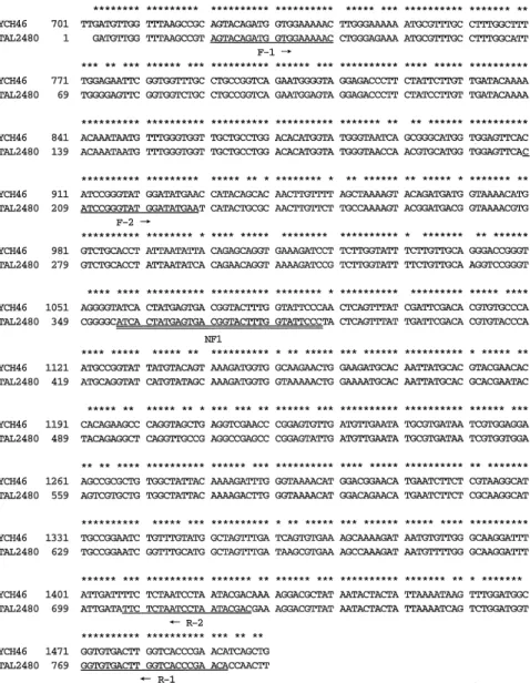
First, we compared the nucleotide sequences of thenanHgene of B. fragilis strain YCH46 (18) with strain TAL2480 (19) to design an oligonucleotide probe that was specific for the nanH gene of B. fragilis. The nucleotide sequences of nanH from both strains were highly conserved and showed 91.1% identity (Fig.1). In the present study, we chose the region at 1057-1088 (numbering in the YCH46 nanH gene) for a B. fragilis-specific probe because (i) this region of more than 30 nucleotides in length is common to both strains, (ii) the guanine plus cytosine content was relatively high (43.8%),
and (iii) this region corresponded to the middle of the F2-R2 annealing sites. Since a sufficient amount of the oligonucleotide probe was easily synthe-sized using a DNA synthesizer and non-radioisotopic labeling is required for use in diagnostic laboratories, we synthesized a digoxigenin-labeled oligonucleotide probe, NH1, based on this region. The nucleotide sequence of NH1 is described in Materials and Methods.
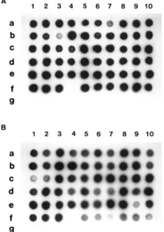
Dot blot hybridization using NH1 was performed against nanH PCR products from sixty strains of B. fragilis and ten other strains of related species. As shown in Fig. 2, positive spots were obtained from all strains of B. fragilis tested except for strain KMS2 (spotted at f-4.). However, strain KMS2, which had initially been identified as B. fragilis, was reidentified as B. vulgatus when we tested this strain using an API20A system (bioMerieux). In addition, none of the products from other species gave positive spots. Based on these findings, it
Fig. 1. Comparison of the nucleotide se-quences of nanH from B. fragilis YCH 46 and TAL2480 using Genetyx-Mac soft-ware Ver8.0.5 (Softsoft-ware Development Co., Ltd., Tokyo, Japan). In the case of strain TAL2480, only a partial nucleotide sequence was available in the DDBJ/ EMBL/Genbank database. The common residues are indicated by asterisks a-bove the nucleotide sequences. The po-sitions of the PCR-primers (underlined) and oligonucleotide probe, NH1 (double underlined), are shown below the se-quence.
T. Kuwahara et al. PCR-dot blot hybridization using nanH
62 T. Kuwahara et al. PCR-dot blot hybridization using nanH
was suggested that (i) the digoxigenin-labeled oligonucleotide probe, NH1, was specific for nanH of B. fragilis and (ii) PCR-dot blot hybridization targeting nanH was useful not only for the
identifi-cation of B. fragilis but also for that of B. vulgatus (PCR with F1-R1 was positive, but dot blot hybridi-zation with NH1 was negative.).
To assess the usefulness of nanH-PCR and the dot blot hybridization assay in clinical specimens, B. fragilisbacteremia model mice were constructed. Three 8-week-old male C57BL/6J mice were in-traperitoneally injected with B. fragilis strain YCH 46, and 0.2 ml blood samples were collected by car-diac puncture at 1, 3 and 6 hours after injection. Ta-ble 1 shows the results of the blood culture. Rapid translocation of B. fragilis cells from the peritoneal cavity to the blood stream was observed. This find-ing might represent the pathogenic potential of this species, but almost all B. fragilis cells appeared to be cleared from the blood stream within 24 hours in healthy mice.
Nested PCR of the nanH gene was performed using each blood sample. Fig. 3 shows the findings of the PCR-dot blot hybridization assay using blood samples from mouse A in Table 1. The 518 bp bands of expected size were detected in the samples at 1, 3 and 6 hours (Fig. 3A), while no band was found in the 0 time control. We confirmed that these were B. fragilis-specific amplification products by dot blot hybridization with digoxigenin-labeled NH1 (Fig. 3B). These findings suggested that PCR-dot blot hybridization based on nanH enables detec-tion of B. fragilis cells in clinical specimens with a cell number as low as 2×10³
cfu/ml, even if the sample tested contains non-viable cells. As shown in Fig. 3, the intensity of the PCR bands was not in proportion to the strength of the hybridization sig-nals. The reason for this might be that the excess amount of template produced a large amount of in-termediate amplification products in the early cy-cles of amplification and reduced the amount of specific bands, or that NH1 directly reacted with genomic DNA contained in blood samples. The latter case would mean that NH1 would enable di-rect detection of B. fragilis cells in blood samples
Fig. 2. Dot blot hybridization using digoxigenin-labeled oligonucleotide probe NH1 against the nanH amplification products generated by primer F1-R1(A) and F2-R2(B). The spots from a-1 to e-10 correspond to B. fragilis strains YCH1 to YCH50. The other spots are as follows : 1, B. fragilis B1 ; 2, B. fragilis B2 ; 3, B. fragilis KMS1 ; 4, B. fragilis KMS2 ; f-5, B. fragilis KMS3 ; f-6, B. fragilis KMS4 ; f-7, B. fragilis KMS 5 ; f-8, B. fragilis TDP-101 ; f-9, B. fragilis ATCC25285 ; f-10, B.
fragilisNCTC9343 ; g-1, B. distasonis ATCC8503 ; g-2, B. eggerthii ATCC27754 ; g-3, B. ovatus ATCC8483 ; g-4, B. thetaiotaomicron ATCC29148 ; g-5, B. uniformis ATCC8492 ; g-6, B. vulgatus ATCC 8482 ; g-7, Porphyromonas asaccharolytica ATCC25260 ; g-8,
Por-phyromonas endodontalis ATCC35406 ; g-9, Porphyromonas
gin-givalis381 ; g-10, Prevotella corporis JCM8529.
Table 1. Results of blood cultures from a bacteremia model mice with B. fragilis.
Experimental mouse Number of B. fragiliscells used in i. p. injec-tion (cfu)
Number of B. fragilis cells detected by blood culture (cfu/ml of blood)
0 ha 1 ha 3 ha 6 ha A Bb Cb 8.0×10⁶ 5.3×10⁶ 5.3×10⁶ 0 0 0 1.0×10⁵ 5.1×10⁵ 2.2×10⁵ 4.3×10³ 5.2×10⁵ 6.6×10³ 2.1×10³ 3.8×10³ 2.5×10³ a The time when the blood samples were collected after i.p. injection, and 0 h means before injection.
b In these experiments, the same culture of B. fragilis was used for injection into two independent C 57/BL/6 J mice.
63
The Journal of Medical Investigation Vol. 48 2001 63
if there was a sufficient number of cells in the sam-ple. It was suggested that the PCR-dot blot hybridi-zation assay using nanH was useful for the detec-tion and quantificadetec-tion of B. fragilis present in clin-ical specimens. Of course, NH1 can be applied for the rapid identification of cultured B. fragilis, but there are many cases in which the culture of blood from a patient is negative even when septicemia is suspected from clinical signs due to the adminis-tration of antibiotics or, particularly in cases of an-aerobic infection, due to storage of anaerobes un-der inappropriate conditions. Furthermore, in al-most all cases, clinical samples do not contain a sufficient number of cells to enable detection using direct hybridization assay. Therefore, an amplifica-tion step is needed before performing the hybridi-zation assay to obtain accurate results.
In the present study, we demonstrated the use-fulness of PCR-dot blot hybridization based on the neuraminidase-encoding gene for specific detec-tion of B. fragilis cells using bateremia model mice.
It was suggested that a combination of the method used in the present study with a traditional culture method would be useful for a clinical survey of the accurate incidence of B. fragilis infection and for differential diagnosis in patients with fever of un-known origin. However, it is necessary to deter-mine whether this procedure is useful in the ical setting by performing tests using various clin-ical samples such as pus, sputum and drainage flu-id in a future study.
ACKNOWLEDGMENTS
The authors thank Drs F. Yoshimura, I. Nakamura, K. Suzuki and K. Tanaka for providing bacterial strains. The work was supported in part by a Grant-in-Aid for Developmental Scientific Research (B) (No.06557020) from the Ministry of Education, Science, Sports and Culture of Japan, and by the Kurozumi Medical Foundation.
REFERENCES
1. Finegold SM : Anaerobic infections in humans : an overview. Anaerobe 1 : 3-9, 1995
2. Patrick S : The virulence of Bacteroides frag-ilis. Rev Med Microbiol 4 : 40-49, 1993
3. Goldstein EJ : Anaerobic bacteremia. Clin In-fect Dis 23 : 97-101, 1996
4. Shah HN, Williams RAD : Dehydrogenase pat-terns in the taxonomy of Bacteroides. J Gen Microbiol 128 : 2955-2965, 1982
5. Booth SJ, van Tassell RL, Johnson JL, Wilkins TD: Bacteriophages of Bacteroides. Rev In-fect Dis 1 : 325-336, 1979
6. Brondz I, Carlsson J, Sjostrom M, Sundqvist G : Significance of cellular fatty acids and sug-ars in defining the genus Porphyromonas. Int J Syst Bacteriol 39 : 314-318, 1989
7. Brondz I, Olsen I, Haapasalo M, van Winkelhoff J : Multivariate analyses of fatty acid data from whole-cell methanolysates of Prevotella, Bacteroides and Porphyromonas spp. J Gen Microbiol 137 : 1445-1452, 1991
8. Mayberry WRD, Lambe W Jr, Ferguson KP : Identification of Bacteroides species by cellu-lar fatty acid profiles. Int J Syst Bacteriol 32 : 21-27, 1982
9. Holland JW, Stauffer LR, and Altemeier WA : Fluorescent antibody test kit for rapid
detec-Fig. 3. Detection of amplification products by nested PCR of the nanH gene of B. fragilis cells in blood samples (A) and results of a specificity test by dot blot hybridization with a digoxigenin-labeled oligonucleotide probe, NH1(B). Lane1, before bacte-rial injection ; lane 2, 1 hour ; lane 3, 3 hours ; lane 4, 6 hours af-ter injection, and M, molecular size marker.
T. Kuwahara et al. PCR-dot blot hybridization using nanH
64 T. Kuwahara et al. PCR-dot blot hybridization using nanH
tion of members of the Bacteroides fragilis and Bacteroides melaninogenicus groups in clinical specimens. J Clin Microbiol 10 : 121-127, 1979 10. Lambe DW, Jr. : Determination of Bacteroides melaninogenicusserogroups by fluorescent anti-body staining. Appl Microbiol 28 : 561-567, 1974 11. Weissfeld AS, Sonnenwirth AC : Rapid detec-tion and identificadetec-tion of Bacteroides fragilis and Bacteroides melaninogenicus by immu-nofluorescence. J Clin Microbiol 13 : 798-800, 1981
12. Miranda AG, Singh KV, Murray BE:DNA fin-gerprinting of Enterococcus faecium by pulsed-field gel electrophoresis may be a useful epi-demiologic tool. J Clin Microbiol 29 : 2752-2757, 1991
13. Stull TL, LiPuma JJ, Edlind TD : A broad-spec-trum probe for molecular epidemiology of bac-teria : ribosomal RNA. J Infect Dis 157 : 280-286, 1988
14. Kostman JR, Alden MB, Mair M, Edlind TD, LiPuma JJ, Stull TL : A universal approach to bacterial molecular epidemiology by polym-erase chain reaction ribotyping. J Infect Dis
171 : 204-208, 1995
15. Saiki RK, Gelfand GT, Stoffel S, Scharf SJ, Higuchi R, Horn GT, Mullis KB, Erlich HA: Primer-directed enzymatic amplification of DNA with a thermostable DNA polymerase. Science 239 : 487, 1988
16. Kuwahara T, Akimoto S, Ugai H, Kamogashira T, Kinouchi T, Ohnishi Y : Detection of Bac-teroides fragilis by PCR assay targeting the neuraminidase-encoding gene. Lett in Appl Microbiol 22 : 361-365, 1996
17. Sambrook J, Frisch EF, Maniatis T : Molecular cloning. A Laboratory Manual, 2nd ed. Cold Spring Habor Laboratory Press, 1989
18. Akimoto S, Ono T, Tsutsui H, Kinouchi T, Kataoka K, Ohnishi Y : Complete sequence of the Bacteroides fragilis YCH46 neuraminidase-encoding gene. Biochem Biophys Res Com-mun 203 : 914-921, 1994
19. Russo TA, Thompson JS, Godoy VG, Malamy MH : Cloning and expression of the Bacteroides fragilis TAL2480 neuraminidase gene, nanH , in Escherichia coli. J Bacteriol 172 : 2594-2600, 1990
65
The Journal of Medical Investigation Vol. 48 2001 65