CASE REPORT
Ruptured Aneurysm Originating from a Posterior Inferior
Cerebellar Artery Communicating Artery:
A Case Report and Review of the Literature
Yuichi SATOH1,2, Kazuhito MATSUZAKI1, Tatsuya HABOSHI1, Kenji SHIMADA1, Hitoshi NIKI3, Mami HANAOKA1, and Koichi SATOH1
1Department of Neurosurgery, Tokushima red cross hospital, Tokushima, Japan, 2Department of Neurosurgery, Tokushima prefectural Miyoshi
hospital, Tokushima, Japan, 3Department of Neurology, Tokushima red cross hospital, Tokushima, Japan
Abstract : This report presents a rare case of such an aneurysm arising from such a communicating artery. A 66-year-old woman presented with a subarachnoid hemorrhage located predominantly in the cisterna magna with intraventricular hemorrhage. Angiography showed hypoplasia of the right posterior inferior cerebellar artery. Its vermian territory was supplied by the communicating artery from the posterior medullary segments of the left posterior inferior cerebellar artery. An aneurysm was on that communicating artery itself at a non-branching site. The aneurysm was trapped the next day. Postoperative computed tomography showed no infarct in the right posterior inferior cerebellar artery territory. Trapping is applicable when other collateral vessels supply the contralateral posterior inferior cerebellar artery territory. J. Med. Invest. 67 : 197-199, February, 2020 Keywords : aneurysm, posterior inferior cerebellar artery, PICA communicating artery, subarachnoid hemorrhage, surgical procedure
INTRODUCTION
Distal posterior inferior cerebellar artery (PICA) aneurysms are very rare and account for 0.28% of all intracranial aneu-rysms (1).
The PICA has many anatomical variations. The vascular territory of the PICA is sometimes supplied by an anterior infe-rior cerebellar artery (AICA) or a contralateral PICA because of hypoplasia or aplasia of an ipsilateral PICA (2). Hlavin et al. first reported a ruptured aneurysm arising from the communicating artery between bilateral PICA distal segments (2). They called it a “PICA communicating artery aneurysm.” Only six cases of PICA communicating artery aneurysms have been reported to date. This aneurysm is extremely rare ; this report describes the seventh case of a PICA communicating artery aneurysm along with a review of the literature.
CASE REPORT
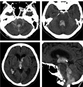
A 66-year-old woman presented with severe headache and vomiting, followed by a decline in consciousness and was emer-gently transported to our hospital. She had a history of diabetes mellitus, hypertension, and dyslipidemia. On admission, the patient was lethargic with a Glasgow coma scale score of 12. Neurological examination disclosed bilateral lower cranial nerve palsies. Computed tomography (CT) on admission demonstrated subarachnoid hemorrhage (SAH) located predominantly in the cisterna magna and intraventricular hematoma (Figure 1). 3D-CT angiography (3D-CTA) showed small aneurysmal dilata-tion that was suspected to originate from the left PICA (data not
shown). Subsequently, digital subtraction angiography (DSA) was performed (Figure 2A-D). A right vertebral angiogram (VAG) showed hypoplasia of the right PICA. The right PICA territory was supplied by the right superior cerebellar artery (SCA), right AICA, and collaterals from the right vertebral artery (VA). On the left VAG, a fine tortuous artery from the posterior medullary segment of the left PICA coursed medially and connected to the cortical segment of the right PICA. A small fusiform aneurysm around 2 mm in diameter arose from the ar-tery interconnecting bilateral PICAs. This arar-tery was identified
The Journal of Medical Investigation Vol. 67 2020
Received for publication June 20, 2019 ; accepted August 29, 2019. Address correspondence and reprint requests to Yuichi Satoh, 815-2, Shima, Ikedacho, Miyoshi, Tokushima 778-0005, Japan and Fax : +81-883-72-6910.
Figure 1. Plain head computed tomography on admission showing subarachnoid hemorrhage located predominantly in the cisterna mag-na and hematoma in the 4th, 3rd, and bilateral lateral ventricles.
198
Y. SATOH, et al. PICA communicating artery aneurysm as a PICA communicating artery. Tortuous arteries from the leftPICAs and left VA were shown to fuse with the communicating artery. Coil embolization of the aneurysm was attempted, but it was difficult to lead the micro catheter to the aneurysm, and the attempt was abandoned. The next day, a midline suboccipital craniotomy was performed. The dura was remarkably tense. There was thick hematoma in the cisterna magna. The caudal loop of the left PICA was identified after hematoma aspiration. The course of the left PICA was traced distally, and its posterior medullary segment was exposed. A small branch was found arising from the arterial segment and coursing caudally along the dorsal aspect of the medulla oblongata. The small aneurysm covered with clot was observed on the arterial branch. The por-tion distal to the aneurysm was dissected, and an anastomosis with the branch from the left VA coursing to the right cerebellar hemisphere was found. The communicating artery was tortuous and did not send perforators around the aneurysm (Figure 3A). The aneurysm was trapped and excised. An intraoperative indo-cyanine green video angiogram showed that the anastomosing vessel from the left VA supplied the distal communicating artery.
After aneurysmal excision, the hematoma in the fourth ventricle was thoroughly evacuated. On pathological examination, the internal elastic lamina was partially disrupted, and findings suggesting a dissecting aneurysm, such as an intramural hema-toma, were not observed. (Figure 3B, C).
Neither symptomatic vasospasm nor hydrocephalus occurred after the operation. DSA performed 27 days after the operation showed disappearance of the aneurysm and patency of the communicating artery (Figure 2E). The patient became alert, but mild truncal ataxia remained, and she was transferred to another hospital for rehabilitation 39 days after the operation.
DISCUSSION
Distal PICA aneurysms are very rare and account for 0.28% and 0.38% of all intracranial aneurysms and ruptured aneu-rysms, respectively (1). Most of these aneurysms arise from the distal PICA trunk (1). Aneurysms arising from the small branches of the PICA are extremely rare. Such PICA branch aneurysms mainly occur on the choroidal branch of the PICA or the PICA communicating artery (2-7).
The PICA has many anatomical variations. Unilateral hypo-plasia and ahypo-plasia of a PICA have been reported (8, 9). A PICA communicating artery develops as a collateral interconnecting bilateral PICAs at a distal portion in the case of hypoplasia or aplasia of a unilateral PICA (2). A PICA communicating artery aneurysm was first reported by Hlavin et al. in 1991 (2). Only six cases of aneurysms of a PICA communicating artery have been reported so far (2-7). These six cases and the present case are listed in Table 1. Of the seven patients, three were male and four were female. Their mean age was 56.9 (ranging from 45 to 68) years and slightly lower than that of patients with distal PICA trunk aneurysms, which was 64 (ranging from 20 to 81) years (1). The shape of aneurysms was saccular in three cases and fusi-form in four cases. An aneurysm occurred from the bifurcation of a PICA and the PICA communicating artery in one case (2), but the others occurred at a nonbranching site of the communicating artery itself. A fusiform aneurysm at a nonbranching site may Figure 2. Preoperative digital subtraction angiograms of the right
vertebral artery (A, B) and of the left vertebral artery(C, D). The right PICA is hypoplastic (A, B). The communicating artery from the posterior medullary segment of the left PICA supplies the vermian territory of the right PICA(C, D). A fusiform aneurysm is noted on the communicating artery (indicated by arrows C, D). Small arteries from the left VA are shown to fuse with the communicating artery distal to the aneurysm(C). Postoperative left vertebral angiogram showing dis-appearance of the aneurysm and patency of the PICA communicating artery (indicated by double arrows E).
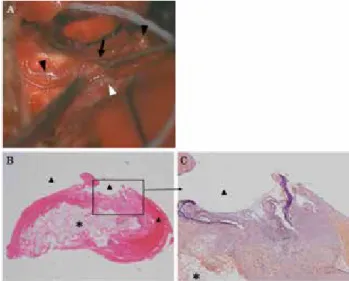
Figure 3. Intraoperative photograph showing the afferent arteries (black triangle head), the efferent artery (white triangle head), and the aneurysm covered with clot and a cottonoid (black arrowhead). Pathological examination of the resected aneurysm (hematoxylin and eosin (B) and elastica van Gieson (C) staining). (C) The internal elas-tic lamina is disrupted.
The asterisk indicates oxidized cellulose used as a hemostatic agent. Triangles indicate the intravascular lumen.
199
The Journal of Medical Investigation Vol. 67 February 2020
suggest a dissecting aneurysm. Pathological examination did not show findings suggesting a dissecting aneurysm in the pres-ent case as far as we could examine. The aneurysm appeared to be a true one in the present case. Sugiyama et al. also reported that a communicating artery aneurysm is a true aneurysm (6). The communicating arteries were tortuous in all cases. Aplasia or hypoplasia of a unilateral PICA or VA was shown in most reported cases (2-7), and, similarly, the contralateral PICA was hypoplastic in the present case. The PICA territory, especially the vermian territory, was mainly supplied through the PICA communicating artery. Sugiyama et al. reported that a collateral vascular network including a PICA communicating artery de-velops to supply the contralateral vermian territory because of insufficient blood supply due to aplasia or hypoplasia of the con-tralateral PICA, AICA, or vertebral artery (6). Udono et al. sug-gested that a PICA communicating artery is a remnant vessel of the embryonic stage (3). The primitive lateral basilar-vertebral anastomoses that communicate with the dorsal side artery of the medulla oblongata are present around the seventh week of em-bryonic development. These findings suggest that hemodynamic stress on primitive vascular walls contributes to the formation of these aneurysms. The size of the aneurysms was small, and the average diameter was 2.67 mm (ranging from 2 to 3 mm) in six cases in which aneurysmal size was accurately described. All patients presented with subarachnoid hemorrhage. A PICA com-municating artery aneurysm might tend to rupture frequently, even though it is small, and there is no report of an unruptured PICA communicating artery aneurysm.
Regarding surgical procedures, neck clipping was performed for three saccular aneurysms, whereas trapping was performed in four cases of fusiform aneurysms. Haga et al. reported treating a fusiform aneurysm with trapping with occipital artery (OA) – PICA bypass (7). In their case, neck clipping was difficult be-cause the aneurysm was fusiform. In addition, the contralateral PICA territory was supplied through the PICA communicating artery, and no blood supply from other arteries was observed. Sugiyama et al. treated a fusiform aneurysm with trapping only. In their case, the contralateral AICA was connected to the distal part of the contralateral PICA. Thus, the collateral circulation of the contralateral PICA territory should be evaluated to select surgical procedures, even though it is difficult to assess if there is sufficient collateral flow. In the present case, the aneurysm was treated with trapping only because the preoperative angio-gram showed small arteries from the left VA fused with the com-municating artery distal to the aneurysm and the right PICA territory was also supplied by the ipsilateral SCA and AICA. As a result, there was no infarction in bilateral PICA territories in the present case. Including present case, there was no ischemic complication in 3 cases treated with trapping (5, 6). Recently, endovascular strategies such as coil embolization have been in-creasingly used for aneurysm treatment, but there is no report of
a PICA communicating artery aneurysm treated with an endo-vascular strategy. In the present case, an endoendo-vascular strategy was attempted, but it was difficult to deliver the micro catheter to the aneurysm because of vascular thinness and tortuousness. Song et al. reported that direct surgery is preferable to an endo-vascular strategy to treat an aneurysm of the distal PICA (10).
CONFLICT OF INTEREST DISCLOSURE
All authors have no conflict of interest.
REFERENCES
1. Horiuchi T, Tanaka Y, Hongo K, Nitta J, Kusano Y, Kobayashi S : Characteristics of Distal Posteroinferior Cerebellar Ar-tery Aneurysms. Neurosurgery 53 : 589-596, 2003 2. Hlavin ML, Takaoka Y, Smith AS : A “PICA
communicat-ing artery” aneurysm : case report. Neurosurgery 29 : 926-929, 1991
3. Udono H, Shiraishi T, Tsuji T, Abe M, Tabuchi K : [True “PICA communicating artery” aneurysm : a case report]. No shinkei geka Neurological surgery 25 : 763-766, 1997 4. Fujiwara K, Ito J, Kanayama S : [Multiple aneurysms of
the PICA communicating artery : a case report]. No shinkei geka Neurological surgery 27 : 177-182, 1999
5. Okuno S, Ohnishi H : Aneurysm of the posterior inferior cerebellar arteries/communicating artery. Cerebrovascular diseases (Basel, Switzerland) 12 : 276-279, 2001
6. Sugiyama S, Fujimura M, Inoue T, Shimizu H, Watanabe M, Tominaga T : Ruptured aneurysm of a posterior inferior cerebellar artery communicating artery. Case report and histological findings. Neurologia medico-chirurgica 52 : 81-83, 2012
7. Haga D, Kuroki T, Andoh S, Nemoto M, Sugo N, Nagao T : Ruptured aneurysm of the PICA communicating ar-tery : a case report. Journal of stroke and cerebrovascular diseases : the official journal of National Stroke Association 23 : 1247-1252, 2014
8. Fujii K, Lenkey C, Rhoton AL, Jr. : Microsurgical anatomy of the choroidal arteries. Fourth ventricle and cerebellopon-tine angles. J Neurosurg 52 : 504-524, 1980
9. Lister JR, Rhoton AL, Jr., Matsushima T, Peace DA : Mi-crosurgical anatomy of the posterior inferior cerebellar artery. Neurosurgery 10 : 170-199, 1982
10. Song J, Park JE, Chung J, Lim YC, Shin YS : Treatment strategies of ruptured posterior inferior cerebellar artery aneurysm according to its segment. Surgical neurology international 8 : 155, 2017
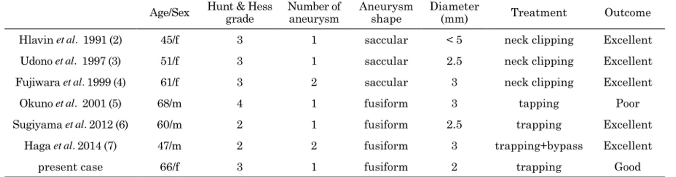
Table 1. Summary of six previously reported cases of PICA communicating artery aneurysms
Age/Sex Hunt & Hess grade Number of aneurysm Aneurysm shape Diameter(mm) Treatment Outcome Hlavin et al. 1991 (2) 45/f 3 1 saccular < 5 neck clipping Excellent Udono et al. 1997 (3) 51/f 3 1 saccular 2.5 neck clipping Excellent Fujiwara et al. 1999 (4) 61/f 3 2 saccular 3 neck clipping Excellent
Okuno et al. 2001 (5) 68/m 4 1 fusiform 3 tapping Poor Sugiyama et al. 2012 (6) 60/m 2 1 fusiform 2.5 trapping Excellent
Haga et al. 2014 (7) 47/m 2 2 fusiform 3 trapping+bypass Excellent present case 66/f 3 1 fusiform 2 trapping Good