滋賀医科大学機関リポジトリびわ庫
59
0
0
全文
(2) Research Articles: Behavioral/Cognitive. Change in brain plasmalogen composition by exposure to prenatal undernutrition leads to behavioral impairment of rats https://doi.org/10.1523/JNEUROSCI.2721-18.2019 Cite as: J. Neurosci 2019; 10.1523/JNEUROSCI.2721-18.2019 Received: 21 October 2018 Revised: 28 July 2019 Accepted: 31 July 2019. This Early Release article has been peer-reviewed and accepted, but has not been through the composition and copyediting processes. The final version may differ slightly in style or formatting and will contain links to any extended data. Alerts: Sign up at www.jneurosci.org/alerts to receive customized email alerts when the fully formatted version of this article is published.. Copyright © 2019 the authors.
(3) 1. Change in brain plasmalogen composition by exposure to prenatal. 2. undernutrition leads to behavioral impairment of rats. 3 4. Abbreviated title: Ethanolamine plasmalogen and behavior. 5 6. Kodai Hino1, Shunya Kaneko1, Toshiya Harasawa1, Tomoko Kimura1, Shiro Takei2,. 7. Masakazu Shinohara3,4, Fumiyoshi Yamazaki5, Shin-ya Morita6, Shumpei Sato5,. 8. Yoshihito Kubo1, Tadaaki Kono1, Mitsutoshi Setou5,7,8, Mina Yoshioka1, Junya Fujino1,. 9. Hiroyuki Sugihara9, Hideto Kojima10, Naoto Yamada11, Jun Udagawa1. 10 11. 1. 12. Medical Science, Otsu 520-2192, Japan. 13. 2. 14. Chubu University, Kasugai, 487-8501, Japan. 15. 3. 16. 650-0017, Japan. 17. 4. 18. Medicine, Kobe, 650-0017, Japan. 19. 5. 20. Anatomy, Hamamatsu University School of Medicine, 1-20-1 Handayama, Higashi-ku,. 21. Hamamatsu, Shizuoka 431-3192, Japan. 22. 6. 23. 520-2192 Japan. Division of Anatomy and Cell Biology, Department of Anatomy, Shiga University of. Department of Environmental Biology, College of Bioscience and Biotechnology,. Division of Epidemiology, Kobe University Graduate School of Medicine, Kobe,. The Integrated Center for Mass Spectrometry, Kobe University Graduate School of. International Mass Imaging Center and Department of Cellular and Molecular. Department of Pharmacy, Shiga University of Medical Science Hospital, Otsu, Shiga. 1.
(4) 24. 7. 25. Higashi-ku, Hamamatsu, Shizuoka 431-3192,) Japan. 26. 8. 27. Block 21 Sassoon Road, Pokfulam, Hong Kong SAR,) China. 28. 9. 29. University of Medical Science, Otsu, Shiga, Japan. 30. 10. 31. Medical Science, Otsu, Shiga, Japan. 32. 11. Preeminent Medical Photonics Education & Research Center, (1-20-1 Handayama,. Department of Anatomy, The University of Hong Kong, (6/F, William MW Mong. Division of Molecular Diagnostic Pathology, Department of Pathology, Shiga. Department of Stem Cell Biology and Regenerative Medicine, Shiga University of. Department of Psychiatry, Shiga University of Medical Science, Otsu 520-2192, Japan. 33 34. Correspondence. 35. Jun Udagawa, Division of Anatomy and Cell Biology, Department of Anatomy, Shiga. 36. University. 37. [email protected]. of. Medical. Science,. Otsu. 520-2192,. Japan.. E-mail:. 38 39. Number of pages: 44. 40. Number of figures and tables: 6 and 5, respectively. 41. Number of words for Abstract, Introduction, and Discussion: 222, 426 and 1496. 42 43. Conflict of Interest: None declared.. 44 45. Acknowledgments. 46. We thank Mr. Kenji Iwabuchi (Department of Stem Cell Biology and Regenerative. 47. Medicine, Shiga University of Medical Science) for assistance in the behavioral test. 2.
(5) 48. MALDI-IMS was conducted via the Imaging Platform supported by the Ministry of. 49. Education, Culture, Sports, Science and Technology (MEXT), Japan and Preppers Co.. 50. Ltd.. 51. 3.
(6) 52. Abstract. 53. Epidemiological studies suggest that poor nutrition during pregnancy influences. 54. offspring predisposition to experience developmental and psychiatric disorders. Animal. 55. studies have shown that maternal undernutrition leads to behavioral impairment, which. 56. is linked to alterations in monoaminergic systems and inflammation in the brain. In this. 57. study, we focused on the ethanolamine plasmalogen of the brain as a possible. 58. contributor to behavioral disturbances observed in offspring exposed to maternal. 59. undernutrition. Maternal food or protein restriction between gestational day (GD) 5.5. 60. and GD 10.5 resulted in hyperactivity of rat male adult offspring. Genes related to the. 61. phospholipid biosynthesis were found to be activated in the prefrontal cortex (PFC), but. 62. not in the nucleus accumbens or striatum, in the offspring exposed to prenatal. 63. undernutrition. Corresponding to these gene activations, increased ethanolamine. 64. plasmalogen (18:0p-22:6) was observed in the PFC using mass spectrometry imaging. A. 65. high number of crossings and the long time spent in the center area was observed in the. 66. offspring exposed to prenatal undernutrition and was mimicked in adult rats via the. 67. intravenous injection of ethanolamine plasmalogen (18:0p-22:6) incorporated into the. 68. liposome. Additionally, plasmalogen (18:0p-22:6) increased only in the PFC, and not in. 69. the nucleus accumbens or striatum. These results suggest that brain plasmalogen is one. 70. of the key molecules to control behavior and its injection using liposome is a potential. 71. therapeutic approach for cognitive impairment.. 72 73. Keywords:. 74. phosphatidylethanolamine; Prefrontal cortex. Hyperactivity;. Maternal. 75 4. undernutrition;. Plasmalogen.
(7) 76. Significance Statement. 77. Maternal undernutrition correlates to developmental and psychiatric disorders. Here, we. 78. found that maternal undernutrition in early pregnancy led to hyperactivity in rat male. 79. offspring and induced gene activation of phospholipid-synthesizing enzyme and. 80. elevation of ethanolamine plasmalogen (18:0p-22:6) level in the prefrontal cortex (PFC).. 81. Intravenous injection of ethanolamine plasmalogen (18:0p-22:6) incorporated into the. 82. liposome maintained crossing activity and was circumscribed to the center area for a. 83. long time period, in prenatally undernourished offspring with aberrant behavior.. 84. Furthermore, the amount of ethanolamine plasmalogen (18:0p-22:6) increased in the. 85. PFC of the rat after injection. Our result suggests that brain plasmalogen is one of the. 86. key molecules to control behavior and that its injection using liposome is a potential. 87. therapeutic approach for cognitive impairment.. 5.
(8) 88. Introduction. 89. Epidemiological studies have linked maternal stress during pregnancy, including. 90. malnutrition, infection, daily life stress, and traumatic events, to the presence of. 91. psychological and developmental disorders in offspring (Hoek et al., 1998; Khashan et. 92. al., 2008; Kinney et al., 2008; Marques et al., 2015; Fineberg et al., 2016; Kundakovic. 93. and Jaric, 2017). A previous study suggested that brain development is disrupted by. 94. prenatal exposure to stress, which alters fetal programming by affecting the epigenome,. 95. such as via changes to DNA methylation and histone modification, and induces. 96. behavioral disturbances (Kundakovic and Jaric, 2017). A postmortem study of the brain. 97. of a patient with schizophrenia suggested decreased Reelin and GAD67 expression due. 98. to the hypermethylation of their promoter regions, which was led by the upregulation of. 99. DNA methyltransferase 1 (DNMT1) genes and may be involved in the etiology of. 100. schizophrenia (Kundakovic, 2014). Meanwhile, a Dutch famine study reported the. 101. relationship between prenatal undernutrition during the first trimester and the increased. 102. incidence of schizophrenia (Brown and Susser, 2008). These reports suggest that. 103. maternal stress may alter brain function through disturbances in the neurotransmission. 104. of certain systems, such as the GABAergic system. In fact, restraint stress on mice. 105. during pregnancy leads to the overexpression of DNMT1 and DNMT3a mRNA, which. 106. is accompanied by the downregulation of Reelin and GAD67 protein levels, as well as. 107. glutamine receptor protein from the hypermethylation of their promoter regions in the. 108. frontal cortex. This cascade of events induced a schizophrenia-like phenotype. 109. observable in behavioral tests performed on the male offspring after birth (Matrisciano. 110. et al., 2013). Prenatal stress is thus associated with a predisposition toward. 111. neurobehavioral disorders. Not only restrained stress, but prenatal caloric restriction has 6.
(9) 112. also been shown to affect the dopamine system and neuronal excitability, resulting in a. 113. decrease in anxiety-like behavior, while protein restriction results in deficits in pre-pulse. 114. inhibition and locomotor activity (Markham and Koenig, 2011; Amaral et al., 2015).. 115. Based on these findings, various neurotransmitter systems, including monoamine,. 116. GABAergic, and glutaminergic systems, appear to be viable therapeutic targets for. 117. treating behavioral disturbances induced by prenatal stress; however, membrane lipids. 118. also seemed to differ between the brains of patients with psychiatric disorders and those. 119. without such disorders, and little is known regarding the underlying mechanisms of this. 120. process (Ghosh et al., 2017). Therefore, we hypothesized that behavioral disturbance. 121. due to early prenatal undernutrition is led by aberrant brain phospholipid metabolism. 122. via fetal programming. In this study, we focused on alterations in the composition of. 123. brain phospholipids that are induced by prenatal undernutrition, and we have identified. 124. a candidate phospholipid to control behavior.. 125. 7.
(10) 126. Materials and methods. 127. Animals. 128. All animal procedures were approved by the Institutional Review Board of the Shiga. 129. University of Medical Science Animal Care and Use Committee (2011-8-1, 2014-3-7,. 130. 2015-12-1, and 2019-4-2). For experiments investigating maternal undernutrition,. 131. 9-week-old male (body weight [BW], 250–280 g) and 8-week-old female (BW, 160–. 132. 190 g) Wistar rats were obtained from CLEA Japan, Inc. (Tokyo, Japan). Six-week-old. 133. male rats were obtained for experiments involving phosphatidylethanolamine (PE). 134. injection. All rats were housed under a 12-h light:dark cycle (lights were turned on at. 135. 08:00) and were allowed to acclimate for greater than 1 week.. 136. Diet. 137. Female rats were acclimated to a standard diet for pregnant rats (AIN-93G), containing. 138. 20% casein for 2 days prior to mating, for which each female was housed with one male. 139. overnight. We defined gestational day (GD)0 as the day when a vaginal plug was. 140. observed. Pregnant rats were randomly assigned to the ad libitum (AL) group, the. 141. food-restriction (F) group, or the isocaloric, low-protein-diet (LPD) group, and. 142. subjected to undernutrition from GD5.5 to GD10.5 or from the day of blastocyst. 143. implantation to the day just before the closure of the neural tube (Fig. 1A) (Erb, 2006).. 144. In humans, this period is comparable to the days from E6.5 to approximately E30,. 145. which is nearing the first half of the first trimester (Bystron et al., 2008; Schoenwolf et. 146. al., 2015). Neural stem cells, and not neurons, exist in the telencephalon because this. 147. period is prior to neurogenesis (Götz and Huttner, 2005; Bystron et al., 2008). The F. 148. group was fed 50% (50F, four dams) or 40% (40F, six dams) of the daily food intake of. 8.
(11) 149. the AL group (eight dams). The rationale is that daily rations fell to no more than 800. 150. kcal during the Dutch famine that occurred between December 1944 and April 1945,. 151. representing 40% of rations (>2000 calories) after June 1945 (Roseboom et al., 2001).. 152. The LPD group (five dams) was fed a diet containing 9% casein. After delivery, pups. 153. were culled to produce litters of eight offspring (four males and four females) per a dam. 154. on postnatal day (P)4. During lactation, the dams were fed CE-2, a standard pellet chow. 155. for rearing and breeding. Subsequently, the offspring were weaned on P28, and. 156. afterward, they were fed CE-2 ad libitum. Male offspring were used in this study. 157. because male humans show a higher risk of neuropsychiatric or neurobehavioral. 158. disorders (e.g., schizophrenia, attention deficit hyperactivity disorder [ADHD], and. 159. autism spectrum disorder [ASD]), than females (Aleman et al., 2003; Werling and. 160. Geschwind, 2013; Arnett et al., 2015). Furthermore, sex differences in ADHD and ASD. 161. may be, in part, genetically mediated (Werling and Geschwind, 2013; Arnett et al.,. 162. 2015). The male offspring experienced handling once a week after weaning, and body. 163. weight of the offspring were measured at 9 and 12 weeks of age.. 164. Preparation of liposomes. 165. Large unilamellar liposomes composed of egg phosphatidylcholine (PC) and. 166. C18:0-22:6 plasmalogen PE (PlsEtn) or C16:0-18:1 diacyl phosphatidylethanolamine. 167. (POPE) were prepared by the extrusion method (Morita et al., 2008). Briefly, a thin film. 168. was obtained by evaporating the lipid chloroform solution and was subsequently. 169. hydrated with saline so that the concentrations of egg PC and PE (18:0p-22:6) (Avanti,. 170. AL) were 8 mg/mL and 2 mg/mL, respectively, for PE liposomes (PELs). Similarly,. 171. mixed solutions of PE (16:0-18:1) and egg PC were prepared at the same concentration. 172. as for the POPE liposomes (POPELs). To produce control liposomes (CLs), PC (10 9.
(12) 173. mg/mL) with no PE was prepared. After five rounds of freezing and thawing, the lipid. 174. suspension was extruded through a polycarbonate filter with 100-nm pore size.. 175. Behavioral test. 176. Effects of prenatal undernutrition on behavior. 177. From June to October, the locomotor activity of the male offspring was evaluated by the. 178. open-field test at 8 weeks of age for the AL (n = 32 from 8 dams), LPD (n = 18 from 5. 179. dams), 50F (n = 13 from 4 dams), and 40F (n = 17 from 6 dams) groups and at 12. 180. weeks for the AL (n = 18 from 5 dams), LPD (n = 19 from 5 dams), and 50F (n = 14. 181. from 5 dams) groups, to examine the impact of maternal undernutrition on behavior.. 182. Behavioral data that included device errors in tracing animals were excluded. The. 183. apparatus, measuring 90 cm in diameter and 45 cm in height, was used to monitor the. 184. behavior of rats, and the behavior was recorded for 10 min under 9 lux of light. Data. 185. were analyzed using the Limelight video tracking system (Actimetrics, IL, USA). The. 186. distance traveled and the time spent in the center was measured under the following. 187. analysis conditions: the open field was divided into the center and peripheral regions so. 188. that 1) the center region was bordered by a concentric circle passing through the. 189. midpoint of the radius of the open field (Condition 1 and 2) the area of the center region. 190. (A1) was the same as that of the peripheral region (A2) (Condition 2). The former. 191. condition was selected to allow crossing behavior to be analyzed.. 192. Effects of plasmalogen (18:0p-22:6) on behavior. 193. To examine the effect of PE (18:0p-22:6), the locomotor activity of male rats was. 194. evaluated before and after PE (18:0p-22:6) injection (for 8- and 14-week-old offspring,. 195. respectively). Rats were assigned to two different groups based on the results of. 196. crossing from the open-field test so that rats with similar locomotor activities were 10.
(13) 197. evenly divided among the groups. A PEL or CL suspension (1 mL/kg BW) was injected. 198. into the tail vein at 14 weeks of age, and the second injection of liposome suspension. 199. was performed 2 days later. The rats in the PEL (n = 6) and CL (n = 7) groups were. 200. subjected to the open-field test or the elevated plus maze test (PEL: n = 6, CL: n = 6) 1. 201. day or 4 days after the second injection, respectively. Behavioral data of one CL rat. 202. acquired by using the elevated plus maze test was excluded because it included device. 203. errors in tracing animals. In the elevated plus maze test, rats were placed in the central. 204. square platform facing the closed arms, and their behavior was recorded for 250. 205. seconds under 8, 10, and 4 lux of light at the central square platform, facing the open. 206. arms and closed arms, respectively (Hino et al., 2019). Time spent in the open and. 207. closed arms was measured in this test. To verify the specific effect of PlsEtn. 208. (18:0p-22:6) on behavior, the alteration of locomotor activity was examined before and. 209. after POPEL, CL, or saline injection. Male rats were assigned to three different groups. 210. based on the results of the crossing analysis, and behavior was evaluated for POPEL (n. 211. = 5), CL (n = 5), and saline injection (n = 4) groups. This experiment was conducted. 212. separately from the PEL injection study. The experimental groups allocated in this study. 213. are listed in Table 2.. 214. Metabolic profiling of plasma and cerebrospinal fluid (CSF). 215. Blood samples were collected from male offspring (AL: four dams and seven litters,. 216. 40F: four dams and six litters) and CSF (AL: four dams and seven litters, 40F: four. 217. dams and five litters) at 9 weeks of age. CSF samples that got mixed with blood were. 218. excluded from the analysis. Rats were anesthetized with sodium pentobarbital solution. 219. (35 mg/kg, intraperitoneally) during the light phase (16:00–18:00). They were placed in. 220. a stereotaxic device (KOPF, CA, USA), and 50 μL of CSF was collected immediately 11.
(14) 221. from the cisterna magna. Then, after decapitation, 5 mL of blood was collected in test. 222. tubes containing EDTA-2Na. The plasma was collected after centrifugation of the blood. 223. at 2,000 × g for 15 min. Samples were stored at −80°C until use.. 224. Hydrophilic metabolites were extracted using the MeOH-CHCl3 method according to. 225. the procedure detailed in previous reports (Tsugawa et al., 2011; Nishiumi et al., 2012).. 226. Fifty μL of plasma or CSF was mixed with 250 μL of a solvent mixture. 227. (MeOH:H2O:CHCl3, 2.5:1:1, v/v/v) containing 20 μL of 0.25 mg/mL 2-isopropylmalic. 228. acid (Sigma-Aldrich, Tokyo, Japan) as the internal standard. The mixture was then. 229. shaken at 37°C for 30 min and centrifuged at 16,000 × g for 5 min at 4°C. Then, 225 μL. 230. of supernatant was mixed with 200 μL of distilled water, and the solution was. 231. centrifuged at 16,000 × g for 5 min at 4°C. The resultant supernatant (250 μL). 232. containing hydrophilic primary metabolites was collected and lyophilized using a freeze. 233. dryer. For oximation, 40 μL of 20 mg/mL methoxyamine hydrochloride (Sigma-Aldrich,. 234. Tokyo, Japan) dissolved in pyridine was mixed with a lyophilized sample, and the. 235. mixture was then shaken at 30°C for 90 min. For derivation, 20 μL of. 236. N-methyl-N-trimethylsilyl-trifluoroacetamide (MSTFA) (GL Science, Tokyo, Japan). 237. was added, and the mixture was shaken at 37°C for 30 min. The mixture was then. 238. centrifuged at 16,000 × g for 5 min at 4°C, and the resultant supernatant was subjected. 239. to gas chromatography–mass spectrometry (GC–MS) analysis.. 240. GC–MS analysis was performed by using a GCMS-QP2010 Ultra device (Shimadzu. 241. Co., Kyoto, Japan) with a fused-silica capillary column (CP-SIL 8 CB low bleed/MS;. 242. 30 m × 0.25 mm inner diameter, film thickness: 0.25 μm; Agilent Co., Palo Alto, CA,. 243. USA). The front inlet temperature was set at 230°C. The flow rate of helium gas. 244. through the column was 39.0 cm/s. The column temperature was held at 80°C for 2 min 12.
(15) 245. and then raised by 15°C/min to 330°C and held for 6 min. The transfer-line and. 246. ion-source temperatures were 250°C and 200°C, respectively. Twenty scans per second. 247. were recorded over the mass range of 85–500 m/z by using the Advanced Scanning. 248. Speed Protocol (ASSP, Shimadzu Co., Kyoto, Japan).. 249. Raw data were exported in netCDF format, and peak detection and alignment were. 250. performed by using MetAlign software (Wageningen UR, The Netherlands). The. 251. resulting data were exported in CSV format and then analyzed with in-house analytical. 252. software (AIoutput), which enabled peak identification and semi-quantification by using. 253. an in-house metabolite library (Tsugawa et al., 2011; Nishiumi et al., 2012). For. 254. semi-quantification, the peak height of a particular ion for each metabolite was. 255. normalized to the peak height of the specified ion of 2-isopropylmalic acid (the internal. 256. standard).. 257. Brain sections. 258. According to previous reports, lesions in the medial PFC cause alterations in the. 259. locomotor activity of rats (Jinks and McGregor, 1997; Fritts et al., 1998). Moreover, the. 260. nucleus accumbens (NAcc) and striatum (CPu) receive input from the PFC and are. 261. associated with locomotor activity and impulsivity (Moreno et al., 2013; Spencer et al.,. 262. 2015; Scofield et al., 2016; Zhu et al., 2016; Dahoun et al., 2017). Hence, sections of. 263. PFC, NAcc, and CPu were subjected to gene expression and phospholipid analyses, and. 264. immunohistochemistry. Male offspring of 9 weeks of age from the maternal. 265. undernutrition experiment and male rats injected with PEL or CL at 14 weeks of age (at. 266. 10 hours after the elevated plus maze test) were anesthetized and euthanized with. 267. sodium pentobarbital solution (100 mg/kg, ip). Brain samples from the AL (four dams. 268. and eight litters), 40F (five dams and eight litters), PEL (n = 4), and CL (n = 4) groups 13.
(16) 269. were immediately dissected out and frozen in dry ice. Cryosections of the brain were cut. 270. at a thickness of 10 μm before use for gene expression and phospholipid analyses.. 271. Greater than eight sections of the prefrontal cortex (PFC) every 200 Pm, greater than six. 272. sections of NAcc (Nucleus accumbens) every 80 Pm, and greater than seven sections of. 273. CPu (Caudate putamen) every 80 Pm were placed on Platinum Pro (Matsunami, Osaka,. 274. Japan) or polyethylene naphthalate membrane slides (Leica Microsystems, Wetzlar,. 275. Germany) for immunohistochemistry or gene expression analysis, respectively. Sections. 276. at 0.8 mm and 2 mm from the frontal end of the cerebral cortex, a section at 0.32 mm. 277. from the anterior end of the NAcc, and a section at 0.48 mm from the anterior end of the. 278. CPu were placed on indium tin oxide (ITO)-coated glass slides (Bruker Daltronics,. 279. Bremen, Germany) for phospholipid analysis.. 280. Gene expression. 281. The PFC, NAcc, and CPu of male offspring in the AL and 40F groups were dissected. 282. and collected from brain sections by a laser microdissection system (LMD6000, Leica. 283. Microsystems, Wetzlar, Germany). Gene expression analysis was performed according. 284. to the protocol described in the previous report (Kimura et al., 2018). Isolated total RNA. 285. was converted to cDNA via reverse transcription (RT) and amplified using the Ovation. 286. PicoSL WTA system V2 (NuGEN Technologies, Inc., San Carlos, CA, USA). The. 287. mRNA expression levels were estimated using quantitative real-time PCR (RT-qPCR). 288. analysis using a LightCycler 480 system (Roche Diagnostics GmbH, Mannheim,. 289. Germany) with SYBR Premix Ex Taq II polymerase (Takara Bio, Kusatsu, Japan). The. 290. RT-qPCR reaction was performed in duplicates, and comparative Cq values of the target. 291. genes (Table 1) normalized to B2m, a reference gene, were compared between the AL. 292. and 40F groups. 14.
(17) 293. Counting microglial cells and activated microglial cells in PFC. 294. In each rat, ten or more brain sections at approximately 4.8 to 2.6 rostral to the bregma. 295. were fixed with 4% paraformaldehyde at room temperature for 30 min, incubated with. 296. rabbit anti-Iba1 antibody (dilution, 1:500; Wako Cat# 019-19741, RRID:AB_839504). 297. and mouse anti-CD11b (dilution, 1:300; Bio-Rad / AbD Serotec Cat# MCA275R,. 298. RRID:AB_321302), and then incubated with goat anti-rabbit IgG (H&L) conjugated. 299. with DyLight 488 (dilution, 1:500; Abcam Cat# ab96895, RRID:AB_10679405) and. 300. goat anti-mouse IgG (H&L) conjugated with DyLight 549 (dilution, 1:500; Rockland. 301. Cat#. 302. 4′,6-diamidino-2-phenylindole (DAPI), and the numbers of all Iba1-expressing. 303. microglial cells and CD11b-labeled activated microglial cells were counted in the. 304. medial PFC by using a fluorescence microscope (IX83, Olympus, Tokyo, Japan). The. 305. density of those cells was then compared between offspring in the AL and 40F groups.. 306. Matrix-assisted. 307. (MALDI-IMS). 308. MALDI-IMS was performed by using the PFC, NAcc, and CPu samples in both. 309. experiments as previously described (Hossen et al., 2015; Sugiyama et al., 2015).. 310. Tissues on ITO-coated glass slides were subjected to matrix application by the. 311. sublimation/deposition method, with 1 g of 9-AA sublimated at 210°C in order for the. 312. deposition thickness to reach 1.0 μm by using the iMLayer device (Shimadzu, Kyoto,. 313. Japan). Experiments were performed by using a mass microscope, a prototype of the. 314. iMScope equipped with a 355-nm Nd:YAG laser (Shimadzu, Kyoto, Japan). Negative. 315. ions from a sample area of 30 Pm × 30 Pm on the PFC, NAcc, and CPu samples were. 316. obtained in a mass range of m/z 400 to 1,000. Adjacent sections of a mouse brain as a. 610-142-121,. laser. RRID:AB_1057533).. Sections. desorption/ionization-imaging. 15. were. mass. stained. with. spectrometry.
(18) 317. reference were laid together with rat brain sections of 40F and AL offspring, and the rats. 318. injected with PEL and CL and were used to correct for differences in peak intensity due. 319. to differences in sample preparation between slides (Fig. 2). The peaks of 52 PE, 16. 320. phosphatidylserine (PS), 14 phosphatidylinositol (PI), 10 lysophosphatidylethanolamine. 321. (lysoPE), and 4 lysophosphatidylinositol (lysoPI) were detected (Taguchi and Ishikawa,. 322. 2010). The peak intensity of each individual phospholipid was corrected using the. 323. average intensity of the corresponding phospholipid of the mouse references. The. 324. average peak intensities of the PFC, NAcc, and CPu, respectively, were compared. 325. between the AL and 40F groups and between the PEL and CL groups.. 326. To identify the peak assigned at m/z 774.5 as the peak of PE (18:0p-22:6), we. 327. performed MALDI tandem mass spectrometry (MS/MS) using the mass microscope. 328. described above (Sugiyama et al., 2015) and identified PE (18:0p-22:6) via. 329. collision-induced dissociation (CID) (Zemski Berry et al., 2014).. 330. Blood cells. 331. Blood was collected from 14-week-old male rats for the PEL and CL injection. 332. experiment. Whole blood cells were washed with saline, applied to a 12-well. 333. flexiPERM® plate (Sarstedt, Tokyo, Japan), and affixed to an ITO-coated glass slide. 334. (Matsunami Glass Industries, Osaka, Japan), of which the surface was coated with. 335. poly-L-lysine (Hossen et al., 2015) to ensure that the blood cells were confluent. Blood. 336. cells were centrifuged to attach the cells to the surface of the glass slide, fixed with. 337. 0.25% glutaraldehyde for 5 min, and rinsed three times with 150 mM ammonium. 338. acetate buffer (pH 7.5). Samples were dried and subjected to MALDI-IMS to examine. 339. the phospholipid content in the blood cells Briefly, negative ions from a sample area of. 340. 10 Pm × 10 Pm were obtained in a mass range of m/z 400 to 1,000. The ratio of the 16.
(19) 341. peak intensity of PE (18:0p-22:6) to the total peak intensity of all lipids was compared. 342. between the PEL and CL groups for the blood cells collected from the rats of these. 343. groups.. 344. Statistical analysis. 345. All data are presented as means ± standard deviations. Differences in body weight and. 346. locomotor activities in the adult offspring between the 40F, 50F, LPD, or AL groups. 347. were identified using one-way analysis of variance (one-way ANOVA) followed by. 348. Dunnett’s multiple comparison test to examine which treatment leads to the disturbance. 349. of the body growth and the behavior in the offspring compared with ad libitum food. 350. access. Analysis of covariance (ANCOVA) was applied to compare changes in. 351. behaviors after liposome injection using the behavioral data at 14 weeks of age as the. 352. dependent variable, data at 8 weeks of age as the covariate, and group allocation (CL. 353. and PEL groups, or CL, POPEL, and saline groups) as the independent variable.. 354. Metabolites in plasma and CSF, gene expression levels, the number of microglia, and. 355. phospholipid levels in the brain were compared between the 40F and AL groups by. 356. using the unpaired Student’s t-test. Phospholipid levels in the blood cells were. 357. compared among CL, PEL, and saline groups using one-way ANOVA followed by. 358. Tukey’s HSD test. Differences were considered significant when p < 0.05 and Cohen’s. 359. d was calculated to assess the effect size. ANCOVA was performed using JMP version. 360. 14.0 software (SAS Institute Inc., Cary, NC, USA). The other statistical analyses were. 361. performed using IBM-SPSS Statistics 22.0 software (IBM-SPSS, Inc., Chicago, IL,. 362. USA).. 363. 17.
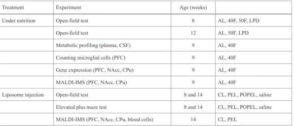
(20) 364. Results. 365. Body weight of male rat offspring. 366. No significant difference was observed between the body weight of male offspring of. 367. 40F and that of AL, 50F, or LPD at 9 weeks of age. However, the body weight of the. 368. male offspring was significantly lower in the 50F and LPD groups (p = 0.06 and 0.10,. 369. respectively) compared with that of the male offspring in the AL group using Dunnett’s. 370. test following one-way ANOVA (p = 0.002, Ʉ ଶ = 0.159 for the main effect) (Table 3).. 371. The body weight of 50F and LPD male offspring became similar to that of AL offspring. 372. at 12 weeks of age (Table 3).. 373. Maternal undernutrition during early pregnancy leads to hyperactivity in rat offspring. 374. To study the effect of nutritional stress during early embryonic stages on postnatal. 375. behavior, the open-field test was performed for adult male offsprings delivered from. 376. dams that underwent food restriction from GD 5.5 to GD 10.5. In this study, the. 377. behavioral tests performed for offspring at 8 weeks of age revealed that the total. 378. distance traveled, the distance traveled in the center area, and the frequency of crossings. 379. were significantly increased for males from the 40F group (p = 0.028, p = 0.036, and p. 380. < 0.001, respectively), the 50F group (p < 0.001, p = 0.004, and p < 0.001, respectively),. 381. and the LPD group (p = 0.026, p = 0.030, and p = 0.016, respectively) compared with. 382. the corresponding findings for males in the AL group, in which dams were fed ad. 383. libitum, using Dunnett’s test following one-way ANOVA (total distance traveled: p <. 384. 0.001, Ʉ ଶ = 0. 209; distance traveled in the center: p = 0.003, Ʉ ଶ = 0.167;. 385. frequency of crossing: p < 0.001, Ʉ ଶ = 0.439 for the main effect; Fig. 1B–D). There. 386. was no significant difference in time spent in the center area among all groups in. 18.
(21) 387. Condition 1 of the open-field test (Fig. 1E), but the time spent in the center area was. 388. longer for both the 40F (p = 0.015) and 50F (p = 0.037) groups compared with the AL. 389. group in Condition 2, where the radius of the center area represented 70% of the open. 390. field (Dunnett’s test following one-way ANOVA: p = 0.011, Ʉ ଶ = 0. 135 for the. 391. main effect; Fig. 1F). Maternal protein restriction during early pregnancy, in part,. 392. contributed to the hyperactivity of the offspring. Increased total distance traveled (LPD:. 393. p < 0.001, 50F: p < 0.001), the distance traveled in the center area (LPD: p < 0.001,. 394. 50F: p = 0.006), the frequency of crossings (LPD: p = 0.001, 50F: p = 0.011), and the. 395. time spent in the center area in Conditions 1 (LPD: p = 0.020, 50F: p = 0.015) and 2. 396. (LPD: p = 0.001, 50F: p = 0.019) were observed even at 12 weeks of age for offspring. 397. from the 50F and LPD groups compared with the AL group (one-way ANOVA with. 398. Dunnett’s test), although this parameter was not examined for the offspring of the 40F. 399. group (Fig. 1G–K). However, locomotor activity did not correspond to the body weight. 400. at 9 and 12 weeks of age (Fig. 1 and Table 3). The p value and effect size for the main. 401. effect in one-way ANOVA of locomotor activities at 12 weeks of age were as follows:. 402. total distance traveled: p < 0.001, Ʉ ଶ = 0. 333; distance traveled in the center: p <. 403. 0.001, Ʉ ଶ = 0.298; frequency of crossing: p = 0.001, Ʉ ଶ = 0.237; time spent in the. 404. center area: p = 0.011, Ʉ ଶ = 0.173 in Condition 1 and p = 0.001, Ʉ ଶ = 0.246 in. 405. Condition 2.. 406. Glyco- and amino-metabolisms are altered in the offspring. 407. In this study, metabolome profiling of the plasma and CSF was performed for offspring. 408. at 9 weeks of age (after behavioral tests) of the 40F group, which displayed the most. 409. severe behavioral changes among the experimental groups (Fig.3-1). The concentration 19.
(22) 410. of glycerol, which is the source of diacylglycerol in phospholipids, was increased in. 411. blood plasma (p = 0.014, d =1.57 ) and CSF (p = 0.001, d = 1.50) in the 40F group. 412. compared with the AL group (Fig. 3A). In plasma, 1,5-anhydro-D-glucitol (p = 0.038, d. 413. = 1.61) and 2-aminoethanol (p = 0.014, d = 1.21) were also increased in the offspring of. 414. the 40F group (Fig. 3A and Fig. 3-1). 2-aminoethanol is converted to O-phosphoethanol. 415. amine, and finally transferred to diacylglycerol or 1-O-alkyl-2-acyl-sn-glycerol to. 416. produce PE (Braverman and Moser, 2012; Vance, 2015).. 417. Microglial cell activation in the PFC is not induced by prenatal undernutrition. 418. The cell densities of Iba1-positive microglial cells and those of both Iba1- and. 419. CD11b-positive activated microglial cells in the PFC were not significantly different. 420. between the AL and 40F groups (Fig. 3B–D). Additionally, no change in the ratio of. 421. activated glial cells to total microglial cells was observed between the AL and 40F. 422. groups (Fig. 3E); therefore, microglial cell activation was not enhanced by prenatal. 423. undernutrition during the early embryonic period.. 424. Expression of genes related to phospholipid biosynthesis is increased in the PFC. 425. of rats exposed to prenatal undernutrition. 426. To examine the modulation of the phospholipid biosynthetic pathway in the PFC, NAcc,. 427. and CPu in offspring (Fig. 4) exposed to maternal undernutrition, gene expression of the. 428. enzymes involved in this pathway was examined. In the brain, dihydroxyacetone. 429. phosphate (DHAP) is the main precursor of phospholipids. DHAP is synthesized from. 430. glucose and serves as a precursor of diacylglycerol, which constitutes the hydrophobic. 431. tail of phospholipids such as PC, PE, and PS (Benjamins et al., 2011). Diacylglycerol. 432. may be, in part, formed from glycerol, mediated through glycerol 3-phosphate (Fig. 4). 433. (Jenkins and Hajra, 1976). Further, DHAP serves as a precursor of the 20.
(23) 434. ether-phospholipid, plasmalogen (Braverman and Moser, 2012). In the first step of the. 435. synthetic pathway of plasmalogen, as well as that for diacyl phospholipids, hexokinase. 436. 1 (Hk1) is one of the key enzymes in the regulation of the carbohydrate metabolic rate,. 437. which converts glucose to glucose 6-phosphate (McKenna et al., 2011). The expression. 438. of Hk1 was enhanced in 40F offspring (p = 0.016, d = 1.68), although. 439. phosphofructokinase (Pfk1), the rate-limiting enzyme in glycolysis (McKenna et al.,. 440. 2011), was not different in the PFC between 40F and control offspring (Fig. 5A, 5B, and. 441. 5-1). Regarding the synthesis of the hydrophobic tail of phospholipids, the gene. 442. expression of the following enzymes related to diacylglycerol and CDP-diacylglycerol. 443. synthesis were elevated in the PFC of the offspring of the 40F group:. 444. glyceronephosphate O-acyltransferase (Gnpat) (p = 0.001, d = 2.32), glycerol kinase (p. 445. = 0.016, d = 1.60), glycerol-3-phosphate transferase (Gpat) 1, 3, and 4 (p = 0.027, 0.032,. 446. and 0.030, and d = 1.46, 1.50, and 1.57, respectively), and phosphatidate. 447. cytidylyltransferase 2 (p = 0.045, d = 1.25). Regarding the synthesis of the hydrophilic. 448. head of phospholipids, the gene expression of ethanolamine kinase for PE (p = 0.033, d. 449. = 1.37), phosphate cytidylyltransferase 1, choline, alpha (Pcyt1a) (p = 0.013, d = 1.68). 450. and phosphate cytidylyltransferase 1, choline, and beta (Pcyt1b) (p = 0.010, d = 1.73). 451. for PC; and phosphatidylserine synthase 1 (Ptdss1) for PS (p = 0.007, d = 1.86) was. 452. elevated in the PFC of the offspring of the 40F group (Fig. 5B). In contrast,. 453. plasmalogen. 454. 1-O-alkyl-2-acyl-sn-glycerol,. 455. 1-O-alkyl-DHAP (Braverman and Moser, 2012). In this pathway, 1-O-alkyl-DHAP is. 456. generated from DHAP by GNPAT and alkylglycerone phosphate synthase (AGPS). 457. (Braverman and Moser, 2012). The genes, AGPS (p = 0.013, d = 1.71) and GNPAT, of. is. synthesized which. from is. 21. ethanolamine produced. from. or fatty. choline alcohol. and and.
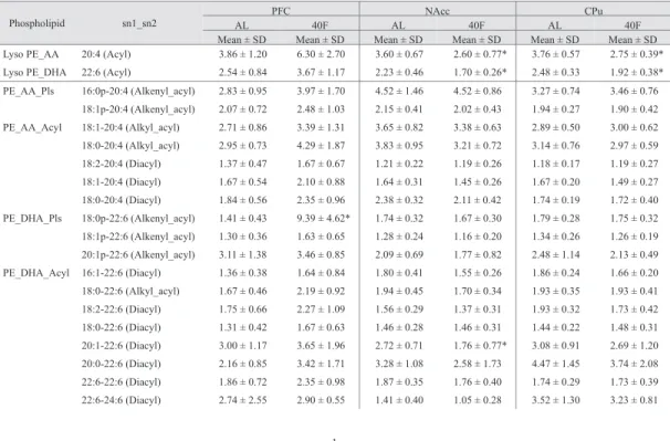
(24) 458. these enzymes were found to be activated whereas the fatty acyl-CoA reductase 1. 459. (Far1) gene expression, which is a potential rate-limiting enzyme (Honsho and Fujiki,. 460. 2017), was not altered in the offspring of the 40F group compared with that of the. 461. offspring of the AL group (Fig. 5B and 5-1). In contrast to the PFC, Gpat1 (p = 0.036, d. 462. = 1.39) and Pcyt1b (p = 0.008, d = 1.65) expression was lower in the offspring of the. 463. 40F group than in the offspring of the AL group in the NAcc and CPu, respectively,. 464. although the expression of the other genes, which showed altered expression in the PFC,. 465. was not changed in the NAcc and CPu (Fig. 5A, B and 5-1). Furthermore, between the. 466. offspring of the AL group and the 40F group, in the PFC, no significant difference was. 467. observed in the expression of the calcium-independent phospholipase A2 (iPla2) gene,. 468. which catalyzes phospholipids (Yeagle, 2016), or that of sphingomyelin synthase 1. 469. (Sgms1) and sphingomyelin phosphodiesterase 3 (Smpd3), which convert PC into. 470. sphingomyelin and ceramide (Vance, 2015), respectively (Fig. 5-1). As described above,. 471. our results indicated that the genes of enzymes related to plasmalogen, as well as diacyl. 472. phospholipids, were activated in the PFC of offspring exposed to prenatal. 473. undernourishment. Alternatively, gene expression was not affected with respect to. 474. aquaporin 9 (Aqp9; a channel permeable to glycerol and water) (Badaut and Regli,. 475. 2004) or major facilitator superfamily domain containing 2A (Mfsd2a; the major. 476. transporter for docosahexaenoic acid [DHA]) (Nguyen et al., 2014). Additionally, the. 477. expression of apolipoprotein E, which is involved in lipid transport (Liao et al., 2017),. 478. was not altered by prenatal undernutrition (Fig. 5-1).. 479. Phospholipid composition is altered in the cerebrum by maternal undernutrition. 480. The peak intensity of m/z 774.5 PE in the PFC of the offspring of the 40F group was. 481. 6.6-fold higher than that of the offspring of the AL group (p = 0.012, d = 2.33; Fig. 5C 22.
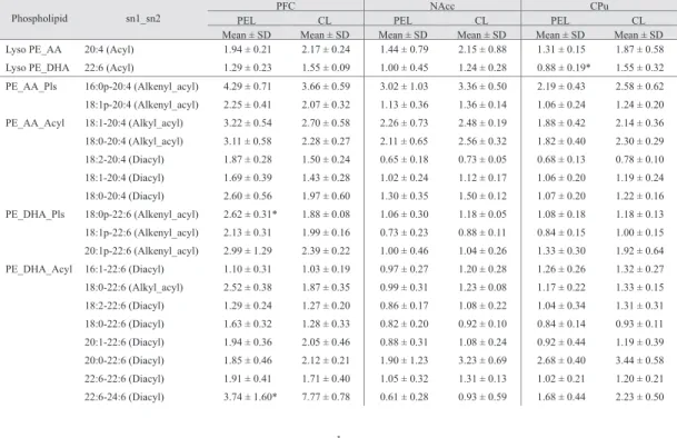
(25) 482. and D). CID of m/z774.5 yielded product ions at m/z 283.2, 327.2, and 464.3; therefore,. 483. m/z 774.5 was identified as PE (18:0p-22:6) (Fig. 5E). Additionally, lysoPE (20:1) in. 484. the PFC of 40F offspring was 1.7-fold that of the AL offspring (p = 0.037, d = 1.36);. 485. however, no significant difference was observed between groups for the other. 486. phospholipids examined (Fig. 5-2). In contrast, the peak intensities of PE (20:1-22:6),. 487. lysoPE (20:4), and lysoPE (22:6) in the NAcc (p = 0.045, 0.034, and 0.031, and d =. 488. 1.30, 1.39, and 1.42, respectively), and of lysoPE (20:4) and lysoPE (22:6) in the CPu. 489. (p = 0.004 and 0.019, and d = 2.07 and 1.57, respectively) were attenuated in the. 490. offspring of the 40F group compared with the offspring of the AL group. However, no. 491. phospholipid showed enhanced peak intensity in the NAcc and CPu of the offspring of. 492. the 40F group (Fig. 5D and 5-2). On the other hand, only PE (18:0p-22:6) in the PFC. 493. varied in amount among PlsEtn examined in this study by prenatal undernutrition (Fig.. 494. 5D, Table 4). From the perspective of the tail forms of the phospholipids, PE. 495. (18:0p-22:6) in the PFC exclusively increased in the offspring of the 40F group among. 496. DHA (22:6)-containing phospholipids, although some phospholipids containing DHA or. 497. arachidonic acid (AA, 20:4) decreased in the NAcc and CPu (Fig. 5D, Table 4).. 498. PlsEtn affects the behavior of adult rats. 499. To examine the effect of PE (18:0p-22:6) on rat behavior, rats were subjected to the. 500. open-field test and elevated plus maze test after intravenous injection of PEL or CL (Fig.. 501. 6A). The age-related decline in the frequency of crossing, as well as the difference in. 502. the time spent in the center area (Condition 2) was reduced in rats in the PEL group. 503. compared with rats in the CL group (Fig. 6B). However, no significant difference was. 504. observed in the results of the elevated plus maze test between the PEL and CL groups. 505. (Fig. 6D). At the same time, no significant difference was observed in the effect of 23.
(26) 506. POPEL, CL, and saline on rat behavior (Fig. 6C and 6E). Four days after the second. 507. injection of PEL (Fig. 6A), the amount of PE (18:0p-22:6) in the PFC (p = 0.019, d =. 508. 3.02), but not that in the NAcc or CPu, was still greater in the PEL group than in the CL. 509. group, while no significant difference was observed in the amount of the other PlsEtn. 510. between these groups, as determined by MALDI-IMS (Fig. 6F and Fig. 6-1). The. 511. increased amount of PE (18:0p-22:6) in the PFC of the PEL group was verified in. 512. another PE injection experiment (Fig. 6-2). Meanwhile, the amounts of PE (22:6-24:6). 513. (p = 0.019, d = 3.02), PE (24:4-22:6) (p = 0.013, d = 3.36), PS (16:0-22:6) (p < 0.001, d. 514. = 9.02), PI (16:1-18:1) (p = 0.004, d = 4.56), and lysoPE (18:1) (p < 0.001, d = 8.58), all. 515. of which are acyl phospholipids, were lower in the PFC in the PEL group than in the CL. 516. group (Fig. 6F). The amounts of PE (16:0-18:1) (NAcc; p = 0.041, d = 2.08, CPu: p =. 517. 0.016, d = 2.68) and lysoPE (20:1) (NAcc; p = 0.012, d = 2.91, CPu: p = 0.033, d =. 518. 2.64) were lower in the NAcc and CPu for the PEL group than in the CL-group. The. 519. amounts of PE (16:1-20:5), PI (18:0-22:4), and plasmalogen PE (18:1p-20:1) were also. 520. decreased in the NAcc of the PEL group (p = 0.008, 0.032, and 0.023, and d = 3.18,. 521. 0.85, and 2.51, respectively), whereas those of lysoPE (18:1), lysoPE (22:6), and. 522. lysoPE (22:4) were decreased in the CPu of that group (p = 0.039, 0.033, and 0.026, and. 523. d = 2.13, 2.55, and 2.42, respectively; Fig. 6F). To verify that increased PE (18:0p-22:6). 524. in the PFC was not ascribed to PE incorporated into the blood cells from liposomes. 525. inside blood vessels, PE (18:0p-22:6) in blood cells was measured by MALDI-IMS.. 526. The ratio of PE (18:0p-22:6) to total lipids for the PEL group did not differ from that for. 527. the CL group (Fig. 6G). Thus, PE (18:0p-22:6) was incorporated at least into the PFC,. 528. but not blood cells, after the injection of PE liposomes. Similar to prenatal. 529. undernutrition, PE (18:0p-22:6) injection did not increase the amount of other PE, PS, 24.
(27) 530. and PI containing DHA or AA (Fig. 6F, Table 5). Furthermore, most phospholipids that. 531. varied in amount after exposure to prenatal undernutrition were not altered by PEL. 532. injection, although lyso PE (22:6) was reduced in both 40F offspring and PEL-injected. 533. rats (Fig. 5D and 6F, Table 5).. 25.
(28) 534. Discussion. 535. The findings of our study suggest that changes in phospholipid composition led by. 536. prenatal undernutrition is associated with hyperactivity in rats, and plasmalogen PE. 537. (18:0p-22:6) injection reproduces a part of hyperactive behaviors. Regarding the cell. 538. membrane, plasmalogens constitute approximately 20% of total phospholipids, both in. 539. the rat cerebral cortex and the human brain (Braverman and Moser, 2012). In humans,. 540. ethanolamine plasmalogen constitute 57% and 84% of the glycerophosphoethanolamine. 541. fraction of the gray and white matter of the frontal cortex, respectively (Braverman and. 542. Moser, 2012). Neurons and myelin are rich in plasmalogens, which decrease membrane. 543. fluidity, increase membrane rigidity, and allow tight packing of phospholipids in the. 544. membrane (Dean and Lodhi, 2017). Plasmalogens play a role in membrane trafficking. 545. and fusion processes, Schwann cell differentiation and function, molecule antioxidation,. 546. and inhibition of neuronal apoptotic signaling. Hence, a deficiency of plasmalogens. 547. induces impairments of neurotransmitter release from synaptosomes to the presynaptic. 548. cleft, myelination and axonal sorting by Schwann cells, and neuronal apoptosis. 549. signaling (Dean and Lodhi, 2017). These reports suggest the clinical importance of. 550. plasmalogens to the nervous system. The amyloid E peptide, which is rich in the brains. 551. of patients with Alzheimer’s disease, reduces AGPS protein stability and decreases. 552. plasmalogen PE levels in patients with Alzheimer’s disease (Han et al., 2001; Grimm et. 553. al., 2011). Recently, Hossain et al. reported that inflammatory stimuli, such as the. 554. administration of lipopolysaccharide (LPS) or polyriboinosinic:polyribocytidylic acid,. 555. reduce plasmalogens in murine glial cells through the activation of NF-NB, which. 556. downregulates Gnpat through increased c-Myc recruitment to the Gnpat promoter. 557. (Hossain et al., 2017). Similar findings have been observed for the murine brain after 26.
(29) 558. aging, exposure to chronic restraint stress, and injection of LPS; furthermore, the. 559. reduction of plasmalogen induced activation of microglial cells and elevated expression. 560. of proinflammatory cytokines (Hossain et al., 2017). In brains from transgenic mice. 561. model of Alzheimer’s disease, and postmortem brain tissues from patients with. 562. Alzheimer’s disease, Gnpat reduction via a similar mechanism has been observed. 563. (Hossain et al., 2017). Likewise, maternal infection, obesity, a high-fat diet, and. 564. restraint stress with bright-light exposure causes microglial cell activation and. 565. proinflammatory cytokine induction in the fetal and postnatal brain of rodents and. 566. monkeys, and maternal stress results in anxiety-like, depressive and aggressive behavior,. 567. and schizophrenia-like behavior in offspring (Bilbo and Tsang, 2010; Grayson et al.,. 568. 2010; Matrisciano et al., 2012; Diz-Chaves et al., 2013; Sasaki et al., 2013; Marques et. 569. al., 2015). In addition, activation of microglia is augmented in the brain including the. 570. anterior and orbitofrontal cortices in young adults with ASD, although the distribution. 571. pattern of activated microglia is similar to that of healthy control subjects, as. 572. determined by positron emission tomography (Suzuki et al., 2013). Maternal obesity. 573. before pregnancy is considered a risk factor for ADHD and ASD in humans (Andersen. 574. et al., 2017). Obesity is involved in elevated inflammatory mediators, e.g. IL-6, which. 575. induces Th17 cell differentiation. IL-17A secreted from Th17 may act to promote ASD. 576. by affecting fetal neurodevelopment (Wong and Hoeffer, 2018). These results suggest. 577. that brain inflammation plays a key role in behavior and that plasmalogen alters brain. 578. function through its anti-inflammatory effects. However, in our study, microglial cell. 579. activation was not altered by maternal undernutrition, at least for the adult offspring.. 580. Further, injection of plasmalogen PE (18:0p-22:6) to adult rats in the 40F group altered. 581. the phospholipid composition and resulted in two characteristic behaviors: frequent 27.
(30) 582. crossing and long time spent in the center area in the open-field test.. 583. Therefore, the hyperactivity of the rat offspring that were exposed to prenatal. 584. undernutrition may be attributable to the phospholipid composition of the brain rather. 585. than a direct effect of undernutrition on inflammatory reactions. Patients with RCDP,. 586. who display plasmalogen deficiency, have psychomotor retardation, and, in severely. 587. affected cases, they display microcephaly and cerebellar atrophy (Berger et al., 2016).. 588. Myelination and neuronal migration are thought to be causes of these features of. 589. patients with RCDP (Berger et al., 2016). RCDP type 1, type 2, and type 3 are caused by. 590. mutations of PEX7, GNPAT, and AGPS (Berger et al., 2016), respectively, all of which. 591. contribute to plasmalogen synthesis. In our study, expression of the latter two genes was. 592. elevated in the PFC. In RCDP fibroblasts with the PEX7 mutation, peroxisome targeting. 593. signal 2 protein, phytanoyl-CoA hydroxylase, and AGPS fail to be imported into the. 594. peroxisome (Yu et al., 2013). The PEX7 homozygous mutation has also been found in. 595. three ASD children whose unaffected siblings were heterozygous or wild-type within. 596. one family (Yu et al., 2013). Moreover, single-nucleotide-polymorphism fine mapping. 597. has shown that GNPAT is a candidate gene for schizophrenia, as is DISC1 (Liu et al.,. 598. 2006). These findings suggest that altered phospholipid metabolism, especially. 599. plasmalogen metabolism, may be involved in a person’s vulnerability to developmental. 600. and psychiatric disorders. Gnpat-knockout mice show delayed migration of granule cell. 601. precursors, enhanced apoptosis in the cerebellum, and hypo- and dysmyelination in the. 602. neocortex, cerebellum, and corpus callosum (Berger et al., 2016). Aberrant myelination. 603. may be one of the key factors in hyperactivity because MR findings suggest altered. 604. myelination in the white matter of adults with ADHD (Wu et al., 2017). Skin fibroblasts. 605. derived from patients with RCDP showed reduced PlsEtn, whereas the total amount of 28.
(31) 606. PE was maintained by an increase in other PEs (Dorninger et al., 2015).. 607. Polyunsaturated fatty acid (PUFA)-containing PlsEtn was reduced in these cells, and. 608. AA-containing, but not DHA-containing, PE species mainly compensated for PlsEtn. 609. deficiency (Dorninger et al., 2015). Similar findings have been observed for. 610. Gnpat-knockout mice; therefore, the ratio among essential PUFAs, as well as the ratio. 611. between PlsEtn and PE, may be critical for brain development, and a shift in these ratios. 612. may be the cause of the psychomotor retardation of patients with RCDP. In contrast, in. 613. our study, the levels of PE (18:0p-22:6) in the PFC and lyso PE (22:6) in CPu were both. 614. altered by exposure to prenatal undernutrition and PE (18:0p-22:6) injection.. 615. Additionally, injection of POPE, which is not a plasmalogen, did not alter rat behavior.. 616. A specific plasmalogen, such as PE (18:0p-22:6), and a specific DHA-containing PE,. 617. such as lyso PE (22:6), coupled with the level of plasmalogen in the brain may have a. 618. function for behavior. Exogenous administration of plasmalogen can be considered as a. 619. potential therapeutic strategy as it results in changes in the phospholipid composition of. 620. the brain. Regarding psychiatric disorders, the levels of PUFAs (e.g., PE22:5n6,. 621. PC20:3n6, and PC22:5n6) were lower in the white matter adjacent to the dorsolateral. 622. PFC of a patient with schizophrenia, while the level of PE20:2n6 was higher, and those. 623. of PE22:5n6, PC20:4n6, and PC22:5n6 were lower, in the white matter of the patient. 624. with bipolar disorder, despite no alterations in the plasmalogen level for both disorders. 625. (Ghosh et al., 2017). A subset combination of the head group (e.g., ethanolamine,. 626. choline, serine, or inositol) and a fatty acid tail, such as DHA or AA, which are. 627. incorporated into the phospholipid, may be critical to evoke the behavioral alterations. 628. that are characteristic of developmental or psychiatric disorders, and thus, could be a. 629. therapeutic target to improve conditions for patients with these diseases. As in one of 29.
(32) 630. the trials, the administration of a PS supplement in a chewable tablet presentation. 631. improved ADHD for children aged 4–14 years (Hirayama et al., 2014). Exogenous PS. 632. can cross the blood–brain barrier (BBB) and function in the brain (Glade and Smith,. 633. 2015). Regarding the delivery of plasmalogen into the brain across the BBB, liposomes. 634. with PC may be one of the preferred carriers, since the intravenous injection of. 635. liposomes has been the preferred delivery route for many previous studies (Vieira and. 636. Gamarra, 2016). However, PE has a cationic head, and the liposome may not be able to. 637. reach the brain because of nonspecific binding to the peripheral tissues and serum. 638. proteins (Vieira and Gamarra, 2016) if the ethanolamine head is exposed on the surface. 639. of the liposome membrane. In our study, the amount of PE (18:0p-22:6) was not altered. 640. in blood cells, suggesting that little of the injected PE was taken into the peripheral. 641. tissues during circulation in the brain before liposomes were captured by the liver.. 642. Plasmalogen is asymmetrically localized to the inner leaflet of the myelin membrane. 643. bilayer (Kirschner and Ganser, 1982), and plasmalogen-rich membranes tend to form. 644. non-lamellar inverse hexagonal structures compared with the membrane, which. 645. exclusively consists of diacyl phospholipids (Dean and Lodhi, 2017). The surface of the. 646. liposomes that were used in our study may be electrically neutral, and their nonspecific. 647. binding to the peripheral tissues and serum proteins may be prevented.. 648. In summary, maternal undernutrition during early pregnancy led to the hyperactivity of. 649. male rat offspring, and the behavioral changes observed may be, in part, caused by an. 650. alteration of the plasmalogen composition in the PFC, which was induced by the. 651. activation of the phospholipid synthetic pathway. Ethanolamine plasmalogen. 652. (18:0p-22:6) appears to play a critical role in behavior. Thus, plasmalogens could be. 653. candidate therapeutic molecules for improving behavioral disorders. Further study of 30.
(33) 654. their complex functions is warranted.. 655 656 657. Funding. 658. This research was supported by JSPS KAKENHI (grant numbers JP 25460242 and. 659. 16K08441 to JU), the grant from the Food Science Institute Foundation. 660. (Ryoushoku-kenkyukai) (to JU), SUMS President’s Grant for Encouragement of Young. 661. Researchers (grant number 1515503ZU to KH), the SUMS grant for the special research. 662. project (grant number 1515503ZZE to JU), the SUMS grant for the integration of basic. 663. and clinical research (grant number 1515503ZO to JU), MEXT/JSPS KAKENHI (grant. 664. number JP15H05898B1 to MS), and Imaging Platform supported by the Ministry of. 665. Education, Culture, Sports, Science and Technology (MEXT) (to MS). FY was. 666. supported by Imaging Platform was supported by MEXT.. 667 668. 31.
(34) 669. References. 670. Aleman A, Kahn RS, Selten JP (2003) Sex differences in the risk of schizophrenia:. 671. evidence from meta-analysis. Arch Gen Psychiatry 60:565-571.. 672. Amaral AC, Jakovcevski M, McGaughy JA, Calderwood SK, Mokler DJ, Rushmore RJ,. 673. Galler JR, Akbarian SA, Rosene DL (2015) Prenatal protein malnutrition. 674. decreases KCNJ3 and 2DG activity in rat prefrontal cortex. Neuroscience. 675. 286:79-86.. 676. Andersen CH, Thomsen PH, Nohr EA, Lemcke S (2017) Maternal body mass index. 677. before pregnancy as a risk factor for ADHD and autism in children. Eur Child. 678. Adolesc Psychiatry.. 679 680 681 682. Arnett AB, Pennington BF, Willcutt EG, DeFries JC, Olson RK (2015) Sex differences in ADHD symptom severity. J Child Psychol Psychiatry 56:632-639. Badaut J, Regli L (2004) Distribution and possible roles of aquaporin 9 in the brain. Neuroscience 129:971-981.. 683. Benjamins J, Murphy E, Seyfried T (2011) Lipids. In: Basic Neurochemistry: Principles. 684. of Molecular, Cellular, and Medical Neurobiology, 8th Edition (Brady S, Siegel. 685. G, Albers RW, Price D, eds), pp81-100: Elsevier Science.. 686 687 688 689 690 691 692. Berger J, Dorninger F, Forss-Petter S, Kunze M (2016) Peroxisomes in brain development and function. Biochim Biophys Acta 1863:934-955. Bilbo SD, Tsang V (2010) Enduring consequences of maternal obesity for brain inflammation and behavior of offspring. FASEB J 24:2104-2115. Braverman NE, Moser AB (2012) Functions of plasmalogen lipids in health and disease. Biochim Biophys Acta 1822:1442-1452. Brown AS, Susser ES (2008) Prenatal nutritional deficiency and risk of adult 32.
(35) 693 694 695. schizophrenia. Schizophr Bull 34:1054-1063. Bystron I, Blakemore C, Rakic P (2008) Development of the human cerebral cortex: Boulder Committee revisited. Nat Rev Neurosci 9:110-122.. 696. Dahoun T, Trossbach SV, Brandon NJ, Korth C, Howes OD (2017) The impact of. 697. Disrupted-in-Schizophrenia 1 (DISC1) on the dopaminergic system: a. 698. systematic review. Transl Psychiatry 7:e1015.. 699 700. Dean JM, Lodhi IJ (2017) Structural and functional roles of ether lipids. Protein Cell. 9:196-206.. 701. Diz-Chaves Y, Astiz M, Bellini MJ, Garcia-Segura LM (2013) Prenatal stress increases. 702. the expression of proinflammatory cytokines and exacerbates the inflammatory. 703. response to LPS in the hippocampal formation of adult male mice. Brain Behav. 704. Immun 28:196-206.. 705. Dorninger F, Brodde A, Braverman NE, Moser AB, Just WW, Forss-Petter S, Brügger B,. 706. Berger. 707. phosphatidylethanolamine. 708. plasmalogens. Biochim Biophys Acta 1851:117-128.. J. (2015). Homeostasis tightly. of. phospholipids. adapts. to. -. changes. The in. level. of. ethanolamine. 709. Erb C (2006) Embryology and Teratology. In: The laboratory rat, 2nd Edition (Suckow. 710. MAW, Steven H. Franklin, Craig L., ed), pp 817-846. Amsterdam; Boston:. 711. Elsevier.. 712. Fineberg AM, Ellman LM, Schaefer CA, Maxwell SD, Shen L, H Chaudhury N, Cook. 713. AL, Bresnahan MA, Susser ES, Brown AS (2016) Fetal exposure to maternal. 714. stress and risk for schizophrenia spectrum disorders among offspring:. 715. Differential influences of fetal sex. Psychiatry Res 236:91-97.. 716. Fritts ME, Asbury ET, Horton JE, Isaac WL (1998) Medial prefrontal lesion deficits 33.
(36) 717. involving or sparing the prelimbic area in the rat. Physiol Behav 64:373-380.. 718. Ghosh S, Dyer RA, Beasley CL (2017) Evidence for altered cell membrane lipid. 719. composition in postmortem prefrontal white matter in bipolar disorder and. 720. schizophrenia. J Psychiatr Res 95:135-142.. 721 722. Glade MJ, Smith K (2015) Phosphatidylserine and the human brain. Nutrition 31:781-786.. 723. Grayson BE, Levasseur PR, Williams SM, Smith MS, Marks DL, Grove KL (2010). 724. Changes in melanocortin expression and inflammatory pathways in fetal. 725. offspring of nonhuman primates fed a high-fat diet. Endocrinology. 726. 151:1622-1632.. 727. Grimm MO, Kuchenbecker J, Rothhaar TL, Grösgen S, Hundsdörfer B, Burg VK,. 728. Friess P, Müller U, Grimm HS, Riemenschneider M, Hartmann T (2011). 729. Plasmalogen. 730. alkyl-dihydroxyacetonephosphate-synthase. 731. processing and is affected in Alzheimer's disease. J Neurochem 116:916-925.. 732 733. synthesis. is by. regulated amyloid. precursor. via protein. Götz M, Huttner WB (2005) The cell biology of neurogenesis. Nat Rev Mol Cell Biol 6:777-788.. 734. Han X, Holtzman DM, McKeel DW (2001) Plasmalogen deficiency in early. 735. Alzheimer's disease subjects and in animal models: molecular characterization. 736. using electrospray ionization mass spectrometry. J Neurochem 77:1168-1180.. 737. Hino K, Kimura T, Udagawa J (2019) Handling has an anxiolytic effect that is not. 738. affected by the inhibition of the protein kinase C pathway in adult prenatal. 739. undernourished male rat offspring. Congenit Anom (Kyoto).. 740. Hirayama S, Terasawa K, Rabeler R, Hirayama T, Inoue T, Tatsumi Y, Purpura M, Jäger 34.
(37) 741. R (2014) The effect of phosphatidylserine administration on memory and. 742. symptoms of attention-deficit hyperactivity disorder: a randomised, double-blind,. 743. placebo-controlled clinical trial. J Hum Nutr Diet 27:284-291.. 744 745. Hoek HW, Brown AS, Susser E (1998) The Dutch famine and schizophrenia spectrum disorders. Soc Psychiatry Psychiatr Epidemiol 33:373-379.. 746. Honsho M, Fujiki Y (2017) Plasmalogen homeostasis - regulation of plasmalogen. 747. biosynthesis and its physiological consequence in mammals. FEBS Lett.. 748. 591:2720-2729.. 749. Hossain MS, Abe Y, Ali F, Youssef M, Honsho M, Fujiki Y, Katafuchi T (2017). 750. Reduction of ether-type glycerophospholipids, plasmalogens, by NF-κB signal. 751. lLeading to microglial activation. J Neurosci 37:4074-4092.. 752. Hossen MA, Nagata Y, Waki M, Ide Y, Takei S, Fukano H, Romero-Perez GA, Tajima S,. 753. Yao I, Ohnishi K, Setou M (2015) Decreased level of phosphatidylcholine. 754. (16:0/20:4) in multiple myeloma cells compared to plasma cells: a single-cell. 755. MALDI-IMS approach. Anal Bioanal Chem 407:5273-5280.. 756 757 758 759. Jenkins BT, Hajra AK (1976) Glycerol kinase and dihydroxyacetone kinase in rat brain. J Neurochem 26:377-385. Jinks AL, McGregor IS (1997) Modulation of anxiety-related behaviours following lesions of the prelimbic or infralimbic cortex in the rat. Brain Res 772:181-190.. 760. Khashan AS, Abel KM, McNamee R, Pedersen MG, Webb RT, Baker PN, Kenny LC,. 761. Mortensen PB (2008) Higher risk of offspring schizophrenia following antenatal. 762. maternal exposure to severe adverse life events. Arch Gen Psychiatry. 763. 65:146-152.. 764. Kimura T, Hino K, Kono T, Takano A, Nitta N, Ushio N, Hino S, Takase R, Kudo M, 35.
(38) 765. Daigo Y, Morita W, Nakao M, Nakatsukasa M, Tamagawa T, Rafiq AM,. 766. Matsumoto A, Otani H, Udagawa J (2018) Maternal undernutrition during early. 767. pregnancy inhibits postnatal growth of the tibia in the female offspring of rats by. 768. alteration of chondrogenesis. Gen Comp Endocrinol 260:58-66.. 769. Kinney DK, Miller AM, Crowley DJ, Huang E, Gerber E (2008) Autism prevalence. 770. following prenatal exposure to hurricanes and tropical storms in Louisiana. J. 771. Autism Dev Disord 38:481-488.. 772. Kirschner DA, Ganser AL (1982) Myelin labeled with mercuric chloride. Asymmetric. 773. localization of phosphatidylethanolamine plasmalogen. J Mol Biol 157:635-658.. 774. Kundakovic M (2014) Chapter 24 - DNA methyltransferase inhibitors and psychiatric. 775. disorders A2 - Peedicayil, Jacob. In: Epigenetics in Psychiatry (Grayson DR,. 776. Avramopoulos D, eds), pp497-514. Boston: Academic Press.. 777 778 779 780. Kundakovic M, Jaric I (2017) The epigenetic link between prenatal adverse environments and neurodevelopmental disorders. Genes 8:104. Liao F, Yoon H, Kim J (2017) Apolipoprotein E metabolism and functions in brain and its role in Alzheimer's disease. Curr Opin Lipidol 28:60-67.. 781. Liu YL, Fann CS, Liu CM, Chen WJ, Wu JY, Hung SI, Chen CH, Jou YS, Liu SK,. 782. Hwang TJ, Hsieh MH, Ouyang WC, Chan HY, Chen JJ, Yang WC, Lin CY, Lee. 783. SF, Hwu HG (2006) A single nucleotide polymorphism fine mapping study of. 784. chromosome 1q42.1 reveals the vulnerability genes for schizophrenia, GNPAT. 785. and DISC1: association with impairment of sustained attention. Biol Psychiatry. 786. 60:554-562.. 787 788. Markham JA, Koenig JI (2011) Prenatal stress: role in psychotic and depressive diseases. Psychopharmacology 214:89-106. 36.
(39) 789. Marques AH, Bjørke-Monsen AL, Teixeira AL, Silverman MN (2015) Maternal stress,. 790. nutrition and physical activity: Impact on immune function, CNS development. 791. and psychopathology. Brain Res 1617:28-46.. 792. Matrisciano F, Tueting P, Maccari S, Nicoletti F, Guidotti A (2012) Pharmacological. 793. activation. 794. schizophrenia-like. 795. Neuropsychopharmacology 37:929-938.. of. group-II. metabotropic. phenotype. induced. glutamate by. receptors. prenatal. stress. corrects in. a. mice.. 796. Matrisciano F, Tueting P, Dalal I, Kadriu B, Grayson DR, Davis JM, Nicoletti F,. 797. Guidotti A (2013) Epigenetic modifications of GABAergic interneurons are. 798. associated with the schizophrenia-like phenotype induced by prenatal stress in. 799. mice. Neuropharmacology 68:184-194.. 800. McKenna M, Dienel G, Sonnewald U, Waagepetersen H, Schousboe A (2011) Energy. 801. metabolism of the brain. In: Basic neurochemistry: principles of molecular,. 802. cellular, and medical neurobiology (Brady S, Siegel G, Albers RW, Price D, eds),. 803. pp 200-231: Elsevier Science.. 804. Moreno M, Economidou D, Mar AC, López-Granero C, Caprioli D, Theobald DE,. 805. Fernando A, Newman AH, Robbins TW, Dalley JW (2013) Divergent effects of. 806. D/Ύ receptor activation in the nucleus accumbens core and shell on impulsivity. 807. and locomotor activity in high and low impulsive rats. Psychopharmacology. 808. 228:19-30.. 809 810. Morita SY, Deharu Y, Takata E, Nakano M, Handa T (2008) Cytotoxicity of lipid-free apolipoprotein B. Biochim Biophys Acta 1778:2594-2603.. 811. Nguyen LN, Ma D, Shui G, Wong P, Cazenave-Gassiot A, Zhang X, Wenk MR, Goh EL,. 812. Silver DL (2014) Mfsd2a is a transporter for the essential omega-3 fatty acid 37.
(40) 813. docosahexaenoic acid. Nature 509:503-506.. 814. Nishiumi S, Kobayashi T, Ikeda A, Yoshie T, Kibi M, Izumi Y, Okuno T, Hayashi N,. 815. Kawano S, Takenawa T, Azuma T, Yoshida M (2012) A novel serum. 816. metabolomics-based diagnostic approach for colorectal cancer. PLoS One. 817. 7:e40459.. 818. Roseboom TJ, van der Meulen JH, Ravelli AC, Osmond C, Barker DJ, Bleker OP. 819. (2001) Effects of prenatal exposure to the Dutch famine on adult disease in later. 820. life: an overview. Mol Cell Endocrinol 185:93-98.. 821. Sasaki A, de Vega WC, St-Cyr S, Pan P, McGowan PO (2013) Perinatal high fat diet. 822. alters glucocorticoid signaling and anxiety behavior in adulthood. Neuroscience. 823. 240:1-12.. 824 825. Schoenwolf GC, Bleyl SB, Brauer PR, Francis-West PH, Larsen WJ (2015) Larsen's human embryology.. 826. Scofield MD, Heinsbroek JA, Gipson CD, Kupchik YM, Spencer S, Smith AC,. 827. Roberts-Wolfe D, Kalivas PW (2016) The nucleus accumbens: mechanisms of. 828. addiction across drug classes reflect the importance of glutamate homeostasis.. 829. Pharmacol Rev 68:816-871.. 830. Spencer RC, Devilbiss DM, Berridge CW (2015) The cognition-enhancing effects of. 831. psychostimulants involve direct action in the prefrontal cortex. Biol Psychiatry. 832. 77:940-950.. 833. Sugiyama E, Masaki N, Matsushita S, Setou M (2015) Ammonium sulfate improves. 834. dDetection of hydrophilic quaternary ammonium compounds through decreased. 835. ion suppression in matrix-assisted laser desorption/ionization imaging mass. 836. spectrometry. Anal Chem 87:11176-11181. 38.
(41) 837. Suzuki K, Sugihara G, Ouchi Y, Nakamura K, Futatsubashi M, Takebayashi K,. 838. Yoshihara Y, Omata K, Matsumoto K, Tsuchiya KJ, Iwata Y, Tsujii M, Sugiyama. 839. T, Mori N (2013) Microglial activation in young adults with autism spectrum. 840. disorder. JAMA Psychiatry 70:49-58.. 841. Taguchi R, Ishikawa M (2010) Precise and global identification of phospholipid. 842. molecular species by an Orbitrap mass spectrometer and automated search. 843. engine Lipid Search. J Chromatogr A 1217:4229-4239.. 844. Tsugawa H, Bamba T, Shinohara M, Nishiumi S, Yoshida M, Fukusaki E (2011). 845. Practical. 846. metabolomics platform for metabolic phenotype analysis. J Biosci Bioeng. 847. 112:292-298.. 848 849. non-targeted. gas. chromatography/mass. spectrometry-based. Vance JE (2015) Phospholipid synthesis and transport in mammalian cells. Traffic 16:1-18.. 850. Vieira DB, Gamarra LF (2016) Getting into the brain: liposome-based strategies for. 851. effective drug delivery across the blood-brain barrier. Int J Nanomedicine. 852. 11:5381-5414.. 853 854. Werling DM, Geschwind DH (2013) Sex differences in autism spectrum disorders. Curr Opin Neurol 26:146-153.. 855. Wong H, Hoeffer C (2018) Maternal IL-17A in autism. Exp Neurol 299:228-240.. 856. Wu ZM, Bralten J, Cao QJ, Hoogman M, Zwiers MP, An L, Sun L, Yang L, Zang YF,. 857. Franke B, Wang YF (2017) White Matter Microstructural Alterations in Children. 858. with. 859. Neuropsychopharmacology 42:572-580.. 860. ADHD:. Categorical. and. Dimensional. Perspectives.. Yeagle PL (2016) Chapter 3 - Biogenesis of membrane lLipids. In: The 39.
(42) 861. Membranes of Cells (3rd Edition), pp 57-71. Boston: Academic Press.. 862. Yu TW et al. (2013) Using whole-exome sequencing to identify inherited causes of. 863. autism. Neuron 77:259-273.. 864. Zemski Berry KA, Gordon WC, Murphy RC, Bazan NG (2014) Spatial organization of. 865. lipids in the human retina and optic nerve by MALDI imaging mass. 866. spectrometry. J Lipid Res 55:504-515.. 867. Zhu Y, Yang D, Ji W, Huang T, Xue L, Jiang X, Chen L, Wang F (2016) The. 868. relationship. 869. hyperactivity disorder: a review. Biomed Res Int 2016:3821579.. between. neurocircuitry. dysfunctions. and. attention. deficit. 870 871. Figure legends. 872 873. Fig. 1 Study design and behavior of rat offspring subjected to prenatal undernutrition.. 874. (A) The experimental schedule is shown. The daily food intake was restricted from. 875. GD5.5 to GD10.5 or from the day of blastocyst implantation to the day just before the. 876. closure of the neural tube. (B–F) Behavioral tests at 8 weeks of age: male rat offspring. 877. exhibited hyperactivity in the LPD (n = 18), 50F (n = 13), and 40F (n = 17) groups. 878. compared with the offspring in the AL group (n = 32). (G–K) Behavioral tests at 12. 879. weeks of age: behavioral disturbances continued at 12 weeks of age in the LPD (n = 19). 880. and 50F (n = 14) offspring compared with the AL offspring (n = 18). The statistical. 881. analysis was conducted using one-way ANOVA with Dunnett’s test. *p < 0.05, **p <. 882. 0.01, *** p < 0.001; A1: the area of the center region, A2: the area of the peripheral. 883. region.. 884 40.
(43) 885. Fig. 2 Preparation of the brain sections for MALDI-IMS (A) Rat brain sections were. 886. aligned between adjacent mouse brain sections (references). (B) Peak intensity of each. 887. phospholipid was corrected using the average peak intensity of the same phospholipid. 888. of the mouse reference sections.. 889 890. Fig. 3 Metabolome profiling of the plasma and CSF, and the observation of microglia in. 891. the PFC. (A) 2-aminoethanol and glycerol increased in the rat offspring exposed to. 892. prenatal undernutrition (plasma: n = 7 in AL and n =6 in 40F; CSF: n = 7 in AL and n =. 893. 5 in 40F) (See also Figure 3-1). (B) The dotted area, which was the middle third of the. 894. box area, was examined. (C) Microglial cell activation in the PFC Iba1-positive. 895. microglia (green), CD11b-positive cells (red), and activated microglia (yellow) are. 896. shown in the PFC of the offspring of the AL and 40F groups. Scale bar, 20 mm. (D) The. 897. densities of microglia and activated microglia were not increased in the PFC of 40F (n =. 898. 8) offspring compared with AL (n = 8) offspring. (E) The ratio of the number of. 899. activated microglia to the total number of microglia was not altered by prenatal. 900. undernutrition. *p < 0.05, **p < 0.01, Student’s t-test.. 901 902. Fig. 4 Biosynthetic pathway of plasmalogens and diacyl phospholipids. The enzymes. 903. related to phospholipid synthesis, with intermediates, are shown. The genes indicated. 904. by underlined bold italic characters were activated. Abbreviations of the. 905. genes are noted as follows: Hk:Hexokinase, GPI:Glucose-6-phosphate isomerase,. 906. Pfk:Phosphofructokinase,. 907. Gpd:Glycerol-3-phosphate dehydrogenase, Far:fatty acyl-CoA reductase, Gk:Glycerol. 908. kinase, Gnpat:Glyceronephosphate O-acyltransferase, Agps:Alkylglycerone phosphate. aldoiase:Fructose-bisphosphate. 41. aldoiase,.
(44) 909. synthase, ADHAPAR:Alkyl/acyl-glycerophosphate acyltransferase, Plpp:Phosphatidic. 910. acid. 911. Plasmenylethanolamine. 912. Chpt:Choline phosphotransferase, Gpat:Glycerol-3-phosphate acyltransferase, Lpcat:. 913. LysoPA-acyltransferase,. 914. Pemt:Phosphoethanolamine N-methyltransferase, Pcyt:Phospate cytidyltransferase,. 915. Pisd:Phosphatidylserine. 916. CDS:CDP-diacylglycerol. 917. Smpd:Sphingomyelin. 918. Pgps:Phosphatidylglycerophosphate. 919. PE:Phosphatidylethanolamine, PC: Phosphatidylcholine, PS: Phosphatidylserine, PI:. 920. Phosphatidylinositol, CL:Cardiolipin, PG, Phosphatidylglycerol, SM:Sphingomyelin.. phosphatase,. Cept:Choline/ethanolamine desaturase,. Ept:Ethanolamine. Chk:choline. kinase,. decarboxylase, synthase, phosphodiesterase,. phosphotransferase, phosphotransferase,. Etnk:Ethanolamine. Ptdss:Phosphatidylserine. kinase,. synthase,. Sgms:Sphingomyelin. synthase,. Pis:Phosphatidylinositol. synthase,. synthase,. Cls:Cardiolipin. synthase,. 921 922. Fig. 5 Gene expression of the enzymes related to phospholipid synthesis and. 923. phospholipid composition of the rat brain. (A) The area examined by gene expression. 924. analysis (B) The ratios of the expression levels of the enzymes in the synthetic pathway. 925. of plasmalogens and diacyl phospholipids in the PFC, NAcc, and CPu for the. 926. 40F-group offspring (n = 8) were compared with those for the AL-group offspring (n =. 927. 8) (See Figure 5-1). (C) The area examined by MALDI-IMS. The dotted areas were. 928. examined in the PFC, NAcc, and CPu. In a section of the PFC, the dotted area is the. 929. middle third of the box area indicated by the solid line. Signal intensity was indicated. 930. by color. (D) The ratios of peak intensities of phospholipids of 40F offspring (n = 7) to. 931. those of AL offspring (n = 7) are shown (See Figure 5-2). (E) The peaks of product ions. 932. by collision-induced dissociation of m/z 774.5 in MALDI tandem mass spectrometry. 42.
図
関連したドキュメント
Furuta, Log majorization via an order preserving operator inequality, Linear Algebra Appl.. Furuta, Operator functions on chaotic order involving order preserving operator
Viscous profiles for traveling waves of scalar balance laws: The uniformly hyperbolic case ∗..
Using the idea of decomposition and aggregation (see related discussions in [10]), we aggregate the states in each weakly irreducible class as one state. This leads to
⑹外国の⼤学その他の外国の学校(その教育研究活動等の総合的な状況について、当該外国の政府又は関
Variational iteration method is a powerful and efficient technique in finding exact and approximate solutions for one-dimensional fractional hyperbolic partial differential equations..
The hierarchy arising from finite state transducers classifies streams by a notion of degree that codifies their intrinsic, invariant infinite pattern.. With ‘intrinsic,
京都 滋賀 大阪 奈良
These findings suggest that maternal exposure to high levels of glyphosate causes ASD-like behavioral abnormalities and abnormal composition of gut microbiota in juvenile off-
