CASE REPORT
Surgical Treatment of Lumbar Herniated Nucleus Pulposus
Combined with Residual Bony Fragment of Apophyseal Ring
Fracture in High-Level Athletes : A Report of Two Cases
Yuto Sugimine1, Kentaro Sakaeda2, Kazuta Yamashita1, Hiroaki Manabe1, Kosuke Sugiura1, Fumio Hayashi1,
Yoshihiro Ishihama1, Yoichiro Takata1, Toshinori Sakai1, Toru Maeda1, Tomohiko Tateishi2, and Koichi Sairyo1
1Department of Orthopedics, Tokushima University Graduate School, Tokushima, Japan, 2Department of Orthopedic Surgery, Doai Memorial Hospital, Tokyo, Japan
Abstract : In this paper, we present 2 cases of herniated nucleus pulposus (HNP) combined with the residual bony fragment of apophyseal ring fracture. HNP typically exists at the narrow lateral recess formed by the bony fragment, and so it has been reported that symptoms may persist. Case 1 was a 22-year-old man, a high-level javelin thrower and Paralympic athlete. He had a history of persistent pain for 4 years due to HNP, and so we performed surgical removal. Case 2 was a 23-year-old male professional baseball player. He was referred to us only 17 days after the onset of pain and presented with muscle weakness, a feature which may prolong the clin-ical course in addition other features such as pain. Thus, we performed a comparatively quick intervention of surgical removal. In both cases, we had excellent clinical outcomes and better function and performance. In conclusion, in cases with HNP and apophyseal ring separation, surgical intervention in the comparatively early phase can be recommended, particularly if the patients desire quick return to their original level of activity. J. Med. Invest. 66 : 358-361, August, 2019
Keywords : High-level athlete, herniated nucleus pulposus, lumbar spine, apophyseal ring fracture
INTRODUCTION
Lumbar apophyseal ring fracture is an avulsion fracture of the posterior apophyseal vertebral endplate. This fracture occurs more commonly in children and adolescents (1-3). Fi-nite element analysis using skeletally immature spine models suggested that the fracture basically appears in the imma-ture spine due to mechanical weakness of the growth plate adjoining the apophyseal body ring (4,5). Generally, patients are treated with conservative management (1) ; however, the clinical features tend to be prolonged and the patient could eventually require surgical intervention to remove the bony fragment (2). Even though conservative treatment would be effective to reduce pain, the residual bony fragment may cause spinal canal stenosis (1) or tightness of the hamstrings (6).
Akhaddar et al. (7) reported that posterior ring apophyseal separation was diagnosed in 87 (5.35%) of 1625 adult patients surgically treated for lumbar disc herniation. Since the sepa-ration typically occurs in children and adolescents, the resid-ual bony fragment of the apophyseal fracture would likely be present.
In this paper, we presented two adult cases of herniated nu-cleus pulposus (HNP) associated with the residual bony frag-ment of apophyseal ring fracture in top athletes, and discuss the management of high-level adult athletes with this comorbid condition.
CASE REPORTS
Case 1
A 22-year-old man who was a javelin thrower noticed low back pain and left buttock pain whenever he practiced shot put or jav-elin throwing about 4 years prior to presentation. He underwent conservative treatment at an outpatient clinic, but his symptoms recurred several times. He was referred to our department for further examination and treatment 4 years after the onset of symptoms. Past medical history was total blindness due to Leb-er’s hereditary optic neuropathy at age 15 years.
On physical examination at our clinic, there was no muscle weakness or sensory disturbance. Straight leg raising test (SLRT) was negative at 80 degrees on both sides but femoral nerve stretch test and Kemp’s sign were positive on the left. There was tenderness in the lower back and posterior aspect of the left thigh. Plain radiography suggested a small bony fragment at the posterior aspect of L4 vertebra, but this was not clear. Magnetic resonance imaging (MRI) revealed lumbar disc herniation (Fig 1). To further evaluate the bony lesion, we performed computed tomography (CT) scanning. CT clearly revealed a residual bony fragment of apophyseal ring fracture at the upper endplate of L4 vertebrae (Fig 1). He was then diag-nosed as having lumbar disc herniation at L3/4 with the osseous remnant of the apophyseal ring fracture at L4.
Because he had been having symptoms for about 4 years, we decided on surgical intervention. We performed micro endoscopic discectomy (MED) and impacted the bony fragment with a bone impactor for reduction (Fig 2). We started conditioning training including lower leg stretching after the surgery. Left thigh pain and numbness had improved in about 2 weeks. After a month, he was allowed to practice throwing and instructed to continue stretching because of tightness of the hamstrings. He returned to competitive sport 3 months after surgery. Presently, 3 years after the surgery, he has been a candidate Paralympic athlete.
The Journal of Medical Investigation Vol. 66 2019
Received for publication February 18, 2019 ; accepted July 25, 2019. Address correspondence and reprint requests to Kazuta Yamashita, MD, PhD, Associate Professor, Department of Orthopedics, Tokus-hima University Graduate School, 3-18-15, Kuramoto, TokusTokus-hima, Tokushima, 770-8503, Japan and Fax : +81-886-33-0178.
359
The Journal of Medical Investigation Vol. 66 August 2019
Case 2
A 23-year-old male professional baseball player felt sudden pain in the right leg. He had previously had low back pain during middle school. MRI revealed lumbar disc herniation at L4/5 and L5/S. The pain worsened over time despite rest and conservative treatment. He developed buttock pain, which together with the leg pain became so severe that he could not sleep well. By 17 days after the initial episode, he was referred to our department for further examination and possible surgery.
On physical examination, manual muscle test showed grade 4 weakness of the flexor hallucis longus (FHL) bilaterally. SLRT was positive at 30 degrees on the right side. Hamstring tightness was observed on both sides. Achilles reflex was hypoactive on the right. MRI revealed lumbar disc bulge at L4/5 and herniation at L5/S. Further CT scan was performed to detect osseous disorder and clearly revealed the osseous remnant of apophyseal ring fracture at S1. These radiologic findings are shown in Fig 3. The duration of the presenting symptoms was about 3 weeks, and so we decided to proceed with surgical intervention. The main rea-son for this decision was the presence of the residual bony frag-ment of the apophyseal ring fracture, which caused narrowing of the lateral recess. This may in turn prolong the clinical course.
We performed transforaminal full endoscopic lumbar discec-tomy (TELD) at L4/5 under local anesthesia, followed by MED at L5/S with bony fragment reduction using a bone impactor under general anesthesia. The procedure was successful with no surgery-related complications such as dural tear or nerve root injury. Immediately after surgery, his symptoms subsided dra-matically. Before surgery, SLRT was positive at 30 degrees on the right side ; however, it improved to negative at 60 degrees on the right side after surgery. After surgery, FHL weakness also improved on both sides.
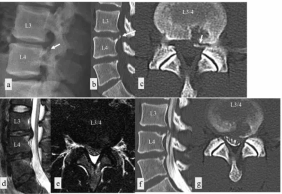
Fig 4 shows the radiologic findings after surgery. The bony Figure 1
Case 1 : Plain radiograph and CT images reveal osseous remnant of apophyseal ring fracture of upper endplate of L4 vertebrae (arrows, a : plane lateral radiograph, b : CT scan sagittal view, c : CT scan axial view).
MRI T2WI shows lumbar disc herniation and left lateral recess stenosis at L3/4 (d : sagittal view, e : axial view). CT myelograms also show lateral recess stenosis at L3/4 (f : sagittal view, g : axial view).
Figure 2
Case 1 : Postoperative bony fragment reduction (a : CT scan sagittal view, b : CT scan axial view)
MRI T2WI shows that lumbar disc herniation was almost removed and stenosis was resolved (c : sagittal view, d : axial view).
360
Y. Sugimine, et al. Bony fragment of apophyseal ring fracturefragment is seen to overhang the spinal canal (a, b) ; however, the fragment was snapped off when it was impacted to avoid pres-sure on the cauda equine and the nerve root. MRI T2 weighted imaging (T2WI) shows that the lumbar disc herniation is almost
completely removed and stenosis is resolved at both L4/5 and L5/s (c, d).
Stretching of the tight hamstrings was performed using an active stretching machine (8), which effectively improves the tightness and allows for better trunk motion. Also, he improved his motor control by training using the concept of Pilates (9). He then returned to competition in the second grade Nippon Pro-fessional Baseball (NPB) league at 2 months after surgery and was able to join the first grade NPB league at 3 months after the surgery. He eventually returned to his original level within the same competitive season. The following year, he played extreme-ly well and was selected as one of the best players.
DISCUSSION
Apophyseal ring fracture is an avulsion vertebral fracture caused by chronic stress and repeated trauma (1-5). The posteri-or apophyseal ring is highly stressed during extension and this can cause weakness of the ring. The weakened posterior area is avulsed following traction stress in flexion (1,2,3,4,5). Conser-vative treatment is usually chosen for patients with apophyseal ring fracture. In general, surgical treatment is considered for symptoms recalcitrant to conservative treatment.
Regarding accuracy of diagnosis, CT is considered the best tool to detect the presence of bony fragment. Kadam et al. (10) reviewed the case of a 15-year-old boy and reported the clinical importance of CT scan to detect the lesion. In both our cases, only plain radiographs and MRIs were obtained. CT scan had not been done at the time they presented to our center. A clear understanding of the osseous condition is essential to ensure a precise management strategy for high-level athletes. Case 1 had a 4-year history of symptoms, which is unlike typical cases of herniation. HNP usually resolves spontaneously (11,12), and does not persist for long (12). Ramos Amador et al. followed 118 HNP cases using MRI, and found that 83% of cases showed spontaneous disappearance of the HNP mass within a year. Thus, we suspected an atypical condition in Case 1, and so performed CT scan. The pain may have persisted because of the HNP in the narrowed lateral recess due to the residual bony fragment. If we suspect the existence of bony fragment during follow up patient of lumbar HNP, we should take CT scan as soon as possible. However, we have to consider that CT also has problem of radiation exposure to patient.
Indications for surgery are important for high-level athletes such as professional players like Case 2 in this report. For Case 1, the decision to perform surgery was relatively easy, because of the longer duration of symptoms (i.e. 4 years). On the other hand, we thoroughly discussed the indications for surgery for Case 2 because of the shorter duration of symptoms (i.e. 17 days). We then decided on acute intervention by surgery for the following reasons :
1. He presented with muscular weakness, and muscle atrophy due to prolonged weakness is rather disadvantageous for pro-fessional athletes.
2. Chronic compression of the nerve root by both the bony fragment and the HNP would have caused the tight ham-strings (6), which in turn would not be good for professional performance.
3. HNP typically exists in a narrow lateral recess due to the bony fragment, so the pain may not subside quickly. In patients with this condition, it was reported that the mean duration of symptoms was more than 1 year (7). Indeed, Case 1 had a 4-year history of pain.
Regarding outcomes, this player returned to the first grade NPB league 3 months after surgery in the same regular season. Figure 3
Case 2 : CT images reveal the osseous remnant of apophyseal ring fracture at S1 (a, b).
MRI T2WI show lumber disc bulge at L4/5 (c) and herniation at L5/S (d).
Spinal canal stenosis at L4/5 (e) and lateral recess stenosis at L5/S (f) are evident on myelograms.
Figure 4
Case 2 : Postoperatively, bony fragment seems to overhang the spinal canal (a, b) ; however, the fragment was snapped off when it was impacted, to avoid pressing on the cauda equina.
MRI T2WI shows lumbar disc herniation is almost removed and stenosis is resolved (c, d).
361
The Journal of Medical Investigation Vol. 66 August 2019
By the next season the following year, his performance was remarkable and at the end of the year he was selected as one of the best players. For this player, we cannot directly compare out-comes with those obtained by conservative treatment ; however, his performance strongly supports the fact that our decision was satisfactory.
Finally, we discuss the indications for surgery in HNP with the residual bony fragment of the apophyseal ring fracture in adults. Basically, HNP can undergo spontaneous reduction (11,12), and thus conservative treatment would be the first choice, even though the HNP typically exists in the narrow later-al recess caused by the bony fragment. Interestingly, Akhaddar et al. (7) reported that HNP combined with apophyseal separa-tion may prolong clinical course. Mean durasepara-tion of symptoms was 16 months, twice longer than that in patients with only HNP (i.e. 8 months).
Taken together, these findings led us to conclude that in cases of HNP with apophyseal ring separation, surgical intervention in the comparatively early phase can be recommended, particu-larly if the patients desire a quick return to their original level of activity.
CONFLICT OF INTERESTS-DISCLOSURE
NothingREFERENCES
1. Higashino K : Long-term Outcomes of Lumbar Posterior Apophyseal End-Plate Lesions in Children and Adolescents. J Bone Joint Surg Am 94 : e74(1-7), 2012
2. Ikata T, Morita T, Katoh S, Tachibana K, Maoka H : Le-sions of the lumbar posterior end plate in children and adolescents. An MRI study. J Bone Joint Surg Br 77(6) : 951-955, 1995
3. Chang CH, Lee ZL, Chen WJ, Tan CF, Chen LH : Clinical significance of ring apophysis fracture in adolescent lumbar
disc herniation. Spine (Phila Pa 1976) 33(16) : 1750-1754, 2008
4. Sairyo K : Three-dimensional finite element analysis of the pediatric lumbar spine. Part1 : pathomechanism of apophy-seal bony ring fracture. Eur Spine J 15 : 923-929, 2006 5. Faizan A, Sairyo K, Goel VK, Biyani A, Ebraheim
N : Biomechanical rationale of ossification of the sec-ondary ossification center on apophyseal bony ring frac-ture : a biomechanical study. Clin Biomech (Bristol, Avon) 22(10) : 1063-1067, 2007
6. Miyagi R, Sairyo K, Sakai T, Tezuka F, Kitagawa Y, Dezawa A : Persistent Tight Hamstrings Following Conservative Treatment for Apophyseal Ring Fracture in Adolescent Athletes : Critical Appraisal. J Medical Invest 61 : 446-451, 2014
7. Akhaddar A, Belfquih H, Oukabli M, Boucetta M : Poste-rior ring apophysis separation combined with lumbar disc herniation in adults : a 10-year experience in the surgical management of 87 cases 14(4) : 475-483, 2011
8. Hasebe K, Okubo Y, Kaneoka K, Takada K, Suzuki D, Sairyo K : The effect of dynamic stretching on hamstrings flexibility with respect to the spino-pelvic rhythm. J Med Invest 63(1-2) : 85-90, 2016
9. Fujitani L, Daiyu Y, Masubuchi Y, Takeda J : Pirates for the low back pain. (in Japanese) Jpn. J Clin Sports Med 10 (33) : 950-960, 2016
10. Kadam G, Narsinghpura K, Deshmukh S, Desai S : Trau-matic lumbar vertebral ring apophysis fracture with disk herniation in an adolescent. Radiol Case Rep 12(2) : 427-430, 2017
11. Henmi T, Sairyo K, Nakano S, Kanematsu Y, Kajikawa T, Katoh S, Goel VK : Natural history of extruded lumbar intervertebral disc herniation. J Med Invest 49(1-2) : 40-3, 2002
12. Ramos Amador A, Alcaraz Mexía M, González Preciado JL, Fernández Zapardiel S, Salgado R, Páez A : Natural history of lumbar disc hernias : does gadolinium enhancement have any prognostic value? Radiologia 55(5) : 398-407, 2013