Mutations
in the Cysteine-Rich
Region
of
Proto-Oncogene
in Patients
Diagnosed
as
Sporadic
Medullary
Thyroid
Carcinoma
the RET
Having
TAKEHIKO KIMURA, KATSUHIKO YOSHIMOTO*, YUTAKA YOKOGOSHI, AND SHIRO SAITO
The First Department of Internal Medicine and "Otsuka Department of Clinical and Molecular Nutrition, School of Medicine, The University of Tokushima, Tokushima 770, Japan
Abstract. Medullary thyroid carcinoma (MTC) and pheochromocytoma appear in either a sporadic or a hereditary form as components of multiple endocrine neoplasia (MEN). Many germline mutations of the RET proto-oncogene have been reported in patients with MEN 2A and 2B, and familial MTC (FMTC). To elucidate the etiological roles in tumorigenesis of sporadic MTCs and pheochromocytomas, mutations in the cysteine-rich region of the RET proto-oncogene were analyzed by using polymerase chain reaction-single strand conformation polymorphism (PCR-SSCP) analysis. Exons 10 and 11 were studied in genomic DNAs from 3 clinically apparent sporadic MTCs, MTCs and pheochromocytomas from 2 patients with MEN 2A,1 with FMTC, 4 with MEN 2B, 3 with neurofibromatosis type 1(NF1),12 sporadic pheochromocytomas and an MTC cell line, TT. All tumors from two patients with MEN 2A and one patient with FMTC had mutations at codon 618 and 634 as well as their leukocytes, reflecting their germline mutations. In this region, no mutations were detected in any tumors from patients with MEN 2B and NF1, and sporadic pheochromocytomas. But mutations were detected and identified in 3 clinically apparent sporadic MTCs and TT cells. A 6 base pair (bp) deletion causing the loss of a cysteine residue at codon 634 and a mutation causing substitution from cysteine to tyrosine at codon 634 were detected in 2 sporadic MTCs as somatic events. In a female patient diagnosed as having sporadic MTC, a mutation at codon 618 was detected not only in tumor tissues, but also in her leukocytes, suggesting a germline mutation of the RET proto-oncogene. In TT cells a heterozygous mutation at codon 634 was detected. These results suggest that RET mutations within a cysteine-rich region may also play an important role in the tumorigenesis of sporadic MTCs, and mutations of RET proto-oncogene should be screened in clinically sporadic cases to exclude hereditary MTCs.
Key words: RET proto-oncogene, Mutation, Sporadic MTC, MEN 2A, Genetic diagnosis
(Endocrine Journal 42: 517-525,1995)
MTC, a tumor arising from the calcitonin-secret-ing parafollicular C-cells, comprises 5-10% of all malignancies arising from the thyroid gland [1]. This tumor appears either as a sporadic nonhered-itary lesion or as a component of three familial
Received: October 8, 1994 Accepted: April 3, 1995
Correspondence to: Dr. Takehiko KIMURA, The First Department of Internal Medicine, School of Medicine, The University of Tokushima, 3-18-15, Kuramoto-cho, Tokushima 770, Japan
syndromes, MEN 2A, MEN 2B and FMTC, which are inherited as an autosomal dominant trait. MEN 2A is variably associated with pheochromocytoma
and primary hyperparathyroidism. MEN 2B is as-sociated with pheochromocytoma, marfanoid habitus, mucosal neuromas, and ganglioneuroma-tosis. FMTC is not associated with other endocrinopathies. Sporadic MTC, a tumor of mod-erate to marked aggressiveness, occurs during the fifth or sixth decade of life as a solitary unilateral thyroid mass without coexisting microscopic C-cell
hyperplasia, associated endocrinopathies,
or a
fa-milial history of MTC.
The loci of the three syndromes, MEN 2A, FMTC
and MEN 2B, link to the chromosome 10g11.2
re-gion, where the RET proto-oncogene has also been
mapped [2, 3]. Germline mutations
of the RET
proto-oncogene have been described in patients
af-fected with MEN 2A, FMTC or MEN 2B [4-10].
The nonconservative substitution of the 5 cysteine
residues located in the extracellular domain
adja-cent to the transmembrane
domain of the RET
protein has been detected in patients with MEN
2A and FMTC [4-6]. In patients with MEN 2B, a
mutation at codon 918 causing the substitution of
a threonine for a methionine in exon 16 of the
ty-rosine kinase domain of the RET protein has been
detected at the germline level [8-10].
There are few data on the pathogenesis of
spo-radic MTC.
Increased
expression
of the RET
proto-oncogene
has been found in MTCs of both
familial and sporadic types as
pheochromocyto-mas [11, 12]. In this study, we screened mutations
of the RET proto-oncogene
in tumors and
leuko-cyte DNA from patients with MEN 2A, MEN 2B,
FMTC, NF1, sporadic
pheochromocytoma
and
MTC, and a TT cell line by the PCR-SSCP method.
We identified germline mutations
in 2 MEN 2A
and 1 FMTC patients as previously reported [4-6].
Interestingly
we demonstrated
nonconservative
mutations in the cysteine-rich extracellular region
adjacent to the transmembrane
domain of the RET
proto-oncogene
in 3 MTCs diagnosed as sporadic
MTCs and TT cells.
Patients
Patient 1 was a 94-year-old man who underwent
gastrectomy because of a gastric ulcer at the age of
75. He was referred to Takamatsu Municipal
Hos-pital for abdominal
pain at the age of 92. The
physical examination was unremarkable.
Because
the serum level of carcinoembryonic antigen (CEA)
was increased (1,230 ng/ml; normal, < 5 ng/ml),
the presence of colon cancer was suspected,
but
fiberoptic colonoscopy revealed no evidence of
co-lon cancer. A computed tomographic
(CT) scan
revealed a mediastinal tumor adjacent to the right
wall of the trachea, and thyroid ultrasonography
revealed a space occupying a region in the right
lobe. The basal serum calcitonin level was 5,981
pg/ml (normal, <100 pg/ml). These findings es-tablished the diagnosis of MTC. He had no family history of MTC or pheochromocytoma. He elect-ed not to undergo surgery, and he remained well until the appearance of a subdural hemorrhage. He died of respiratory failure from acute bronchop-neumonia at the age of 94. An autopsy revealed that the tumor was well demarcated, and its size was 5 x 4.3 x 3.5 cm. The tumor consisted of
spin-dle-shaped cells with eosinophilic cytoplasm. Amyloid was demonstrated among the fibrous stro-ma. Immunohistochemical staining for calcitonin and CEA revealed abundant signals in the cyto-plasm of the neoplastic C-cells within the tumor mass. Calcitonin staining of the thyroid tissue out-side the carcinoma revealed no other foci of MTC or C-cell hyperplasia in either lobe. No regional lymph nodes or distant metastases were detected. No pathologic abnormalities of the adrenal, par-athyroid and pituitary glands were evident. Patient 2 was a 48-year-old woman. On a
regu-lar physical examination, a thyroid nodule was found and she was referred to Takamatsu Munici-pal Hospital for further evaluation. Her history was unremarkable except for acute appendicitis at the age of 33. There was no family history of MTC or pheochromocytoma. Her serum CEA level was increased (90 ng/ml). Thyroid ultrasonography and CT scan showed a solitary mass in the left lobe, and scanning with radioactive iodine showed a cold nodule in the left lobe. She underwent left thyroid lobectomy. Histological diagnosis was MTC (3 x 1.5 x 1.2 cm) with regional lymph node metastases. The tumor was immunohistochemi-cally positive for calcitonin and CEA, and there was marked interstitial deposition of amyloid. She was diagnosed as having sporadic MTC.
Subse-quently, she received radiation therapy to the neck. She has had no signs of recurrence for the past 7 years, and no clinical symptoms or other data sug-gest the presence of pheochromocytoma.
Patient 3 was a 36-year-old woman, whose thy-roid tumor was first revealed by a physical examination. There was no family history of MTC or pheochromocytoma, and her two sisters and three children were healthy. At admission to Tokushima Municipal Hospital, her serum levels of calcitonin and CEA were 1,854 pg/ml and 96.3 ng/ml, respectively. Since a CT scan and ultra-sonography revealed one tumor (2.5 x 1.8 cm in size) on the left lobe of the thyroid, subtotal
thy-roidectomy was performed. A histological exami-nation revealed that the tumor was MTC with regional lymph node metastases, and the
noncan-cerous region of the thyroid showed findings of chronic thyroiditis but no apparent C-cell hyper-plasia. There were no clinical or laboratory findings consistent with pheochromocytoma. She was di-agnosed as having sporadic MTC. She has remained in good health without recurrence for 4 years since the operation.
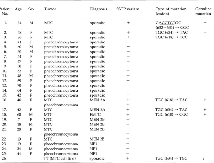
from the tissues, leukocytes, and paraffin-embed-ded tissues, as described [13, 14]. DNAs were extracted from 3 clinically apparent sporadic MTCs, MTCs and pheochromocytomas from 2 patients with MEN 2A, MTC from 1 patient with FMTC, MTCs and pheochromocytomas from 4 patients with MEN 2B, pheochromocytomas from 3 patients with NF1, and 12 sporadic pheochromocytomas
(Table 1). A human medullary thyroid carcinoma cell line, TT (CRL 1803) was obtained from ATCC.
Materials and Methods
DNA preparation
Tissues were obtained at surgery or autopsy, and a diagnosis of MTC was confirmed by histological examinations of the tumors. DNA was isolated
PCR-SSCP
PCR amplification was performed by using the oligonucleotide primers described by Donis-Keller
et al. [5]: 5'-GCGCCCCAGGAGGCTGAGTG-3' and
5'-CGTGGTGGTCCCGGCCGCC-3' for exon 10;
5'-CCTCTGCGGTGCCAAGCCTC-3' and
5'-CACCGGAAGAGGAGTAGCTG-3' for exon 11.
Amplified DNA fragments were all of the size
ex-pected (exon 10,187 bp; exon 11,234 bp). PCR
proceeded in a Program Temp Control System
PC-700 (ASTEC, Fukuoka,
Japan) with 50 ng of
genomic DNA in a total volume of 5 p1 containing
1.5 ,uCi of [a-32P]dCTP (3,000 Ci/mmol;10
mCi/
ml). Thirty-five cycles consisted of 30 sec at 94 °C
for denaturation,
30 sec at 68 °C for exon 10, or at
65 °C for exon 11 for annealing, and 1 min at 72 °C
for extension. The reaction mixture (5 µl) was
heat-ed with 95 ,ul of dye (95% formamide/20
mM
EDTA/0.05% bromophenol blue/0.05% xylene
cy-anol), then 1 p1 of the mixture was applied to a 5%
polyacrylamide
gel containing 45 mM Tris-borate
(pH 8.3) and 4 mM EDTA. Glycerol (5% or 10%)
was added when specified, as described [14].
Elec-trophoresis proceeded at 30 W for 4-6 h at room
temperature
or for 6-10 h at 4 °C. The gel was
dried and exposed to X-ray film with intensifying
screens at - 70 °C for 12 to 24 h.
Sequencing analysis
Abnormal bands detected by PCR-SSCP were excised from the dried gel, placed in 100 p1 of dis-tilled water, and incubated at 37 °C. An aliquot of the supernatant was then used as a template for the PCR reaction. PCR products were cloned into the pCRTMII vector with a TA Cloning kit (Invitro-gen Co., San Diego, CA). Plasmid DNAs were sequenced with a SequenaseTM Version 2.0 DNA Sequencing Kit (Amersham, Buckinghamshire, UK) according to the manufacturer's instructions, then resolved on a 5% denaturing polyacrylamide gel. The accuracy of our sequencing data in cloned DNAs was confirmed by analyzing at least 10 in-dependent clones.
Reverse Transcription (RT)-PCR procedure
Total RNA from TT cells was prepared by using acid guanidinium thiocyanate-phenol-chloroform extraction [15]. cDNA was made from 2 jig of total RNA with M-MLV reverse transcriptase (Promega, Madison, WI) and random hexamers. The cDNA was then amplified by PCR in 30 cycles
with 2 primers
[5'-GGGGGATTAAAGCTGGC-TAT-3' (exon 10) and
5'-TGGCTTGTGGGC-AAACTTGT-3' (exon 11) (codon 600-667)]. The PCR conditions were as follows: 1 min denatur-ation at 94 °C,1 min annealing at 60 °C and 1 min
extension at 72 °C. DNA was electrophoresed on a 10% polyacrylamide gel, followed by ethidium bromide staining. The gels were photographed with an ultraviolet transilluminator.
Results
Screening of point mutations of the RET
proto-oncogene by PCR-SSCP and sequencing
PCR-SSCP of exon 11 from genomic DNA of the
sporadic MTC of patient 1, as shown in Fig. 1A,
disclosed an extra band with an altered migration
relative to those amplified from leukocytes of the
same patient. Sequencing revealed a 6 by deletion
including codons 632 to 634 in exon 11 (Fig. 2A).
The deletion caused a loss of the cysteine residue
at codon 634 from glutamine (codon 632)-leucine
(codon 633)-cysteine (codon 634) (GAGCTGTGC,
deleted 6 by are underlined).
PCR-SSCP of exon
11 from the MTC of patient 2, as shown in Fig. 1B,
disclosed an extra band relative to those amplified
from her leukocytes.
As shown in Fig. 2B, codon
634 of TGC for cysteine was mutated to TAC for
tyrosine by a G to A transition.
The mutation of
patient 1 destroyed a Alul restriction site, and the
mutation of patient 2 created a new Rsal
restric-tion site, and restricrestric-tion analyses of PCR products
also confirmed the results (data not shown).
PCR-SSCP of exon 10 from the MTC and the leukocytes
of patient 3 revealed common extra bands relative
to those amplified from leukocytes from a healthy
subject (Fig. 1C). As shown in Fig. 2C, codon 618
of TGC for cysteine was mutated to TCC for serine
by a G to C transversion in both MTC and
leuko-cytes of this patient.
In these three MTCs, the other alleles of exon 11
(patients 1 and 2) or exon 10 (patient 3) of the RET
proto-oncogene had normal sequences, and no
de-tectable variations in exon 10 (patients 1 and 2) or
exon 11 (patient 3) were revealed by PCR-SSCP.
Direct sequencing of codon 918 of the RET
proto-oncogene revealed normal sequences in these 3
MTCs.
We analyzed a panel of genomic DNAs of
tu-mors from 2 patients with MEN 2A,1 with FMTC,
4 with MEN 2B, 3 with NF1, and 12 sporadic
pheo-chromocytomas
for mutations in exons 10 and 11.
PCR-SSCP and sequencing revealed germline
mu-tations from cysteine (TGC) to tyrosine (TAC) at
Fig. 1. PCR-SSCP of exons 11 (A and B) and 10 (C) of the RET proto-oncogene. Panels A, B and C show the PCR-SSCP profiles of patients 1, 2, and 3, respectively. A. Lane 1, leukocytes from a healthy subject; lane 2, MTC from patient 1; and lane 3, leukocytes from patient 1. B. Lane 1, leukocytes from a healthy subject; lane 2, MTC from patient 2; and lane 3, leukocytes from patient 2. C. Lane 1, leukocytes from a healthy subject; lane 2, MTC from patient 3; and lane 3, leukocytes from patient 3. Electrophoresis was performed in a 5% polyacrylamide gel without glycerol at room temperature. Arrow heads denote the bands with altered migration relative to controls.
Fig. 2. Nucleotide sequence analysis of the RET proto-oncogene from genomic DNA. DNA fragments that showed a mobility shift on SSCP, and DNA from normal leukocytes were PCR-amplified. The PCR products were cloned into the pCRTMII vector and sequenced. A. Sequence of the variant allele at exon 11 of the RET proto-oncogene from a MTC of patient 1. The left panel shows the normal sequence of codons 630-637. The right panel shows the 6 by deletion. Deleted bases are indicated by asterisks in the left panel. B. Sequence of the variant allele at exon 11 of the RET proto-oncogene from MTC of patient 2. The panel shows the G to A transition at codon 634. The mutated base is indicated by an asterisk. C. Sequence of the variant allele at exon 10 of the RET proto-oncogene from an MTC of patient 3. The left panel shows the normal sequence of codons 614-621. The right panel shows the G to C transversion at codon 618. The mutated base is indicated by an asterisk.
codon 618 and 634 in 2 patients with MEN 2A, and to arginine (CGC) at codon 618 in one patient with FMTC. The results are summarized in Table 1. These PCR reactions were repeated several times for each sample. In all cases PCR-SSCP and se-quence analyses confirmed the results. No extra bands in PCR-SSCP of exons 10 and 11 were de-tected in tumors from patients with MEN 2B and sporadic pheochromocytomas. These results were also confirmed by directly sequencing the genom-ic DNAs.
In TT cells, we detected a heterozygous muta-tion at codon 634 (TGC for cysteine to TGG for tryptophan) of the RET proto-oncogene by PCR-SSCP analysis and nucleotide sequencing (data not shown). The mutation created a new HhaI restric-tion site.
Expression of both awild-type and a mutated RET
allele in TT cells
A fragment of the appropriate size (203 bp) re-sulted from RT-PCR of RNA from TT cells by using primers located in exons 10 and 11. The nucle-otide sequence of RT-PCR product was confirmed by directly sequencing. The new HhaI restriction site produces the 106 and 97 by fragment in the mutant allele, but not in the wild-type allele. Re-striction analysis of RT-PCR fragments from TT cells showed digested and undigested fragments, and these results imply expression of both the wild-type and the mutated allele of RET proto-oncogene in TT cells (Fig. 3).
Discussion
There have been a few reports describing genet-ic changes in sporadic MTCs. One is the loss of heterozygosity (LOH) on chromosomes 1p and/or 22q in a few MTCs, and no consistent LOH in any chromosome 10 probes tested [16-19]. Another is the low mutation frequency of the ras or p53 genes [16, 20]. We also reported that LOH on informa-tive loci, or mutations of the ras, p53 or gsp genes in sporadic MTCs were undetectable [14, 21-23]. Thus, the precise gene(s) responsible for sporadic MTC and the mechanism relating to its structural alterations to development of the tumor have yet to be defined.
Germline mutations of the RET proto-oncogene have been described in patients with MEN 2A, FMTC and MEN 2B [4-10]. The RET proto-onco-gene encodes a protein structurally related to
transmembrane receptors with cytoplasmic
ty-rosine-kinase domains, but its putative ligand has not yet been identified [24, 25]. Direct evidence for the involvement of the RET proto-oncogene in human tumors has been obtained in human thy-roid papillary carcinomas [26]. Recently, Santoro et al. reported that mutations of the RET proto-oncogene play a critical role in tumor formation as a consequence of the activation of the RET proto-oncogene kinase [27]. Substitution from cysteine to other amino acids in MEN 2A is considered to disrupt normal disulfide bonds, and leads to acti-vated homodimers of RET protein. And MEN 2B mutation, substitution from methionine to
threo-Fig. 3. HhaI restriction enzyme digestions of PCR fragments amplified cDNA from TT cells. A fragment including part of exons 10 and 11 was amplified by 2 primers described in Materials and Methods. M, eX174 HaeIII-digested DNA fragments used as molecular weight markers; lane 1, undigested fragment (203 bp); lane 2, HhaI digested fragments (203, 106 and 97 bps).
nine at codon 918, alters the substrate specificity of RET protein without dimerization.
We confirmed that tumor DNAs of MTCs and pheochromocytomas from 2 patients with MEN 2A and one with FMTC had mutations of cysteine res-idues in the extracellular cysteine-rich region. In these patients, these mutations were also found in their leukocyte DNA, indicating germline muta-tions of the RET proto-oncogene. In the genetic forms of MTC, a germline mutation of the RET proto-oncogene may result in the initial hyperpro-liferation of C-cells. Subsequent genetic steps, possibly at other chromosome loci, presumably re-sult in selected clonal transformation.
With respect to sporadic MTCs, Donis-Keller et al. [5, 9] have found only one mutation in 16 MTCs by analysing exons 10 and 11 of the RET
proto-oncogene. This caused a 6 by deletion that removed the cysteine residue at codon 630, but clinical data from the patient with this deletion were not reported. Although Eng et al. [10] detect-ed a codon 918 mutation (ATG to ACG) in 5 of 13 sporadic MTCs, no mutations in exon 10 or 11 were detected in 13 sporadic MTCs. Blaugrund et al. also identified codon 918 mutations in 6 of 15 clin-ically sporadic cases of MTC [28]. Mutations of the RET proto-oncogene in sporadic MTCs were mainly detected at codon 918 rather than within the cysteine-rich region. But in this study, three MTCs and TT cells had mutations at cysteine resi-due in the cysteine-rich region. The TT cell line was established from a 77-year-old patient with a sporadic MTC [18, 29]. In TT cells, we detected a point mutation at codon 634 and expression of both the mutant allele and wild-type allele of the RET proto-oncogene by restriction analysis of RT-PCR products. These results were concordant with a recent report [27]. Our one sporadic MTC patient had a 6 by deletion, which resulted in the loss of a cysteine residue at codon 634, distinct from loss of that at codon 630 reported by Donis-Keller et al. [5, 9]. According to a study of 118 families with in-herited MTC, mutations detected in patients with MEN 2A and FMTC were restricted to 5 cysteine residues (codons 609, 611, 618, 620 and 634), of which the most frequent events were at codon 634 [6]. Therefore, the somatic 6 by deletion including codon 634 and the mutation causing a substitution from cysteine to tyrosine at codon 634 in sporadic MTCs in patients 1 and 2 may play an important role in the tumorigenesis of sporadic MTCs.
A 6 by deletion in patient 1 may be due to the
accumulation of DNA damage or incompleteness
of DNA repair accompanying
aging, but the
rela-tion between
molecular
events and the aging
process is controversial.
As his MTC lesion was
asymptomatic
and had no regional lymph node
metastases, it might be a slowly progressive type.
Several investigators have screened for mutations
in exon 10 and 11 of the RET gene in sporadic
MTCs and failed to find somatic events in these
regions [5, 10, 28, 30]. Our two patients do have
somatic mutations within the cysteine-rich region,
but the frequency could not be discussed because
of the limited number of our patients.
Although
difference in the frequency of mutations
may be
attributed to racial difference, a large scale study
should be conducted.
We found a conversion of the cysteine residue
to serine at codon 618 in a case of clinically
appar-ent sporadic MTC (patiappar-ent 3). Since this patiappar-ent
was diagnosed as having sporadic type due to the
apparent absence of any family history of MTC,
signs of associated endocrine lesions or bilateral
thyroid involvement of MTC, she was not screened
until diagnosis.
Analysis of the DNAs from her
leukocytes revealed the same mutation as well as
the MTC. These results indicated that the patient
in fact had hereditary MTC. Because the genetic
change was probably transmitted to her offspring,
DNA analysis for the detection of the mutation
should be carried out to identify subjects at risk,
but unfortunately
we could not obtain their
con-sent. Olson et al. have reported that information
about family history, clinical presentation, and
his-topathologic
examination
are not sufficient
to
exclude hereditary forms of MTC [31]. The case of
a germline mutation at codon 618 in a clinically
sporadic MTC patient was reported by Blaugrund
et al. [28]. In future, genetic markers such as
mu-tations of the RET proto-oncogene should be used
as an aid in distinguishing
sporadic from
heredi-tary MTC.
Acknowledgments
We thank Drs. Masaru Tsuyuguchi, Isao Morim-oto, Sumiya Eto, Ryoyu Takeda, Ryuichiro Soma, Kozo Hashimoto, Tsuneo Ogawa, Haruo Sumitani, Kunio Ogasawara, Ryuichi Yamasaki, Hideo Ta-kahashi, Kazuto Kameyama, Keiko Miya, Hidetaka
Hone, Toshihiko Inoue, and Emiko Hosoi for
pro-viding tumors.
We thank Drs. Toshiaki Sano,
Hisajiro Kumagai, and Hiroshi Morizumi for
patho-logical diagnosis. We thank Miss Chisato Tanaka
and Miss Maki Moritani for technical assistance.
We are grateful to Professor Mitsuo Itakura for
continuous support.
This work was supported
in
part by a Grant-in-Aid for Scientific Research from
the Ministry of Education, Science and Culture of
Japan to S. S., and by a grant from Otsuka
Phar-maceutical Factory, Inc., for Otsuka Department of
Clinical and Molecular Nutrition, School of
Medi-cine, The University of Tokushima.
References
1. DeLellis RA (1993) The pathology of medullary roid carcinoma and its precursors. Monogr Pathol
(United States) 35: 72-102.
2. Gardner E, Papi L, Easton DF, Cummings T, son CE, Kaplan M, Love DR, Mole SE, Moore JK, Mulligan LM, Norum RA, Ponder MA, Reichlin S, Stall G, Telenius H, Telenius-Berg M, Tunnacliffe A, Ponder BAJ (1993) Genetic linkage studies map
the multiple endocrine neoplasia type 2 loci to a small interval on chromosome 10g11.2. Hum Mol Genet 2: 241-246.
3. Mole SE, Mulligan LM, Healey CS, Ponder BAJ, Tunnacliffe A (1993) Localization of the gene for multiple endocrine neoplasia type 2A to a 480 kb region in chromosome band 10g11.2. Hum Mol Genet 2: 247-252.
4. Mulligan LM, Kwok JBJ, Healey CS, Elsdon MJ, Eng C, Gardner E, Love DR, Mole SE, Moore JK, Papi L, Ponder MA, Telenius H, Tunnacliffe A, der BAJ (1993) Germ-line mutations of the RET proto-oncogene in multiple endocrine neoplasia
type 2A. Nature 363: 458-460.
5. Donis-Keller H, Dou S, Chi D, Carlson KM, Toshima K, Lairmore TC, Howe JR. Moley JF, Goodfellow P,
Wells SA Jr (1993) Mutations in the RET oncogene are associated with MEN 2A and FMTC. Hum Mol Genet 2: 851-856.
6. Mulligan LM, Eng C, Healey CS, Clayton D, Kwok JBJ, Gardner E, Ponder MA, Frilling A, Jackson CE, Lehnert H, Neumann HPH, Thibodeau SN, Ponder
BAJ (1993) Specific mutations of the RET oncogene are related to disease phenotype in MEN
2A and FMTC. Nature Genet 6: 70-74.
7. Tsai M-S, Ledger GA, Khosla S, Gharib H, Thibodeau SN (1994) Identification of multiple docrine neoplasia, type 2 gene carriers using linkage analysis and analysis of the ret proto-oncogene. J
Clin Endocrinol Metab 78:1261-1264.
8. Hofstra RMW, Landsvater RM, Ceccherini I, Stulp RP, Stelwagen T, Luo Y, Pasini B, Hoppener JWM, van Amstel HKP, Romeo G, Lips CJM, Buys CHC (1994) A mutation in the RET proto-oncogene ciated with multiple endocrine neoplasia type 2B and sporadic medullary thyroid carcinoma. Nature
367: 375-376.
9. Carlson KM, Dou S, Chi D, Scavarda N, Toshima K, Jackson CE, Wells SA Jr. Goodfellow PJ, Keller H (1994) Single missense mutation in the
tyrosine kinase catalytic domain of the RET protooncogene is associated with multiple
crine neoplasia type 2B. Proc Natl Acad Sci USA 91: 1579-1583.
10. Eng C, Smith DP, Mulligan LM, Nagai MA, Healey CS, Ponder MA, Gardner E, Scheumann GFW, son CE, Tunnacliffe A, Ponder BAJ (1994) Point mutation within the tyrosine kinase domain of the
RET proto-oncogene in multiple endocrine neoplasia type 2B and related sporadic tumors. Hum Mol Genet 3: 237-241.
11. Santoro M, Rosati R, Grieco M, Berlingieri MT, D'Amato GL, de Franciscis V, Fusco A (1990) The ret proto-oncogene is consistently expressed in man pheochromocytomas and thyroid medullary
carcinomas. Oncogene 5:1595-1598.
12. Miya A, Yamamoto M, Morimoto H, Tanaka N, Shin E, Karakawa K, Toyoshima K, Ishizaka Y, Mori T, Takai S (1992) Expression of the ret
oncogene in human medullary thyroid carcinomas and pheochromocytomas of MEN 2A. Henry Ford Hosp Med J 40: 215-219.
13. Yoshimoto K, Iizuka M, Iwahana H, Yamasaki R, Saito H, Saito S, Sekiya T (1989) Loss of the same
alleles of HRAS1 and D115151 in two independent pancreatic cancers from a patient with multiple
docrine neoplasia type 1. Cancer Res 49: 2716-2721. 14. Yoshimoto K, Iwahana H, Fukuda A, Sano T, Saito S, Itakura M (1992) Role of p53 mutations in crine tumorigenesis: Mutation detection by polymerase chain reaction-single strand
tion polymorphism. Cancer Res 52: 5061-5064. 15. Chomczynski P, Sacchi N (1987) Single-step method
of RNA isolation by acid guanidinium phenol-chloroform extraction. Anal Biochem 162:
156-159.
16. Okazaki M, Miya A, Tanaka N, Miki T, Yamamoto M, Motomura K, Miyauchi A, Mori T, Takai S (1989)
Allele loss on chromosome 10 and point mutation of ras oncogenes are infrequent in tumors of MEN
2A. Henry Ford Hosp Med J 37:112-115.
17. Yang K-P, Nguyen CV, Castillo SG, Samaan NA (1990) Deletion mapping on the distal third region of chromosome 1p in multiple endocrine neoplasia
type IIA. Anticancer Res 10: 527-534
18. Nelkin BD, Nakamura Y, White RW, de Bustros AC, Herman J, Wells SA Jr, Baylin SB (1989) Low
incidence of loss of chromosome 10 in sporadic and hereditary human medullary thyroid carcinoma. Cancer Res 49: 4114-4119.
19. Khosla S, Patel VM, Hay ID, Schaid DJ, Grant CS, van Heerden JA, Thibodeau SN (1991) Loss of
erozygosity suggests multiple genetic alterations in pheochromocytomas and medullary thyroid nomas. J Clin Invest 87:1691-1699.
20. Yana I, Nakamura T, Shin E, Karakawa K, Kurahashi H, Kurita Y, Kobayashi T, Mori T, Nishisho I, Takai S (1992) Inactivation of the p53 gene is not required for tumorigenesis of
lary thyroid carcinoma or pheochromocytoma. Jpn J Cancer Res 83:113-1116.
21. Kubo K, Yoshimoto K, Yokogoshi Y, Tsuyuguchi M, Saito S (1991) Loss of heterozygosity on
mosome 1p in thyroid adenoma and medullary carcinoma, but not in papillary carcinoma. Jpn J Cancer Res 82:1097-1103.
22. Yoshimoto K, Iwahana H, Fukuda A, Sano T, Katsuragi K, Kinoshita M, Saito S, Itakura M (1992) ras mutations in endocrine tumors: Mutation
tection by polymerase chain reaction-single strand conformation polymorphism. Jpn J Cancer Res 83:
1057-1062.
23. Yoshimoto K, Iwahana H, Fukuda A, Sano T, Itakura M (1993) Rare mutations of the Gs alpha subunit gene in human endocrine mors: Mutation detection by polymerase chain reaction-primer-introduced restriction analysis. Cancer 72:1386-1393.
24. Takahashi M, Buma Y, Iwamoto T, Inaguma Y, Ikeda H, Hiai H (1988) Cloning and expression of
the ret proto-oncogene encoding a tyrosine kinase with two potential transmembrane domains.
Oncogene 3: 571-578.
25. Takahashi M, Buma Y, Hiai H (1989) Isolation of ret proto-oncogene cDNA with an amino-terminal signal sequence. Oncogene 4: 805-806.
26. Grieco M, Santoro M, Berlingieri MT, Melillo RM, Donghi R, Bongarzone I, Pierotti MA, Della Porta
c, Fusco A, Vecchio c (1990) PTC is a novel ranged form of the ret proto-oncogene and is
frequently detected in vivo in human thyroid lary carcinoma. Cell 60: 557-563.
27. Santoro M, Carlomagno F, Romano A, Bottaro DP, Dathan NA, Grieco M, Fusco A, Vecchio c, Matoskova B, Kraus MH, Di Fiore PP (1995) vation of RET as a dominant transforming gene by
germline mutations of MEN 2A and MEN 2B. ence 267: 381-383
28. Blaugrund JE, Johns MM Jr. Eby YJ, Ball DW, Baylin SB, Hruban RH, Sidransky D (1994) RET oncogene mutations in inherited and sporadic medullary thyroid cancer. Hum Mol Genet
1897.
29. Leong SS, Horoszewicz JS, Shimaoka K, Friedman M, Kawinski E, Song MJ, Ziegel R, Chu TM, Baylin
S, Mirand EA (1981) A new cell line for study of human medullary thyroid carcinoma. In: Andreoli M, Monaco F, Robbins J (eds) Advances in Thyroid Neoplasia. Field Educational Italia, Rome: 95-108.
30. Quadro L, Panariello L, Salvatore D, Carlomagno F, Del Prete M, Nunziata V, Colantuoni V, Di Giovanni c, Brandi ML, Mannelli M, Gheri R, Verga U, Libroia A, Berger N, Fusco A, Grieco M, Santoro
M (1994) Frequent RET protooncogene mutations in multiple endocrine neoplasia type 2A. J Clin Endocrinol Metab 79: 590-594.
31. Olson JE, Hughes J, Alpern HD (1992) Family bers of patients with sporadic medullary thyroid carcinoma must be screened for hereditary disease. Surgery 112:1074-1079.