O R I G I N A L A R T I C L E – T H O R A C I C O N C O L O G Y
High STMN1 Expression is Associated with Cancer Progression and Chemo-Resistance in Lung Squamous Cell Carcinoma
Pinjie Bao, MS1, Takehiko Yokobori, MD, PhD1,2, Bolag Altan, PhD3, Misaki Iijima, MD, PhD1, Youko Azuma, MD, PhD1, Ryoichi Onozato, MD, PhD1, Toshiki Yajima, MD, PhD1, Akira Watanabe, MD, PhD1, Akira Mogi, MD, PhD1, Kimihiro Shimizu, MD, PhD4, Toshiteru Nagashima, MD, PhD4, Yoichi Ohtaki, MD, PhD4, Kai Obayashi, MD4, Seshiru Nakazawa, MD, PhD4, Tuya Bai, MS1, Reika Kawabata-Iwakawa, PhD5, Takayuki Asao, MD6, Kyoichi Kaira, MD, PhD3, Masahiko Nishiyama, MD, PhD5, and Hiroyuki Kuwano, MD, PhD1
1Department of General Surgical Science, Gunma University Graduate School of Medicine, Gunma, Japan;2Research Program for Omics-Based Medical Science, Division of Integrated Oncology Research, Gunma University Initiative for Advanced Research, Gunma, Japan;3Department of Oncology Clinical Development, Graduate School of Medicine, Gunma University, Gunma, Japan;4Department of Thoracic and Visceral Organ Surgery, Graduate School of Medicine, Gunma University, Gunma, Japan;5Department of Molecular Pharmacology and Oncology, Graduate School of Medicine, Gunma University, Gunma, Japan;6Big Data Center for Integrative Analysis, Gunma University Initiative for Advance Research, Gunma, Japan
ABSTRACT
Background. Known as a microtubule-destabilizing pro- tein, STMN1 (gene symbol: STMN1) regulates the dynamics of microtubules, cell cycle progress, and chemo- resistance against taxane agents. It is highly expressed in various human cancers and involved in cancer progression as well as poor prognosis.
Methods. Expression of STMN1 was examined by immunohistochemistry using FFPE tissue sections from 186 patients with lung squamous cell carcinoma (LSCC).
Analysis of STMN1 suppression was performed for STMN1small interfering RNA (siRNA)-transfected LSCC cell lines to determine the change in proliferation, invasive and apoptosis abilities, and paclitaxel sensitivity.
Results. The cytoplasmic STMN1 expression in LSCC was higher than in normal tissues. The high expression was sig- nificantly associated with vascular invasion (P=0.0477) and poor prognosis. In addition, the proliferating and
invasive abilities were decreased, and the apoptosis ability and paclitaxel sensitivity were increased in STMN1-sup- pressed LSCC cells compared with control cells.
Conclusion. The results suggest that STMN1 is a prognostic factor that also is associated with caner progression and chemo-resistance. Therefore, STMN1 could be a predictor for poor prognosis and a potential therapeutic target in LSCC.
Lung cancer remains the leading cause of cancer-related mortality globally. Non-small cell lung cancer (NSCLC) is the most commonly diagnosed lung malignancy, account- ing for approximately 85% of cases.1Lung squamous cell carcinoma (LSCC), a subtype of NSCLC, is the second most frequently diagnosed subtype, accounting for approximately 30% of total lung malignancies.1
During the last decade, the treatment of lung adeno- carcinoma has improved significantly with the introduction of targeted therapies such as the epidermal growth factor receptor-tyrosine kinase inhibitors and the anaplastic lym- phoma kinase inhibitiors.2,3 However, in LSCC, no such molecular targets have been identified, limiting the devel- opment of new therapies. Therefore, an urgent need exists to discover reliable prognostic biomarkers for LSCC and develop improved targeted molecular therapies.
Findings show that STMN1 (oncoprotein 18 and LAP18) induces microtubule de-polymerization by sequestering tubulinand stimulation of catastrophes.4High STMN1 expression has been associated with cancer Electronic supplementary material The online version of this
article (doi:10.1245/s10434-017-6083-0) contains supplementary material, which is available to authorized users.
ÓSociety of Surgical Oncology 2017 First Received: 23 January 2017
T. Yokobori, MD, PhD e-mail: [email protected] DOI 10.1245/s10434-017-6083-0
proliferation, resistance to taxanes such as paclitaxel, and a poorer prognosis in a variety of human cancers.5In addi- tion, STMN1 plays an important role in various biologic processes that regulate cytoskeleton formation, cell cycle progression, mitotic division, cellular migration, and invasion, all of which are critical in tumorigenesis. As studies show, STMN1 may act as a proliferation marker and integrate diverse signaling pathways such as tumor suppressor p27, p53, and phosphatidylinositol-3 kinase (PI3K/Akt) pathways.6–8
To improve the prognosis for LSCC patients, STMN1 may be important as a cancer biomarker and therapeutic target. In this study, we performed immunohistochemistry of clinical LSCC samples to investigate the clinical sig- nificance of STMN1 and evaluated the relationship between STMN1 and Ki67. A STMN1 suppression anal- ysis was performed to determine the effects of STMN1 expression on cellular proliferation, invasion, apoptosis, and paclitaxel sensitivity in LSCC cell lines.
MATERIALS AND METHODS Clinical Samples and Cell Lines
Surgical sections were obtained from 186 LSCC patients (169 men and 17 women) who had undergone potentially curative surgery at the Integrative Center of General Sur- gery of Gunma University between April 1999 and January 2013, and had been managed in accordance with institu- tional guidelines and the Helsinki Declaration after written informed consent had been obtained from all participants.
All the patients were classified into either a discovery cohort, whose samples were obtained from General Sur- gical Science, or a validation cohort, whose samples were obtained from Thoracic Visceral Organ Surgery. The median age of the 80 patients in the discovery cohort was 62 years (range, 44–84 years), and the median follow-up period was 791 days (range, 13–4869 days). The median age of the 106 patients in the validation cohort was 66 years (range, 48–85 years), and the median follow-up period was 570 days (range, 23–3360 days). No patients received chemotherapy or radiotherapy before surgery.
Human LSCC cell lines EBC1, H520, RERF-LC-AI, and LK-2 were maintained in RPMI-1640 medium con- taining 10% fetal bovine serum supplemented with 100 U/
ml penicillin and streptomycin sulfate and cultured in a humidified 5% carbon dioxide (CO2) incubator at 37°C.
Immunohistochemistry
Paraffin-embedded blocks of the specimens were cut into 2-lm-thick sections and mounted on glad slides. The protocol of the immunohistochemistry experiment is
described in previous research.9 The mouse monoclonal anti-STMN1 antibody (Santa Cruz Biotechnology, Santa Cruz, CA, USA) and mouse monoclonal anti-human Ki67 antibody (DAKO, Carpinteria, CA, USA) were diluted respectively at 1:200 and 1:2000 in phosphate-buffered saline (PBS) containing 0.1% bovine serum albumin.
Negative control specimens were incubated without pri- mary antibody, and no detectable staining was evident.
The immunohistochemical slides were scanned and evaluated by two experienced researcher groups. The level of STMN1 immunoreactivity was defined as in Fig. S1.
The expression levels were evaluated by two groups of independent researchers, whose evaluations reached a consensus for all the samples. For Ki-67, a median of 8%
was used as the cutoff point to identify the high and low groups according to a previous paper.10
Immunofluorescence
The sections were prepared, and endogenous peroxidase was blocked as described earlier. The sections were then boiled in citrate buffer (pH 6.4) for 15 min in a microwave.
Nonspecific binding sites were blocked by incubation with protein block serum-free reagent (DAKO, Carpinteria, CA, USA) for 30 min, and the sections were incubated with the primary antibodies against STMN1 (1:200) and Ki67 (1:2000) for 1 h at room temperature. Multiplex covalent labeling (STMN1, cyanine 3; Ki67, fluorescein) with tyramide signal amplification (OpalTM 3-Plex Kit; Perk- inElmer, Waltham, MA, USA) was performed according to the manufacturer’s protocol. All sections were counter- stained with DAPI stain (Waltham, MA, USA) and examined under an All-in-One BZ-X710 fluorescence microscope (KEYENCE Corporation, Osaka, Japan).
Protein Extraction and Western Blot Analysis
Total protein was extracted from each lung squamous cell carcinoma cell line including EBC1, H520, LK-2, and RERF-LC-AI with PROPREP protein extraction solution (iNtRON Biotechnology, Kyungki-Do, Korea). The pro- tocol of Western blot analysis was same as described in a previous paper.11
STMN1 RNA Interference
For the study, STMN1-specific small interfering RNA (siRNA) and scrambled siRNA as a negative control were purchased from Bonac Corporation (Fukuoka, Japan).
Using an in vitro electroporation protocol, RNA interfer- ence assays for the target cell lines EBC-1 and H520 were performed. In brief, the cells were suspended in Opti-MEM I (Life Technologies, Carlsbad, CA, USA) without serum
at a density of 19107cells/ml. Then siRNA was added to the cell suspension at a concentration of 1.5 mmol. Next, 100ll of the cell suspension was transferred to a 2-mm gap cuvette electrode and then subjected to electroporation using an electroporator (CUY21EDIT II; BEX Co., Ltd., Tokyo, Japan). The conditions for electroporation were as follows: one pulse of 250 V with a 10-ms duration and a 40-ms interval, followed by one pulse of 20 V with a 50-ms duration and a 50-ms interval at 940lF capacity with reversed polarity. After incubation for 20 min, med- ium was added. After 48 h of incubation, an RNA interference assay was performed.
Proliferation and Chemo-Sensitivity Assay
Proliferation and chemo-sensitivity assays were per- formed using cells that had been transfected with siRNA
targeting STMN1 transcripts. For the proliferation assay, EBC-1 and H520 cells were plated at approximately 5000 cells per well on 96-well plates with 100ll of medium.
The water-soluble tetrazolium (WST)-8 assay (Dojindo Laboratories, Tokyo, Japan) was used to quantify cell viability according to the manufacturer’s protocol. The absorbance values were read using a microtiter plate reader (Molecular Devices, Sunnyvale, CA, USA).
For the chemo-sensitivity assay, EBC-1 and H520 cells were plated at approximately 19104 cells per well on 96-well plates with 100ll of medium in each well. Various concentrations (0, 0.1, 1, 10, 100, or 1000 nmol) of paclitaxel (Sawai Seiyaku, Osaka, Japan) were added for 48 h. Cell viability was quantified by WST-8. Each experiment was performed with 12 replicate wells for each concentration, and experiments were repeated twice.
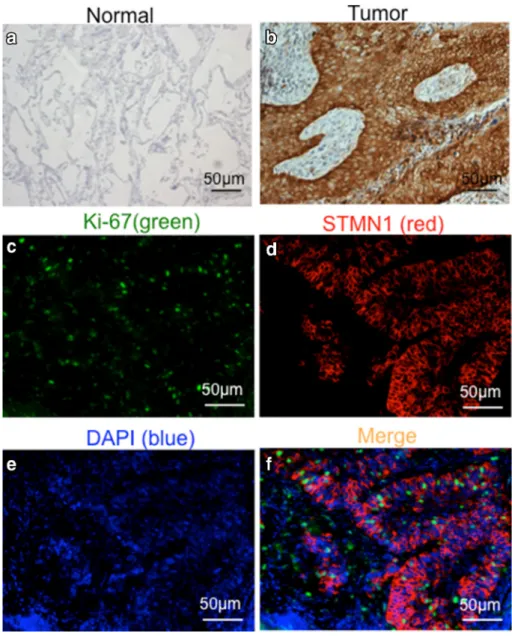
FIG. 1 Immunohistochemical analysis of STMN1 in surgical specimens. Representative immunohistochemical staining of STMN1 inanormal tissue andbcancer tissue of the same case (magnification9200).c–f Immunofluorescence staining of Ki67 (green), STMN1 (red), and nucleus (DAPI) and merged image of STMN1 and Ki67 with 50-lm scale bar
Invasion and Apoptosis Assays
The EBC1 cells were treated with STMN1 siRNA and then incubated for 48 h. Matrigel invasion assays were performed using 24-well BioCoat Matrigel invasion chambers (Corning International, NY, USA) according to the manufacturer’s instructions. The upper chambers were seeded with EBC1 cells (39104) in 300ll of fetal bovine serum (FBS)-free medium and incubated for 12 h. After the non-transfected cells were removed, the isolated transfected cells were stained with a Diff-Quik staining kit (Sysmex, Hyogo, Japan).
The EBC1 cells transfected with STMN1 siRNA were resuspended, after which the cell suspension containing 59104cells per 100ll was stained with Annexin V-PI as per the manufacturer’s protocol (Tali Apoptosis Kit [A10788]–Annexin V Alexa Flour 488, Waltham, MA, USA, and propidium iodide) and analyzed by the Tali Image-Based Cytometer (Invitrogen). The apoptotic cells showed a green fluorescence.
RNA Extraction and Quantitative Real-Time PCR
Total RNA was extracted from cells with the miRNA- easy Kit (Qiagen, Hilden, Germany). The measurements and protocol were the same as described in a previous paper.11
Statistical Analysis
Statistically significant differences were analyzed with paired t test analysis, the Wilcoxon test for continuous variables, the chi-square test, and repeated-measures anal- ysis of variance (ANOVA). Survival curves were generated according to the Kaplan–Meier method, and statistical significances were determined using the log-rank test. Uni- and multivariate survival analyses were performed using the Cox’s proportional hazards model. Results were con- sidered statistically significant when the relevantP value was lower than 0.05. All statistical analyses were per- formed using JMP 9 software (SAS Institute, Cary, NC, USA).
RESULTS
High Expression Level of STMN1 in LSCC
Analysis of STMN1 expression was performed by immunohistochemistry on a discovery cohort and a vali- dation cohort in combination totaling 186 LSCC samples.
Expression of STMN1 was higher in LSCC tumor cells than in normal cells in the same sample (Fig.1a and b). A
high level of STMN1 was expressed by 114 samples, and a low level of STMN1 was expressed by 72 samples. The result was validated via RNA sequence analysis using the TCGA online data set (Fig. S2).
Clinicopathologic Factors of LSCC and Expression of STMN1 in LSCC
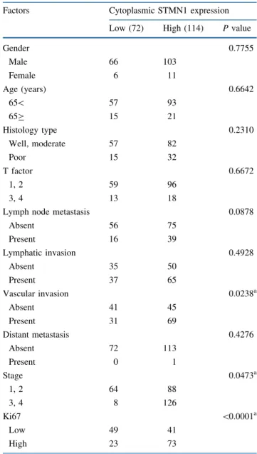
Expression of STMN1 was significantly correlated with vascular invasion (P=0.0238) and stage (P=0.0473), and a correlation trend was seen with lymph node metas- tasis (P =0.0878) (Table1). Additionally, we found that STMN1 expression was significantly associated with Ki67 expression (P\0.0001) (Table1) and could be frequently collocated (Fig.1c–f).
TABLE 1 STMN1 expression and clinicopathologic factors
Factors Cytoplasmic STMN1 expression
Low (72) High (114) Pvalue
Gender 0.7755
Male 66 103
Female 6 11
Age (years) 0.6642
65\ 57 93
65C 15 21
Histology type 0.2310
Well, moderate 57 82
Poor 15 32
T factor 0.6672
1, 2 59 96
3, 4 13 18
Lymph node metastasis 0.0878
Absent 56 75
Present 16 39
Lymphatic invasion 0.4928
Absent 35 50
Present 37 65
Vascular invasion 0.0238a
Absent 41 45
Present 31 69
Distant metastasis 0.4276
Absent 72 113
Present 0 1
Stage 0.0473a
1, 2 64 88
3, 4 8 126
Ki67 \0.0001a
Low 49 41
High 23 73
aP\0.05
STMN1 Expression Was a Prognostic Marker for 186 LSCC Patients and for 57 LSCC Patients Without Lymph Node Metastasis
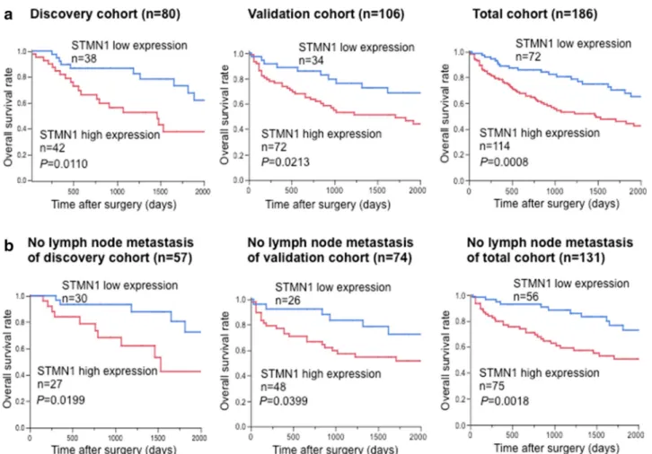
Kaplan-Meier analysis showed that high expression of STMN1 was strongly associated with a poor overall sur- vival (OS) for each cohort. In the discovery cohort, 42 cases were classified as high expression of STMN1 and the remaining 38 cases with low expression of STMN1. The OS of the STMN1 high-expression group was significantly shorter than that of the STMN1 low-expression group (P=0.0110) (Fig.2a, left panel), with a similar result obtained from the validation cohort (P=0.0213) (Fig.2a, center panel) and the total cohort (P=0.0008) (Fig.2a, right panel). Of the 57 patients without lymph node metastases in the discovery cohort, 30 expressed a high level of STMN1 but had a shorter OS than the 27 patients who expressed a low level of STMN1 (P=0.0199) (Fig.2b), with the same expression observed in the
validation cohort (P=0.0399) and the total cohort (P=0.0018) (Fig.2b; Fig. S3).
STMN1 Expression Independently Predicted Poor Prognosis
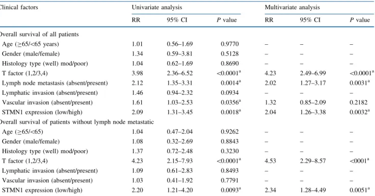
As shown in Table2, both the univariate analysis and the multivariate regression analysis showed that high expression of STMN1 was an independent prognostic factor for both the 186 LSCC patients (P=0.0032) and the 131 LSCC patients without lymph node metastasis (P=0.0051).
STMN1 Regulated Proliferation Potency and Increased Paclitaxel Sensitivity
We evaluated the protein and mRNA levels of STMN1 expression in EBC-1, H520, Lk-2, and RERF-LC-AI cell lines using Western blotting and reverse transcription-polymerase
FIG. 2 Survival analysis of 186 lung squamous cell carcinoma (LSCC) patients according to the expression level of cytoplasmic STMN1.aThe overall survival rates of the STMN1 high-expression group were significantly lower than those of the low-expression group from each cohort: discovery cohort (P=0.0110), validation cohort
(P=0.0213), and total cohort (P=0.0008). bThe STMN1 high- expression patients without lymph node metastasis had a poorer prognosis than the STMN1 low-expression group from each cohort:
discovery cohort (P=0.0199), validation cohort (P=0.0399), and total cohort (P=0.0018)
chain reaction (RT-PCR) and found STMN1 strongly expressed in the EBC-1 and H520 cell lines (Fig.3a). The STMN1-suppression experiment was performed by transfect- ingSTMN1siRNA1 orSTMN1siRNA2 in the EBC-1 and H520 cell lines and confirmed by Western blotting and RT- PCR (Fig.3b and c). The ability of the cell lines to proliferate was significantly lower in the siRNA-treated cell lines than in the control cell lines (Fig.3d). TheSTMN1siRNA-transfected cells also were treated with paclitaxel (0, 0.1, 1, 10, 100, and 1000 nmol) for 48 h. The findings showed that the viability of theSTMN1siRNA groups was significantly lower than that of the control groups (Fig.3e).
Additionally, co-expression analysis of the available TCGA data on lung squamous cell carcinoma was per- formed via the cBioPortal website. We identified 75 genes (Spearman score, [0.44) that correlated with STMN1 mRNA expression. To clarify the functional role of these genes, we performed Ingenuity Pathway Analysis (IPA) and the Database for Annotation, Visualization and Inte- grated Discovery (DAVID) analyses, which showed that 4 genes were related to cell growth, 13 genes were related to cell proliferation, 10 genes reportedly had biomarker applications, and 8 genes (includingTUBA1B andTYMS) contributed to drug efficacy (Table S2).
DISCUSSION
This study demonstrated that high expression of STMN1 was associated with poorer prognosis for LSCC patients, particularly patients without lymph node metastasis.
Expression of STMN1 was significantly associated with all clinicopathologic factors such as vascular invasion, advanced stage, and Ki67 expression in surgically resected LSCC. Consistent with clinical outcome, in vitro silencing of STMN1using siRNA inhibited the invasion and prolif- eration of cell lines compared with controls.
Typically, LSCC grows rapidly and enters the lymphatic vessels to reach the lymph nodes, from where it spreads into surrounding and distant tissues. However, in our study, the patients without lymph node metastasis but high STMN1 expression had a worse prognosis in all the inde- pendent cohorts. Similar results have been reported for esophageal squamous cell carcinoma.12
Our study demonstrated for the first time that STMN1 high expression is an independent risk factor for LSCC patients without lymph node metastasis. For the patients whose LSCC had spread to the lymph nodes (n=22), no statistically significant prognostic effect was observed, but this may have been due to the small sample size.
TABLE 2 Uni- and multivariate analyses of clinicopathologic factors affecting overall survival rates
Clinical factors Univariate analysis Multivariate analysis
RR 95% CI Pvalue RR 95% CI Pvalue
Overall survival of all patients
Age (C65/\65 years) 1.01 0.56–1.69 0.9770 – – –
Gender (male/female) 1.34 0.59–3.81 0.5128 – – –
Histology type (well) mod/poor) 1.04 0.62–1.69 0.8690 – – –
T factor (1,2/3,4) 3.98 2.36–6.52 \0.0001a 4.23 2.49–6.99 \0.0001a
Lymph node metastasis (absent/present) 2.12 1.35–3.31 0.0014a 2.02 1.27–3.17 0.0031a
Lymphatic invasion (absent/present) 1.46 0.94–2.32 0.0934 – – –
Vascular invasion (absent/present) 1.61 1.03–2.53 0.0356a 1.32 0.85–2.09 0.2182
STMN1 expression (low/high) 2.09 1.31–3.45 0.0018a 2.04 1.26–3.38 0.0032a
Overall survival of patients without lymph node metastatic
Age (C65/\65) 1.04 0.47–2.04 0.9262 – – –
Gender (male/female) 1.08 0.32–2.69 0.8843 – – –
Histology type (well) mod/poor) 1.37 0.72–2.48 0.3230 – – –
T factor (1,2/3,4) 4.23 2.15–7.93 \0.0001a 4.53 2.29–8.57 \0001a
Lymphatic invasion (absent/present) 1.09 0.61–2.83 0.8493 – – –
Vascular invasion (absent/present) 1.03 0.41–1.92 0.7791 – – –
STMN1 expression (low/high) 2.20 1.21–4.20 0.0093a 2.34 1.28–4.49 0.0051a
RRrelative risk,CIconfidence interval
aP\0.05
The current study showed that high STMN1 expression correlated with vascular invasion in LSCC, as reported for diffuse type gastric cancer13 and hepatocellular carci- noma.14 Consistently, it also was demonstrated that STMN1 together with its microtubule-destabilizing activity contributes to epithelial-mesenchymal transition (EMT), which stimulates the malignant potential in cancer cells.15 As published for bladder cancer,16 breast cancer,17 and cervical cancer,18we also found that a crucial relationship exists between protein levels of STMN1 and the prolifer- ation marker Ki67 in LSCC patients, and that the invasion ability in vitro possibly is due to its activity on microtubules.
In the immunohistochemistry analysis of two indepen- dent patient cohorts, high STMN1 expression was correlated with poor OS. Moreover, the multivariate
analysis of OS showed high STMN1 expression to be an independent prognostic factor. Previous studies also have shown that high STMN1 expression is related to prognosis in various cancers.15 Therefore, STMN1 is expected to serve as a prognostic-predictive marker for LSCC.
Recent studies have suggested that STMN1 high expression is correlated with the resistance to microtubule- acting drugs in cultured cells19,20 and in patients with endometrial cancer21or breast cancer.22High expression of STMN1 has been shown to decrease markedly the binding of and sensitivity to paclitaxel but not to affect sensitivity to chemotherapeutic drugs that do not target micro- tubules.19 Paclitaxel binding with beta-tubulin subunits of microtubules stabilize the microtubule polymer and protect it from disassembly by comparing the content of poly- merized microtubules as a function of STMN1 FIG. 3 STMN1 function analysis of human lung squamous cell
carcinoma (LSCC) cell lines.aEBC-1, H520, LK-2, and RERF-LC- AI cells were prepared and subjected to Western blotting with STMN1 antibody, withb-actin used as a loading control. Intensity was measured using Image Quant LAS 4000. Expressions ofSTMN1 in EBC-1, H520, LK-2, and RERF-LC-AI cells were evaluated by quantitative reverse transcription-polymerase chain reaction (RT- PCR) and normalized byGAPDHexpression.b, cExpression level of
STMN1 in EBC-1 followed by detection of H520 treated withSTMN1 siRNA1 or siRNA2. Next, suppression of STMN1 expression was confirmed by a Western blotting experiment and an RT-PCR experiment.dProliferation of EBC-1 and H520 treated with siRNA was inhibited after treatment with siRNA.ePaclitaxel sensitivity of EBC-1 and H520 cells treated withSTMN1siRNA1 and siRNA2 was induced compared with the control and parent cells.RERFRERF-LC- AI,Pparent,NCnegative control
expression.23 We discovered that suppression of STMN1 could increase the paclitaxel sensitivity. Because only a few patients were treated with taxanes, we could not def- initely relate the STMN1 expression to the taxane agent sensitivity in the clinical samples. However, our database analysis clarified that theSTMN1 mRNA expression was related to cell proliferation-correlated genes and the paclitaxel-efficacy genesTYMSandTUBA1B(Table S3).
jdmA few recent studies have consistently concluded that high expression of STMN1 may be related to pacli- taxel resistance in non-small lung cancer patients.24,25High STMN1 expression might be a predictor for paclitaxel resistance in LSCC patients, and a combination of pacli- taxel and anti-STMN1 drugs may provide a prospective therapy against various cancers in the future.
CONCLUSION
This study showed that STMN1 expression is associated with a shorter OS in LSCC. In addition, high STMN1 expression positively correlated with Ki67 accumulation and contributed to proliferation, progression, and paclitaxel resistance in LSCC. Therefore, we conclude that evaluation of STMN1 expression in LSCC may be a useful prognostic marker and a promising candidate for targeted therapy.
ACKNOWLEDGMENTS Masahiko Nishiyama received a research grant from Yakult Honsha Co. Ltd. Grants-in-Aid for Sci- entific Research from the Japan Society for the Promotion of Science (JSPS) (Grant Nos. 26461969, 15K10129 and 15K10085). The work was supported in part by Uehara Zaidan, Gunma University Initiative for Advanced Research (GIAR). We thank Ms. Yukie Saito, Ms.
Tomoko Yano, Ms. Yuka Matsui, and Ms. Ayaka Ishida, and Ms.
Ayaka Ishikubo for their excellent assistance.
REFERENCES
1. Scagliotti G, Govindan R. Targeting angiogenesis with multi- targeted tyrosine kinase inhibitors in the treatment of non-small cell lung cancer.Oncologist.2010;15:436–46.
2. Takano T, Fukui T, Ohe Y, et al. EGFR mutations predict sur- vival benefit from gefitinib in patients with advanced lung adenocarcinoma: a historical comparison of patients treated before and after gefitinib approval in Japan. J Clin Oncol.
2008;26:5589–95.
3. Mengoli MC, Barbieri F, Bertolini F, Tiseo M, Rossi G. K-RAS mutations indicating primary resistance to crizotinib in ALK- rearranged adenocarcinomas of the lung: report of two cases and review of the literature.Lung Cancer.2016;93:55–8.
4. Howell B, Larsson N, Gullberg M, Cassimeris L. Dissociation of the tubulin-sequestering and microtubule catastrophe-promoting activ- ities of oncoprotein 18/stathmin.Mol Biol Cell.1999;10:105–18.
5. Gavet O, Ozon S, Manceau V, Lawler S, Curmi P, Sobel A. The stathmin phosphoprotein family: intracellular localization and effects on the microtubule network.J Cell Sci.1998;111(Pt 22):3333–46.
6. Baldassarre G, Belletti B, Nicoloso MS, et al. p27(Kip1)-stathmin interaction influences sarcoma cell migration and invasion.
Cancer Cell.2005;7:51–63.
7. Schimmack S, Taylor A, Lawrence B, et al. Stathmin in pan- creatic neuroendocrine neoplasms: a marker of proliferation and PI3 K signaling.Tumour Biol.2015;36:399–408.
8. Sonego M, Schiappacassi M, Lovisa S, et al. Stathmin regulates mutant p53 stability and transcriptional activity in ovarian cancer.
EMBO Mol Med.2013;5:707–22.
9. Altan B, Yokobori T, Mochiki E, et al. Nuclear karyopherin- alpha2 expression in primary lesions and metastatic lymph nodes was associated with poor prognosis and progression in gastric cancer.Carcinogenesis.2013;34:2314–21.
10. Shimizu K, Ohtaki Y, Altan B, et al. Prognostic impact of stathmin 1 expression in patients with lung adenocarcinoma. J Thorac Cardiovasc Surg.2017;S0022-5223(17)30646-3 11. Bai T, Yokobori T, Altan B, et al. High STMN1 level is asso-
ciated with chemo-resistance and poor prognosis in gastric cancer patients.Br J Cancer.2017;116:1177–85.
12. Akhtar J, Wang Z, Jiang WP, Bi MM, Zhang ZP. Stathmin overexpression identifies high risk for lymphatic metastatic recurrence in pN0 esophageal squamous cell carcinoma patients.
J Gastroenterol Hepatol.2014;29:944–50.
13. Jeon TY, Han ME, Lee YW, et al. Overexpression of stathmin1 in the diffuse type of gastric cancer and its roles in proliferation and migration of gastric cancer cells.Br J Cancer.2010;102:710–18.
14. Hsieh SY, Huang SF, Yu MC, et al. Stathmin1 overexpression associated with polyploidy, tumor-cell invasion, early recurrence, and poor prognosis in human hepatoma. Mol Carcinogen.
2010;49:476–87.
15. Lu Y, Liu C, Xu YF, et al. Stathmin destabilizing microtubule dynamics promotes malignant potential in cancer cells by epithelial-mesenchymal transition. HBPD INT Hepatobil Pan- creat Dis Int.2014;13:386–94.
16. Hemdan T, Linden M, Lind SB, et al. The prognostic value and therapeutic target role of stathmin-1 in urinary bladder cancer.Br J Cancer.2014;111:1180–7.
17. Arnedos M, Drury S, Afentakis M, et al. Biomarker changes associated with the development of resistance to aromatase inhibitors (AIs) in estrogen receptor-positive breast cancer.Ann Oncol.2014;25:605–10.
18. Howitt BE, Nucci MR, Drapkin R, Crum CP, Hirsch MS.
Stathmin-1 expression as a complement to p16 helps identify high-grade cervical intraepithelial neoplasia with increased specificity.Am J Surg Pathol.2013;37:89–97.
19. Alli E, Bash-Babula J, Yang JM, Hait WN. Effect of stathmin on the sensitivity to antimicrotubule drugs in human breast cancer.
Cancer Res.2002;62:6864–9.
20. Watanabe A, Suzuki H, Yokobori T, et al. Stathmin1 regulates p27 expression, proliferation and drug resistance, resulting in poor clinical prognosis in cholangiocarcinoma. Cancer Sci.
2014;105:690–6.
21. Werner HM, Trovik J, Halle MK, et al. Stathmin protein level, a potential predictive marker for taxane treatment response in endometrial cancer.PloS One.2014;9:e90141.
22. Kuang XY, Chen L, Zhang ZJ, et al. Stathmin and phospho- stathmin protein signature is associated with survival outcomes of breast cancer patients.Oncotarget.2015;6:22227–38.
23. Fojo AT.The Role of Microtubules in Cell Biology, Neurobiol- ogy, and Oncology.Humana Press, Totowa, 2008.
24. Powrozek T, Mlak R, Krawczyk P, et al. Retrospective analysis of second-line chemotherapy outcomes with paclitaxel or doc- etaxel in correlation with STMN1 polymorphism in advanced non-small cell lung cancer patients. Clin Transl Oncol.
2016;18:33–9.
25. Sun RF, Liu ZG, Wang LM, et al. Overexpression of stathmin is resistant to paclitaxel treatment in patients with non-small cell lung cancer.Tumor Biol.2015;36:7195–204.