an Important Role in Cue‑Dependent Fear Memory Consolidation
著者 征矢 晋吾
著者別表示 Soya Shingo journal or
publication title
博士論文本文Full 学位授与番号 13301甲第4445号
学位名 博士(医学)
学位授与年月日 2016‑06‑30
URL http://hdl.handle.net/2297/46455
doi: 10.1523/JNEUROSCI.1130-13.2013
Creative Commons : 表示 ‑ 非営利 ‑ 改変禁止 http://creativecommons.org/licenses/by‑nc‑nd/3.0/deed.ja
Behavioral/Cognitive
Orexin Receptor-1 in the Locus Coeruleus Plays an Important Role in Cue-Dependent Fear Memory Consolidation
Shingo Soya,
1Hirotaka Shoji,
2Emi Hasegawa,
1Mari Hondo,
1Tsuyoshi Miyakawa,
2Masashi Yanagisawa,
3Michihiro Mieda,
1and Takeshi Sakurai
11
Department of Molecular Neuroscience and Integrative Physiology, Faculty of Medicine, Kanazawa University, Kanazawa, Ishikawa 920-8640, Japan,
2
Division of Systems Medical Science, Institute for Comprehensive Medical Science, Fujita Health University, Toyoake, Aichi 407-1192, Japan, and
3
International Institute for Integrative Sleep Medicine, University of Tsukuba, Tsukuba 305-8575, Japan
The noradrenergic (NA) projections arising from the locus ceruleus (LC) to the amygdala and bed nucleus of the stria terminalis have been implicated in the formation of emotional memory. Since NA neurons in the LC (LC-NA neurons) abundantly express orexin receptor-1 (OX1R) and receive prominent innervation by orexin-producing neurons, we hypothesized that an OX1R-mediated pathway is involved in the physiological fear learning process via regulation of LC-NA neurons. To evaluate this hypothesis, we examined the phenotype of Ox1r
⫺/⫺mice in the classic cued and contextual fear-conditioning test. We found that Ox1r
⫺/⫺mice showed impaired freezing responses in both cued and contextual fear-conditioning paradigms. In contrast, Ox2r
⫺/⫺mice showed normal freezing behavior in the cued fear-conditioning test, while they exhibited shorter freezing time in the contextual fear-conditioning test. Double immunolabeling of Fos and tyrosine hydroxylase showed that double-positive LC-NA neurons after test sessions of both cued and contextual stimuli were significantly fewer in Ox1r
⫺/⫺mice. AAV-mediated expression of OX1R in LC-NA neurons in Ox1r
⫺/⫺mice restored the freezing behavior to the auditory cue to a comparable level to that in wild-type mice in the test session. Decreased freezing time during the contextual fear test was not affected by restoring OX1R expression in LC-NA neurons. These observations support the hypothesis that the orexin system modulates the formation and expression of fear memory via OX1R in multiple pathways. Especially, OX1R in LC-NA neurons plays an important role in cue-dependent fear memory formation and/or retrieval.
Introduction
Orexins (Sakurai et al., 1998) are implicated in regulation of sleep/wakefulness, energy homeostasis, and the reward system (Sakurai, 2007; Sakurai and Mieda, 2011). Orexin-immuno- reactive fibers are observed in almost the entire neuraxis (Peyron et al., 1998; Date et al., 1999; Nambu et al., 1999), with especially dense fibers in monoaminergic nuclei in the brainstem (Date et al., 1999; Nambu et al., 1999; Peyron et al., 2000). The distribu- tion of the two orexin receptors is consistent with these projec- tion sites (Marcus et al., 2001).
It has been thought that orexin-2 receptor (OX2R) plays a pivotal role in sleep–wake regulation (Hondo et al., 2010; Sakurai and Mieda, 2011). Metabolic and feeding regulation by orexins is also suggested to be regulated through an OX2R-mediated path-
way (Funato et al., 2009). However, very limited information is available regarding the physiological role of the orexin-1 receptor (OX1R).
OX1R is abundantly expressed in the locus ceruleus (LC), which plays important roles in many functions via its widespread projections (Bailey et al., 2000; McGaugh and Roozendaal, 2002).
The LC contains tyrosine hydroxylase (TH)-positive noradren- ergic (NA) neurons. Because orexin neurons send rich projec- tions to the LC, and OX1R is expressed in Vmat2-positive neurons in the LC (Mieda et al., 2011), the physiological function of OX1R might be closely related to the function of NA neurons in the LC (LC-NA neurons). Consistently, orexin potently ex- cited LC-NA neurons (van den Pol et al., 2002).
There is much evidence suggesting the importance of the NA system in emotional memory formation. Conditioned fear stress caused a robust increase in the firing rate of NA neurons (Chen and Sara, 2007) and induced Fos expression in the LC (Ishida et al., 2002). LC-NA neurons project to the lateral amygdala (LA), an important structure for emotional memory (Bush et al., 2010).
These observations suggest that NA input from the LC to the LA is one of the key factors in fear memory formation.
Orexin neurons receive input from the limbic system (Winsky-Sommerer et al., 2004; Sakurai et al., 2005; Yoshida et al., 2006). These observations raise the possibility that orexin neurons may be activated by emotional information transmitted
Received March 13, 2013; revised July 18, 2013; accepted Aug. 6, 2013.
Author contributions: T.S. designed research; S.S., H.S., M.H., and T.S. performed research; E.H., T.M., M.Y., M.M., and T.S. contributed unpublished reagents/analytic tools; S.S. and T.S. analyzed data; S.S. and T.S. wrote the paper.
This work was funded primarily by the government of Japan through its “Funding Program for Next Generation of World-leading Researchers” (grant to T.S.). PRSx8 was provided by Dr. Kwang-soo Kim. We thank Dr. Wendy Gray for reading this manuscript.
Correspondence should be addressed to Dr. Takeshi Sakurai, Department of Molecular Neuroscience and Inte- grative Physiology, Faculty of Medicine, Kanazawa University, Kanazawa, Ishikawa 920-8640, Japan. E-mail:
[email protected] or [email protected].
DOI:10.1523/JNEUROSCI.1130-13.2013
Copyright © 2013 the authors 0270-6474/13/3314549-09$15.00/0
The Journal of Neuroscience, September 4, 2013 • 33(36):14549 –14557 • 14549
from the amygdala, and in turn send excitatory output to the LC, and this connection plays an important role in modulating emo- tional memory.
We found here that Ox1r
⫺/⫺mice showed decreased freezing behavior to both cued and contextual stimuli during both the conditioning and test periods, suggesting that OX1R is involved in evoking fear-related behavior. In contrast, Ox2r
⫺/⫺mice re- sponded normally with freezing behavior in the cued fear- conditioning test. We also found that Fos expression in LC-NA neurons after exposure to the fearful context was significantly lower in Ox1r
⫺/⫺mice than in wild-type mice. Importantly, we found that freezing behavior recovered to a level comparable to that in wild-type mice in the test period of cued fear conditioning when OX1R expression in LC-NA neurons was restored, suggest- ing that OX1R-mediated regulation of LC-NA neurons plays a significant role in the acquisition and consolidation of cue- dependent fear memory.
Materials and Methods
Animals. All experimental procedures were approved by the Animal Ex- periment and Use Committee of Kanazawa University (AP-111947), and were thus in accordance with National Institutes of Health guidelines.
We used Ox1r
⫺/⫺mice in which the Ox1r-coding region in exon 1 of the Ox1r gene is disrupted by inserting a tau-LacZ cassette (Hondo et al., 2010). Ox1r
⫺/⫺mice used in the experiments were obtained from the mating of heterozygous Ox1r
⫹/⫺mice. Ox2r
⫺/⫺mice were previously reported (Willie et al., 2003). Exon 1 of the Ox2r gene was replaced in-frame with the nlacZ cassette. Ox2r
⫺/⫺mice used in the experiments were obtained from the mating of heterozygous Ox2r
⫹/⫺mice. Geno- typing these mice was done by PCRs with tail DNA according to the method described previously (Willie et al., 2003). These mice were back- crossed to wild-type C57BL/6J mice for ⬎ 10 generations. Their litter- mates of each genotype were used as wild-type control. Mice were maintained under a strict 12 h light/dark cycle in a temperature- and humidity-controlled room and fed ad libitum.
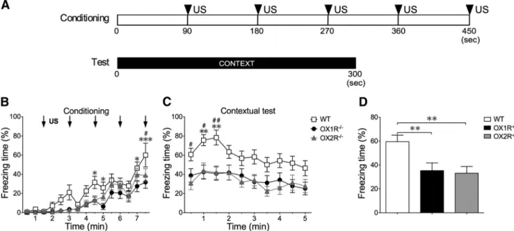
Behavioral experiments. All experiments were performed during the light phase (13:00 –16:00) using male 12- to 14-week-old mice. Before the experiments, these mice were isolated for 2 weeks. The experimenters were blinded to the genotypes until all data had been gathered and ana- lyzed. Behavioral experiments in this study were basically performed according to modifications of previously described protocols (Miyakawa et al., 2003). The behavior of mice was recorded using a charge coupled device video camera and analyzed with a video-tracking system Com- pACT VAS version 3.0x (Muromachi Kikai). For the cued fear- conditioning test, mice were placed in a conditioning chamber (15 ⫻ 12 ⫻ 13 cm, acrylic walls with a gridded floor and bright lighting) for 90 s before giving a conditioned stimulus (CS), a 2900 Hz, 80 dB tone that lasted 30 s immediately, followed by the presentation of an uncondi- tioned stimulus (US), a mild foot shock of 0.3 mA for 2 s, on the training day. Five consecutive trials of training were performed (see Fig. 1A). After an additional stay for 90 s in the chamber, the mouse was returned to its home cage. Mice were tested 24 h after the training. Cued fear test was conducted in a chamber with a different context (13 ⫻ 20 ⫻ 11 cm, a plastic cage surrounded with a white screen and bright lighting). Freezing behavior was scored during the testing session. Freezing behavior is de- fined as the complete absence of any movement except for respiration and heartbeat. Freezing behavior is measured by an automated scoring system (Muromachi Kikai), which recognizes complete absence of movement lasting for longer than 2 s as freezing. Testing was performed with the same procedure as in the conditioning period, but without application of US (Fig. 1A).
For the contextual fear-conditioning test, mice were placed in the conditioning chamber for 90 s before giving a US, a mild foot shock of 0.3 mA for 2 s, on the training day. Five sequential foot shocks at 90 s intervals were applied. The mice were tested 24 h after the conditioning.
They were put in the same conditioning chamber for 5 min without presentation of CS (see Fig. 2A). We used different mice for the cued test
and contextual test. Data are presented as mean ⫾ SEM. The apparatus was cleaned with 30% ethanol solution, and then with water and dried after each trial to eliminate possible odor cues left by previous subjects.
To measure the ultrasonic vocalizations of mice, we used an ultrasonic vocalization (USV) analyzer (Muromachi Kikai), following the manu- facturer’s instructions.
Construction of recombinant adeno-associated virus vectors. The com- mon structure of the adeno-associated virus (AAV)-2 ITR-containing plasmids used in this study is ITR-promoter-cDNA encoding a protein- WPRE-poly (A)
⫹signal-ITR. cDNA encoding rat Ox1r fused with EYFP at the C terminal was inserted downstream of the PRSx8 promoter (AAV-PRSx8-OX1R-EYFP; see Fig. 4A) (Hwang et al., 2001). We used AAV with PRSx8 promoter carrying ChR2 (NA-ChR2) for the control group (AAV-PRSx8-ChR2-EYFP; Fig. 4A), because ChR2 has a similar seven transmembrane structure to that of OX1R. AAV-2 vectors with a mutant form of the cap gene (Zhong et al., 2008) were produced using a triple-transfection, helper-free method, and purified as described previ- ously (Sasaki et al., 2011). In brief, 293A cells (Invitrogen), cultured in 10 100 ⫻ 20 mm cell culture dishes per viral vector, were transfected with pHelper (Stratagene), pACG-2-Y730F (containing a mutant form of the cap gene of AAV2, provided by Dr. Arun Srivastava of the University of Florida), and pAAV-PRSx8-OX1R-EYFP or pAAV-PRSx8-ChR2-EYFP, using a standard calcium phosphate method. Three days later, the cells were collected, pelleted, and resuspended in freezing buffer (10 ml of 0.15
M